1132
Clinical utility of breast DWI in the assessments of breast lesions using different b values1Diagnostic Imaging and Nuclear Medicine, Kyoto University Graduate School of Medicine, Kyoto, Japan, 2Clinical Innovative Medicine, Institute for Advancement of Clinical and Translational Science, Kyoto University Hospital, Kyoto, Japan, 3Breast Surgery, Kyoto University Graduate School of Medicine, Kyoto, Japan
Synopsis
We have 1) assessed the clinical utility of DWI in the diagnosis of breast lesions using DW-based BI-RADS lexicons and lesion conspicuity and 2) investigated the diagnostic performance of breast lesions based on DW-based BI-RADS category by 3 radiologists using breast DW images with b=800 or b=1500 s/mm2. Agreement in DW-based BI-RADS categories tended to be higher in b1500 compared to b800 DW images. Diagnostic performance based on b800 and b1500 DW images was not uniform among 3 readers, which highlight the necessity and importance of the standardization on reading protocols and experience from DWI readers.
Introduction
The Breast Imaging-Reporting and Data System (BI-RADS) has been established to standardize breast imaging reporting and encourage consistency in breast imaging interpretations between radiologists when creating their reports. Although not part of BI-RADS, breast diffusion-weighted imaging (DWI) has been increasingly used for the diagnosis and monitoring of breast lesions. Optimal b values have been proposed, however, the reported b values vary widely within the literature1,2, hence, further investigation including diagnostic performance using different b values is warranted. Thus, the purpose of our study is 1) to assess the clinical utility of DWI in the diagnosis of breast lesions using adjusted BI-RADS lexicons and lesion conspicuity and 2) to investigate the diagnostic performance of breast lesions based on adjusted BI-RADS category by 3 radiologists using breast DW images with b=800 or b=1500 s/mm2.Materials & Methods
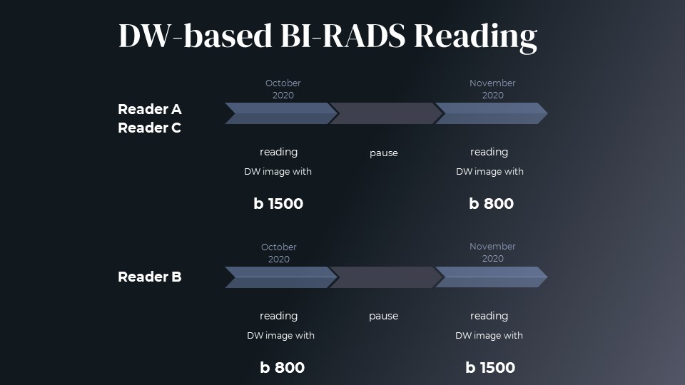
Patient enrollment & DWI setting: This prospective study was approved by the institutional review board and included 118 women who were suspected of breast tumors. After excluding patients receiving neoadjuvant treatment or cases of no abnormal findings, 45 patients with 45 breast lesions (24 malignant, 21 benign) were further analyzed. Breast MRI was performed using a 3-T system (MAGNETOM Prisma, Siemens) equipped with a dedicated 18-channel breast array coil. Diffusion-weighted images were acquired using 5 b values of 0, 200, 800, 1000, 1500 sec/mm2; repetition time/echo time, 6900/49ms; flip angle, 90°; FOV, 330×187mm; matrix, 162×92; slice thickness, 3.0 mm; acquisition time, 2.32 min.DW image interpretation: Two independent radiologists (readers A and B) first assessed DW (b=800) images, while one radiologist (reader C) assessed DW (b=1500) images blinded to DCE-MRI and the other b value images, according to an adjusted BI-RADS lexicon for lesion classification (DW-based BI-RADS reading)3. Readers A and B assessed DW (b=1500) images, while reader C assessed DW (b=800) images the following month (figure 1). Lesion conspicuity was also assessed using a semi-quantitative score, ranging from 4 (excellent) to 0 (not visible). Interobserver agreement of adjusted BI-RADS lexicons were calculated by using kappa statistics. Lesion conspicuity was compared using the Wilcoxon test between b800 and b1500 DW images. The diagnostic performance was evaluated using a ROC curve analysis.
Results
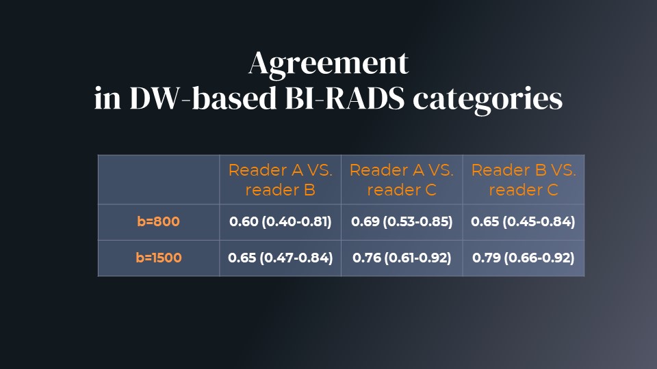
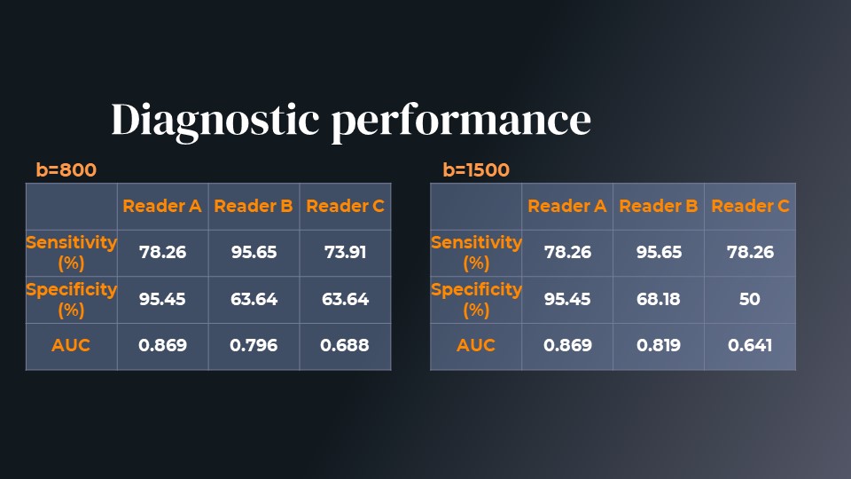
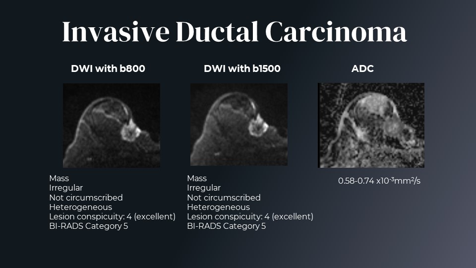
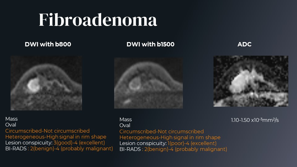
Agreement in adjusted BI-RADS categories tended to be higher in b1500 compared to b800 DW images (table 1). The representative cases are shown in figures 2 and 3. All three readers had the same imaging findings for the malignant tumor (fig.2: invasive ductal carcinoma), while the findings were different among 3 readers for the benign tumor (fig.3: fibroadenoma). Agreements in the evaluation of adjusted BI-RADS lexicons among 3 readers were variable both for mass (0.43-0.67), and non-mass lesions (0.00-0.75). Lesion conspicuity tended to be lower with b800 compared to b1500 (2.7vs.2.8, 2.2vs.2.2, 2.9vs.3.2 for reader A, B, and C, respectively), however, it didn't reach a significant difference. Diagnostic performance was not uniform as shown in table 2. AUC for b800 and b1500 was equivalent for reader A, superior with b800 for reader C and superior with b1500 for reader B. Reading time was variable between b800 and B1500 DW images (153vs.101, 127vs.173, 119vs.119 sec for reader A, B, and C, respectively)Discussion
Agreement in adjusted BI-RADS categories tended to be higher with b1500 than b800 DW images. While b800 was recommended by EUSOBI (Consensus article)1, b1000-1500 has been advocated by Korean radiologists2. b800 has the advantage to be versatile and less demanding in terms of MRI scanner performance and SNR, but higher diagnostic performance is expected (due to higher weighting toward diffusion hindrance by tissue features) when higher b values can be reached while maintaining good image quality and CNR. Higher lesion conspicuity might be observed with b1500 compared to b800, which suggests potentially higher CNR with DW images with b1500. Differences in diagnostic performance (AUC), as well as agreements in adjusted BI-RADS lexicons, were found between readers, especially for some non-mass lesions, suggesting the need for standardization in reporting breast DWI findings.Conclusion
Diagnostic performance based on b800 and b1500 DW images was not uniform among 3 readers. Besides b values, attention should be given to standardization on reading protocols and experience from DWI readers.Acknowledgements
This work was supported by the JSPS KAKENHI Grant number18K15588. We would like to thank Dr. Denis Le Bihan for his valuable advice.
References
1. Baltzer P, Mann RM, Iima M, et al. Diffusion-weighted imaging of the breast-a consensus and mission statement from the EUSOBI International Breast Diffusion-Weighted Imaging working group. Eur. Radiol. 2020;30(3):1436–1450.
2. Lee SH, Shin HJ, Moon WK. Diffusion-Weighted Magnetic Resonance Imaging of the Breast: Standardization of Image Acquisition and Interpretation. Korean J. Radiol. 2020. Available at: http://dx.doi.org/10.3348/kjr.2020.0093.
3. Kishimoto AO, Kataoka M, Iima M, et al. The comparison of high-resolution diffusion weighted imaging (DWI) with high-resolution contrast-enhanced MRI in the evaluation of breast cancers. Magnetic Resonance Imaging. 2020; 71:161-169.
Figures