1126
Evaluation of the efficacy of therapy for breast cancer using DWI and DCE-MRI based on acquired radial golden-angle compressed sensing1CTMRI, The Fourth Hospital of Hebei Medical University, Shijiazhuang,Hebei, China, 2MR Scientific Marketing,Siemens Healthcar, Beijing, China
Synopsis
In this study, we studied the feasibility of GRASP based DCE technique and readout-segmented DWI technique in early prediction and evaluation of breast cancer response to neoadjuvant chemotherapy. We found the combination of these two techniques demonstrated great potential in evaluating the response of breast cancer to neoadjuvant therapy.
Objective
To evaluate the feasibility of early prediction and evaluation of breast cancer response to neoadjuvant therapy (NAT) using the combination of readout-segmented-EPI based diffusion-weighted-imaging (RS-DWI) technique and radial golden-angle compressed sensing acquisition based Golden angle radial sparse (GRASP-DCE) technique.Methods
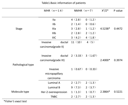
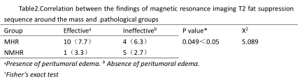
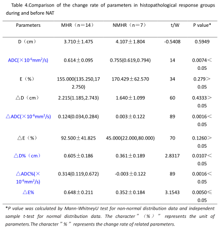
From July 2020 to December 2020, twenty-one patients (age range: 27-65; mean age 30.7±5.67) with breast mass-like lesions after NAT underwent breast MRI scanning with the GRASP-DCE and RS-DWI sequences at a 3T MR scanner (MAGNETOM Vida, Siemens Healthcare, Erlangen, Germany). All patients underwent three times of these two examinations, one before (EX1) and one after the six procedures of neoadjuvant therapy (EX2) and one after the eight procedures of neoadjuvant therapy (EX3). After operation, according to the new-modified pathological grading system of Miller & Payne, all patients were divided into two groups: major histological response (MHR) and non-major histological response (NMHR) group. Without knowing the pathological results, two doctors with 3 and 10 years' experience in breast imaging diagnosis evaluated and analyzed these quantitative parameters before and after NAT: value of the longest diameter of the tumor (Diameter,D), the mean ADC value of the tumor, the existence of peritumoral edema in T2WI images (effective or ineffective about neoadjuvant therapy, i.e. negative conversion rate), early enhancement rate (E value). The difference and the percentage change of each parameter were calculated to evaluate the pathological reaction. An independent sample t-test and Mann-Whitney U test were used to compare the differences between two groups. Multivariate logistic regression was used to identify independent factors for evaluating the pathological reaction. The diagnostic performance of the diagnosis equation was evaluated using the receiver operating characteristic (ROC) curve. The diagnostic efficacies of GRASP-DCE and RS-DWI were compared independently or combined.Results & Discussion
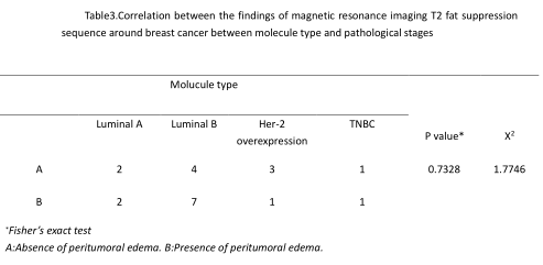
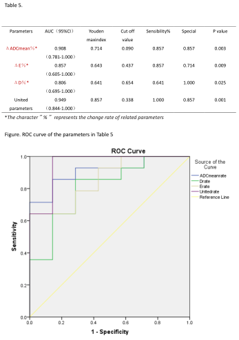
Among the 21 patients, 14 patients were classified as MHR group,7 patients after NAT were classified as NMHR group. No significant difference was found among D value, ADCmean value, E value and △E value between the two groups before NAT (P > 0.05). After NAT, D value decreased significantly in both MHR group and NMHR group (P < 0.05). After NAT, significantly differences between two groups were found in ΔADCmean% (P=0.0016<0.05,W=89), ΔD% (P=0.0107<0.05,t=-2.8317), ΔE (P=0.000<0.05,t=8.275), ΔE% (P=0.0052<0.05,t=-3.1543). The negative conversion rate of peritumoral edema in T2WI was higher than that in the control group (P=0.04<0.05,X2=5.089), but there was no significant difference in the negative conversion rate between the two groups (P=0.7328 > 0.05). The AUC values of ΔADCmean%, ΔE% and ΔD% for MHR group were 90.82%, 85.71% and 80.61% respectively, while the joint prediction parameter AUC was 0.949. Logistic multivariate regression model showed that ΔADCmean% could be used as an independent predictor of NAT efficacy.DCE has been used to non-invasively assess the microcirculatory perfusion and vascular permeability of the tumor[1],which has shown promising results in evaluating treatment response to chemoradiotherapy in breast cancer[2].In literatures’ reports, the diagnostic performance of DCE-MRI derived parameters may be influenced by the temporal and spatial resolution of images. Thus a high image quality is very important in improving the performance of DCE images[3]. A recently described acquisition technique combining a continuously acquired radial k-space trajectory with golden-angle sampling and sparse parallel reconstruction employing compressed sensing offers simultaneous high spatial and high temporal resolution as well as motion robustness to DCE[4]. DWI with sequence of multi-shot readout segmentation of long variable echo-trains (RESOLVE) can improve image quality of the small and complex anatomic structure with less susceptibility artifacts, blurring from T2 signal decay and distortion [5]. This raises the possibility of diagnosing with a higher accuracy.Through the analysis of quantitative parameters of GRASP-DCE sequence and ADC values based on RS-EPI sequence, the combination of these two techniques demonstrated more potential in evaluation of breast cancer response to neoadjuvant chemotherapy.
Conclusion
The quantitative parameters of DCE-MRI based on GRASP sequence, when combined with ADC values based on RS-EPI sequence, showed more potential in early prediction and evaluation of breast cancer response to neoadjuvant chemotherapy. The larger early enhancement rate of tumorous lesions and the higher ADC value indicates that the probability of MHR is higher.Acknowledgements
No acknowledgement found.References
[1] Verma S, Turkbey B, Muradyan N, Rajesh A, Cornud F, Haider MA, Choyke PL, Harisinghani M. Overview of dynamic contrast-enhanced MRI in prostate cancer diagnosis and management. AJR Am J Roentgenol. 2012 Jun;198(6):1277-88. doi: 10.2214/AJR.12.8510. PMID: 22623539; PMCID: PMC6309691.
[2] Drisis S, Metens T, Ignatiadis M, Stathopoulos K, Chao SL, Lemort M. Quantitative DCE-MRI for prediction of pathological complete response following neoadjuvant treatment for locally advanced breast cancer: the impact of breast cancer subtypes on the diagnostic accuracy. Eur Radiol. 2016 May;26(5):1474-84. doi: 10.1007/s00330-015-3948-0. Epub 2015 Aug 27. PMID: 26310583.
[3] Hao W, Zhao B, Wang G, Wang C, Liu H. Influence of scan duration on the estimation of pharmacokinetic parameters for breast lesions: a study based on CAIPIRINHA-Dixon-TWIST-VIBE technique. Eur Radiol. 2015 Apr;25(4):1162-71. doi: 10.1007/s00330-014-3451-z. Epub 2014 Oct 17. PMID: 25323603.
[4]. Rosenkrantz AB, Geppert C, Grimm R, et al. Dynamic contrast-enhanced MRI of the prostate with high spatiotemporal resolution using compressed sensing, parallel imaging, and continuous golden-angle radial sampling: preliminary experience. J Magn Reson Imaging. 2015; 41(5):1365–1373. [PubMed: 24833417]
[5] Schob S, Meyer HJ, Dieckow J, Pervinder B, Pazaitis N, Hohn AK, et al. Histogram Analysis of Diffusion Weighted Imaging at 3T is Useful for Prediction of Lymphatic Metastatic Spread, Proliferative Activity, and Cellularity in Thyroid Cancer. Int J Mol Sci. 2017; 18(4). https://doi.org/10.3390/ijms18040821 PMID: 28417929; PubMed Central PMCID: PMC5412405.
Figures