1122
Correction of Artifacts Induced by B0 Inhomogeneities with RPG on a Breast Diffusion Phantom1Radiology, University of California San Diego, La Jolla, CA, United States, 2University of California San Diego, La Jolla, CA, United States, 3National Institute of Standards and Technology, Boulder, CO, United States
Synopsis
Diffusion-weighted imaging (DWI) has shown potential to diagnose and monitor breast cancer, but is limited in part due to geometric distortion artifacts that affect echo-planar imaging. Generating distortion-free images is a key step towards its clinical implementation. Here, we investigated the effectiveness of artifact-reduction data collection strategies (i.e. parallel imaging DWI and reduced field-of-view DWI) in combination with reverse polarity gradient post-processing in correcting for geometric distortion artifacts.
Introduction
Multiparametric breast MRI is a valuable imaging technique for the screening, staging, and treatment response in breast cancer.1 Diffusion weighted imaging (DWI), typically acquired with echo-planar imaging (EPI), has been incorporated into clinical breast MRI protocols because of its potential to assess cancerous tissues without exogenous contrast agents.1 However, its widespread use has been limited due to relatively low spatial resolution and distortion artifacts that affect EPI.2 In breast imaging, distortion artifacts are pronounced due to off-isocenter scanning and anatomical complexity. Therefore, generating distortion-free images is a key step towards its clinical implementation.Acquisition strategies to reduce the geometric distortion artifacts that arise from susceptibility differences at air-tissue boundaries in EPI have been developed (i.e. parallel imaging3 and reduced field-of-view EPI4). Similarly, post-processing methods such as the reverse polarity gradient (RPG) method can retrospectively correct for B0 inhomogeneities by estimating the displacement field that minimizes the geometric difference between positive and negative phase-encoding (PE) trajectory volumes. RPG has demonstrated improved localization of breast, brain, and prostate cancers,5-7 and can theoretically be used with any DWI strategy. However, its performance in correcting distortion artifacts in combination with several other EPI data collection strategies has not been evaluated in the breast. Thus, the purpose of this work was to evaluate how well RPG performs in correcting distortion artifacts following different artifact-reduction data collection methods.
Methods
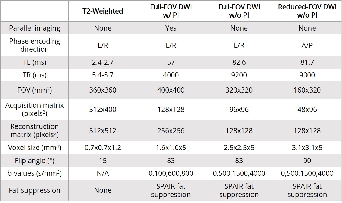
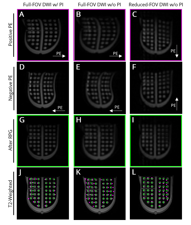
A breast phantom8 designed by UCSF and the National Institute of Standards and Technology was scanned using a breast array coil at two different 3T MRI scanners (MR750, GE Healthcare, Milwaukee, Wisconsin, USA). DWI pulse sequences included full field-of-view (FOV) DWI with parallel imaging (PI), and full-FOV EPI and reduced-FOV EPI without PI, with parameters as shown in Table 1. The breast phantom contained a polycarbonate plate with 42 circular perforations (10mm in diameter, 15mm center-to-center spacing, Fig. 1). Positive and negative PE trajectories were acquired for all DWI sequences and used to correct distortion artifacts using RPG.5 Regions of interest (ROIs) around each perforation on the grid were drawn using MATLAB (R2019b, MathWorks, Natick, Massachusetts, USA). Reference geometric centers of all perforations were defined on T2-weighted data, while the centers in DWI data were defined as the voxel of highest signal intensity within each ROI. Distortion magnitude in PE and frequency-encoding (FE) directions were calculated with respect to reference points, before and after RPG distortion correction.Statistics were performed using R programming (R Foundation for Statistical Computing, Vienna, Austria). Three-way repeated measures analysis of variance (ANOVA) was used to evaluate differences in PE and FE distortions across scanners, DWI sequences, and before and after distortion correction. Individual comparisons were performed using Tukey’s pairwise tests. Significance was set at p<0.05.
Results
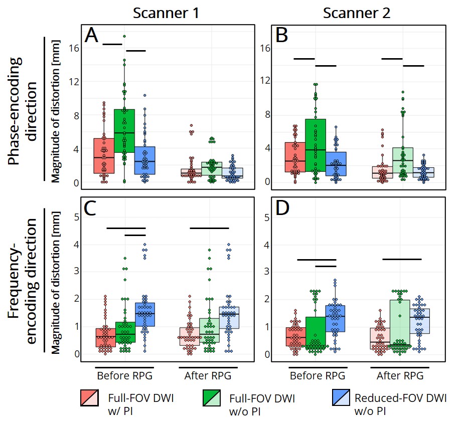
For both PE and FE directions, initial distortions for each scan were comparable across scanners, except for full-FOV without PI, which was larger (p<0.05) in the PE direction (Fig. 2, Table 2). As expected, the distortion in the FE direction was significantly less than that in the PE direction. For both scanners, full-FOV DWI without parallel imaging presented the largest (p<0.05) initial distortions in the PE direction, while those for full-FOV with PI and reduced FOV without PI were significantly less, as anticipated. Interestingly, reduced-FOV images had the largest (p<0.05) initial distortion in the FE direction compared to both full-FOV DWI datasets.RPG improved distortion in the PE direction (p<0.05), but its effects were negligible in the FE direction (p>0.05) across all scan types. After RPG, the magnitude of the distortion in the PE direction was not consistently reduced across both scanners specifically in full-FOV DWI without PI.
Discussion and Conclusions
Initial distortion in the PE direction was similar between reduced-FOV without PI and full-FOV with PI as both strategies reduce the readout duration, thus reducing the magnitude of the distortion artifact. In contrast, full-FOV without PI presented the largest distortions before and after RPG. Interestingly, the distortion in the FE direction was largest in reduced-FOV without PI both before and after RPG, which was not observed in full-FOV with PI. This can be attributed to differences in TE between both sequences, as longer readout durations result in larger distortion artifacts. Unfortunately, PI could not be used in combination with reduced-FOV and back-to-back collection of positive and negative PE data.Overall, both scanners displayed similar trends regarding distortion magnitudes and extent of RPG correction; however, small differences were still present, despite being acquired with the same scanning parameters and software version. A potential reason may be the optimization of RPG parameters used, which tests combinations of input parameters to maximize mutual information between corrected positive and negative PE volumes.
A combination of data collection and post-processing strategies may be necessary to produce distortion-free EPI-DWI images, but may be limited by the technologies and resources available at each institution. Future work will focus on evaluating the effects of different artifact-reduction data collection methods and RPG on breast DWI estimates.
Acknowledgements
GE Healthcare and the California Breast Cancer Research ProgramReferences
1. Mann RM, Cho N, Moy L. Breast MRI: State of the Art. Radiology. 2019;292:520-536.
2. Shi R-y, Yao Q-y, Wu L-m, Xu J-r. Breast lesions: Diagnosis using diffusion weighted imaging at 1.5T and 3.0T—Systematic review and meta-analysis. Clin Breast Cancer. 2018;18:e305–e320.
3. Bammer R, Keeling SL, Augustin M, et al. Improved diffusion-weighted single-shot echo-planar imaging (EPI) in stroke using sensitivity encoding (SENSE). Magn Reson Med. 2001;46:548-54.
4. Peng Y, Li Z, Tang H, et al. Comparison of reduced field-of-view diffusion-weighted imaging (DWI) and conventional DWI techniques in the assessment of rectal carcinoma at 3.0T: Image quality and histological T staging. J Magn Reson Imaging. 2018; 47:967-975.
5. Holland D, Kuperman JM, Dale AM. Efficient correction of inhomogeneous static magnetic field-induced distortion in Echo Planar Imaging. Neuroimage. 2010;50:175-83.
6. Rakow-Penner RA, White NS, Margolis DJA, et al. Prostate diffusion imaging with distortion correction. Magn Reson Imaging. 2015;33:1178-1181.
7. Rodríguez-Soto AE, Park H, Holland D, et al. Correction of artifacts induced by B0 inhomogeneities in breast MRI using reduced field-of-view echo-planar imaging and enhanced reverse polarity gradient method. medRxiv 2020;03.31.20048900.
8. Keenan KE, Wilmes LJ, Aliu SO, et al. Design of a breast phantom for quantitative MRI. J Magn Reson Imaging. 2016; 44:610-619.
Figures