1120
Application of Two Deep Learning Networks for Diagnosis of Breast Cancer on MRI: Automatic Detection Using Mask R-CNN Followed by Classification Using ResNet501Department of Radiation Oncology, Rutgers-Cancer Institute of New Jersey, Robert Wood Johnson Medical School, New Brunswick, NJ, United States, 2Department of Radiological Sciences, University of California, Irvine, CA, United States, 3Department of Radiology, The First Affiliate Hospital of Wenzhou Medical University, Wenzhou, China, 4Department of Medical Imaging, Taichung Tzu-Chi Hospital, Taichung, Taiwan, 5Department of Radiology and Research Institute of Radiological Science, Severance Hospital, Yonsei University College of Medicine, Seoul, Korea, Republic of
Synopsis
Mask R-CNN and ResNet50 were implemented to search the entire breast MRI to identify suspicious lesions, and then to further evaluate their likelihood of malignancy. The dataset included 103 malignant and 73 benign lesions in 153 patients. In detection phase using Mask R-CNN, 101 malignant, 48 benign, and 130 normal enhancing tissues were detected as suspicious. When putting them into ResNet50 for characterization, 99 cancers were correctly diagnosed as malignant, and only 16 benign lesions and 16 normal enhancing tissues remained as likely malignant. The true positive rate was 99/103=96%, and many detected false positives were dismissed during classification phase.
Introduction
Breast MRI is a well-established imaging modality for diagnosis of breast cancer and screening in high-risk women. After the dataset is acquired, the first task is to identify suspicious abnormality areas, and then to further characterize them for diagnosis. Since many sequences with thin slices are acquired to cover the entire breast with hundreds of images, radiologists need to spend time to carefully evaluate the entire dataset. Therefore, the reading is usually done with the assistance of computer-aided diagnosis (CAD) software to display important information on the workstation for evaluation. In recent years, deep learning algorithms have been applied to perform many clinical tasks, including automatic search of abnormality and diagnostic classification. We have previously developed a fully-automatic detection model using Mask Reginal-Convolutional Neural Network (R-CNN), but many enhanced areas were detected as false positives [1]. However, this is normal in clinical evaluation, and the identified areas should be furthered characterized and dismissed easily, thus not drastically affecting the overall diagnostic accuracy. We have also developed a diagnostic model using ResNet50 model to predict the malignancy probability of the detected lesion [2]. In this study, these two networks were combined to test its diagnostic validity in clinical practice, to investigate its potential as a fully-automatic CAD system for breast MRI without any human intervention.Methods
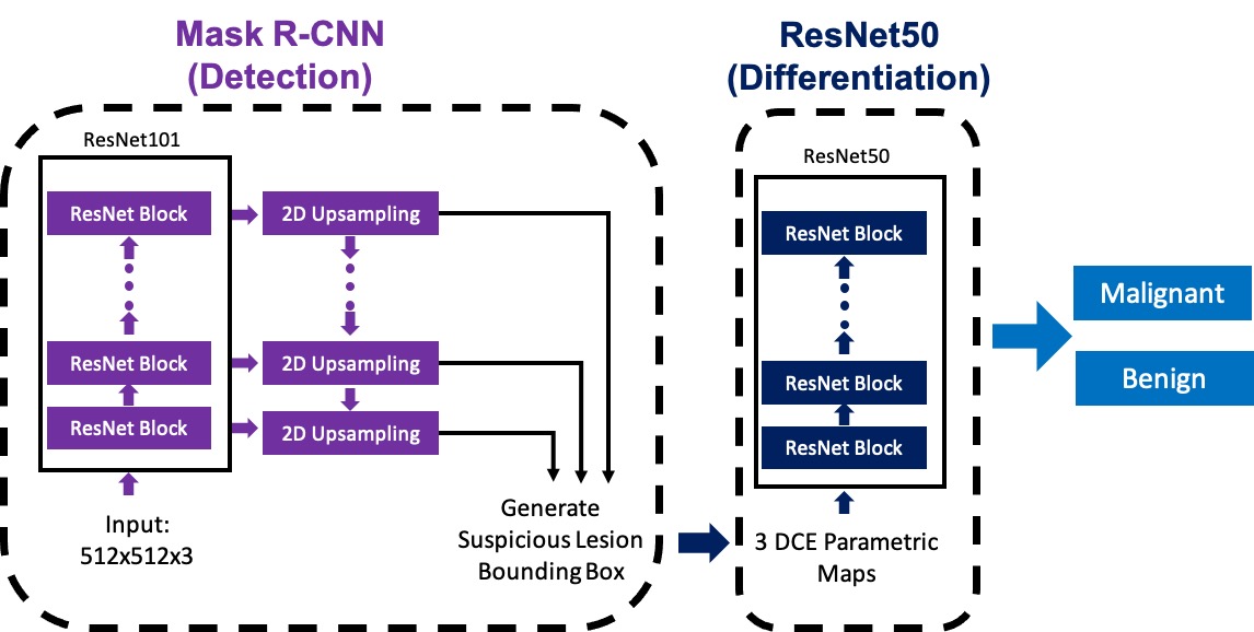
This dataset had a total of 153 patients, including 91 patients with a total of 103 pathologically confirmed malignant cancers (mean age 52±11), and 62 patients with a total of 73 benign lesions diagnosed by pathological confirmation or follow-up (mean age 45±9). The MRI was performed using a GE 3.0T system. DCE was acquired using 6 frames, one pre-contrast (F1) and 5 post-contrast (F2-F6). All slices were evaluated using a pre-trained Mask R-CNN framework to detect suspicious lesions. The architecture is shown in Figures 1 and 2. The ResNet101 was selected as the backbone network to build Feature Pyramid Network (FPN) and ImageNet was used as initial parameter values. The number of input channel is 3. The pre-contrast image, and the subtraction images of the diseased breast and the contralateral normal breast were used as inputs, where the symmetry could be used to eliminate the false detection coming from parenchymal enhancements. The outputs of FPN were sent to three sub-networks: bounding box regression network, object classification network, and mask network. This Mask R-CNN model was trained and tested using two different datasets before [1]. To decrease random false positive predictions, only lesions > 5mm, i.e. found in 4 or more consecutive slices were considered. After the suspicious lesions were identified, the differential diagnosis was made using three heuristic DCE parametric maps generated according to: the early wash-in signal enhancement (SE) ratio [(F2-F1)/F1]; the maximum SE ratio = [(F3-F1)/F1]; the wash-out slope [(F6-F3)/F3]. The cropped maps containing the suspicious lesion areas were put into the ResNet50 to assess the malignancy probability, by using a previously trained model [2].Results
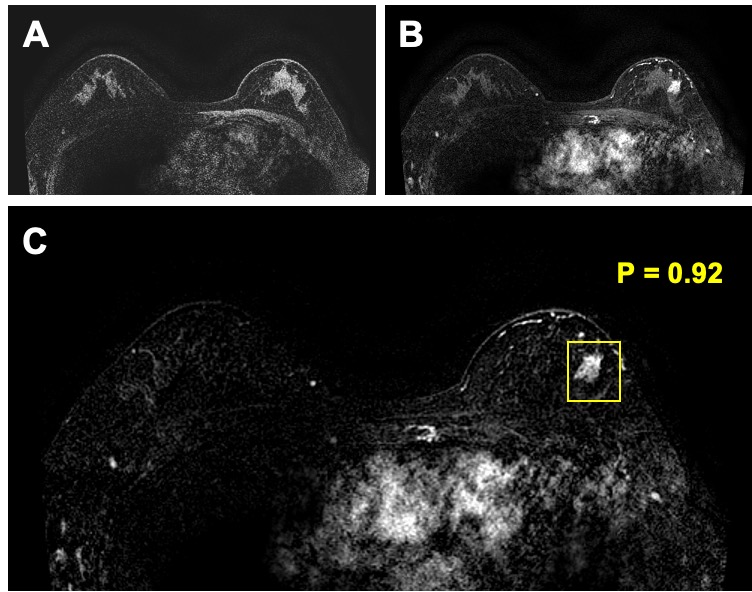
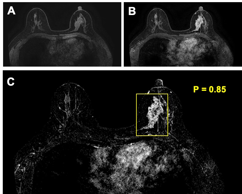
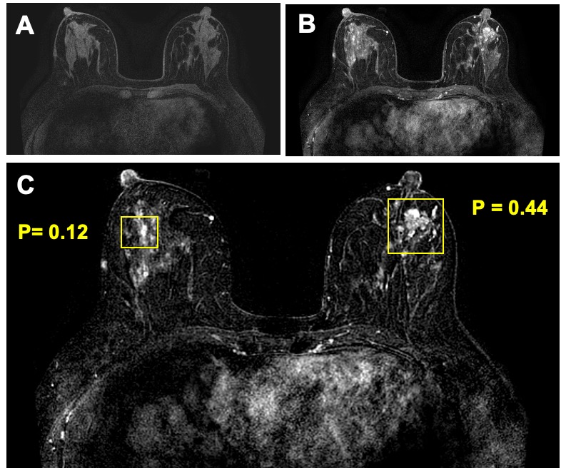
The deep learning analysis was performed using each slice as an independent input, and the results were combined to give per-lesion based prediction. For identification using Mask R-CNN, in 103 malignant lesions, 101 were correctly detected and only 2 small lesions were missed. In 73 benign lesions, 48 were identified by Mask R-CNN. Also, 130 normal areas were detected as suspicious. All these detected areas were put into the ResNet50 to evaluate the malignancy probability. The results from all slices in a lesion were combined to generate per-lesion diagnosis, based on the highest probability using the threshold of ³ 0.5 as malignant. In the malignancy group, 99/101 detected lesions were correctly diagnosed as malignant. In the benign group, 16/48 detected lesions were diagnosed as malignant. In the detected 130 normal areas, only 16 were mis-diagnosed as malignant. Therefore, combing the detection and diagnosis results together, 99/103 malignant cancers were diagnosed as true positive (TN), and only 4 were missed (FN), 2 not detected and 2 diagnosed as benign. There were a total of 32 false positive (FP) cases, 16 from confirmed benign cases and 16 from normal enhanced tissues. Three case examples are demonstrated in Figures 3 to 5.Discussion
Many studies have investigated the value of machine learning, including radiomics and deep learning, for differentiation of benign and malignant lesions. These studies mainly focused on characterization of already identified abnormal lesions. The detection was a much more challenging task, especially in MRI where many images were acquired to cover the entire breast. In this study, we implemented a fully automatic deep learning method, by first applying the Mask R-CNN to search the entire set of MR images and identify suspicious lesions, and then followed by applying ResNet50 to further characterize the lesions and give diagnosis as malignant or benign. The results show that by combining these two deep learning networks together, 99 of 103 cancers were correctly identified and diagnosed, so with a true positive (TP) rate of 99/103=96%. Only 16 benign lesions and 16 normal enhancing tissues were false positives in 153 patients, with false positive (FN) rate of 32/153 = 20.9%. Therefore, although Mask R-CNN will identify many suspicious areas, when ResNet50 is utilized to characterize them and predict the malignancy probability, many wrongly identified areas will be dismissed, which can reduce the false positive while maintaining a high true positive detection. These results illustrate the potential of Mask R-CNN detection followed by ResNet50 classification for automatic detection and further characterization of identified lesions to develop a fully-automatic computer-aided diagnosis system for breast MRI.Acknowledgements
No acknowledgement found.References
[1] Zhang Y, Chan S, Park VY, et al. Automatic Detection and Segmentation of Breast Cancer on MRI Using Mask R-CNN Trained on Non–Fat-Sat Images and Tested on Fat-Sat Images. Academic Radiology E-pub before print. https://doi.org/10.1016/j.acra.2020.12.001
[2] Zhou J, Zhang Y, Chang KT, et al. Diagnosis of Benign and Malignant Breast Lesions on DCE-MRI by Using Radiomics and Deep Learning With Consideration of Peritumor Tissue. J Magn Reson Imaging. 2020 Mar;51(3):798-809. doi: 10.1002/jmri.26981.
Figures