1098
Combination study of spectral CT and DCE-MRI quantitatively predicting vascular invasion of rectal cancer preoperatively
Wan Dong1, Ailian Liu1, Anliang Chen1, Yuhui Liu1, and Lizhi Xie2
1Radiology, the First Affiliated Hospital of Dalian Medical University, Dalian, China, 2GE Healthcare, MR Research, Beijing, China
1Radiology, the First Affiliated Hospital of Dalian Medical University, Dalian, China, 2GE Healthcare, MR Research, Beijing, China
Synopsis
Vascular tumor thrombus has been proved to be closely related to the poor prognosis of rectal cance, of which the current gold standard for the diagnosis of is postoperative pathology. In this work, we explored the feasibility of spectral CT and DCE-MRI in quantitatively predicting vascular invasion of rectal cancer. Results showed that the arterial NIC combined with Ktrans can differentiate the vascular invasion from normal status more accurately. Therefore, spectral CT combined with DCE-MRI may serve as a feasible and non-invasive way in predicting vascular invasion of rectal cancer preoperatively, that is of great significance for clinical diagnosis.
Introduction
According to the 2018 world cancer statistics [1], the incidence and mortality of colorectal cancer ranked fourth and second among all malignant tumors, respectively, and kept rising. Colorectal cancer is mostly found in the rectum. Invasion of tumor cells into lymph nodes or blood vessels is a key step in the process of metastasis [2]. Spectral CT technology includes separation of base substances, with iodine-water based substance pairs being the most common. It is very sensitive to the iodine contrast agent in the lesion, which can improve the contrast and increase the detection rate of the lesion [3]. Its quantitative value is iodine content (IC). DCE-MRI is a functional sequence, which can not only provide the morphological characteristics of the tumor, but also obtain the hemodynamic quantitative parameters of the lesion tissue perfusion or microcirculation permeability through post-processing of the image, so as to objectively evaluate the enhancement of the lesion. DCE-MRI includes Ktrans, Kep and Ve quantitative parameters. At present, it is widely used in the research of prostate cancer and cervical cancer[4]. Here, we explored whether spectral CT combined with DCE-MRI can identify vascular invasion of rectal cancer.Materials and Methods
Data of 32 patients with rectal cancer ( age range from 44 to 89 years, mean age: 63.34 ± 10.18; 12 women) were retrospectively collected. The cases were divided into two groups (group A: 17 cases with vascular invasion, and group B: 15 cases without vascular invasion). All patients underwent pelvic MR scans (including DCE sequence and some routine scans) on a 3.0T MR scanner (GE Signa HDxt, America), and pelvic spectral CT scans. Scanning parameters for the DCE-MRI were as follows: using LAVA fast volume scan sequence, FOV=40mm×32mm, TR/TE=3.5/1.5ms, slice thick/gap=3.6/0mm, NEX=0.69, matrix=256×192, scanning time=4min, 40 periods are scanned totally. Scanning parameters for the spectral CT were as follows: using gemstone spectral imaging (GSI) mode, tube voltage=80kVp/140kVp, tube current=230-445mA, slice thick/gap=5/5mm, detector width=80mm, tube rotation time =0.6s, pitch=0.992:1, FOV=50mm×50mm, the arterial, venous, and delayed images were obtained 30 s, 60 s, and 120 s after the contrast medium injection. Iodine concentration (IC) and quantitative parameter value of DCE-MRI (Ktrans, Kep and Ve) were measured by two radiologists. Three ROIs were placed at the largest level of the lesion and thier mean value were calculated as the value of the whole lesion (Figure 1). NIC = iodine concentration of lesion / iodine concentration of common iliac artery at the same level. The intra-class correlation coefficient was used to test the consistency between measurements by the two radiologists. The Mann Whitney U test was used to analyze the difference of each parameter value between the two groups. Logistic regression was used to combine significantly different parameter values, and got predicted probabilities. The ROC curve was used to test potency of the predictive value in distinguishing the two groups.Results
Measurements by the two radiologists were in good agreement (ICC=0.885), and the average of them was taken for subsequent analysis. The value of arterial NIC (ANIC), intravenous NIC (VNIC), Ktrans and Kep were significantly higher in group A than group B (P<0.05, respectively) (Table 1). Ktrans combined with ANIC has the highest efficacy, with area under the curve (AUC) of 0.875, sensitivity of 86.7%, and specificity of 88.2% (Table 2), and the predicted probability was significantly lower in group A than group B (Figure 2).Discussion and Conclusion
The advantage of spectral CT is that it can accurately reflect the IC of the tumor, and its evaluation of the substance composition is better than the conventional simple CT value measurement. The higher the malignant degree of the tumor, the higher the IC. Ktrans represents the capillary permeability-surface area product per unit volume, which depends on blood flow, vascular endothelial cell permeability and endothelial cell surface area. Kep is the flow rate of contrast medium from extracellular space to intravascular flow, which is closely related to vascular wall permeability. In our work, the ANIC-Ktrans combined value were significantly lower in the group of vascular invasion than the other. And the ROC curve indicated that the combined value can differentiate the them with a high accuracy. In summary, spectral CT combined with DCE-MRI may serve as a feasible and non-invasive way in predicting vascular invasion of rectal cancer preoperatively, that is of great significance for clinical diagnosis.Acknowledgements
No acknowledgement foundReferences
[1] Bray F, Ferlay J, Soerjomataram I et al. Global cancer statistics 2018: GLOBOCAN estimates of incidence and mortality worldwide for 36 cancers in 185 countries[J]. CA Cancer J Clin, 2018, 68(6): 394-424.[2] Bayar S, Saxena R, Emir B et al. Venous invasion may predict lymph node metastasis in early rectal cancer[J]. Eur J Surg Oncol, 2002, 28(4): 413-417.[3] Graser A, Johnson TRC, Chandarana H et al. Dual energy CT: preliminary observations and potential clinical applications in the abdomen[J]. Eur Radiol, 2009, 19(1): 13-23.[4] Liu Bing, Sun Zhen, Ma Wan-Ling et al. DCE-MRI Quantitative Parameters as Predictors of Treatment Response in Patients With Locally Advanced Cervical Squamous Cell Carcinoma Underwent CCRT[J]. Front Oncol, 2020, 10: 585738.Figures
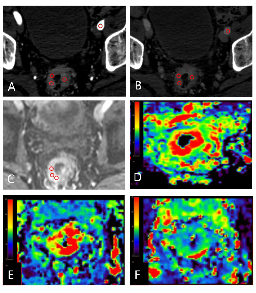
Figure.1 A 52-year-old male with vascular invasion of rectal cancer. A-B are the arterial phase and venous phase of spectral CT, respectively. C is the original DCE image. D-F are the pseudo-color graphs of Ktrans, Kep and Ve, respectively. Three ROIs were placed on the largest section of the lesion.
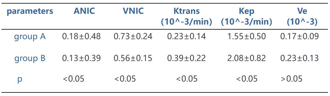
Table.1 The mean value and difference test of each parameter in the two groups.
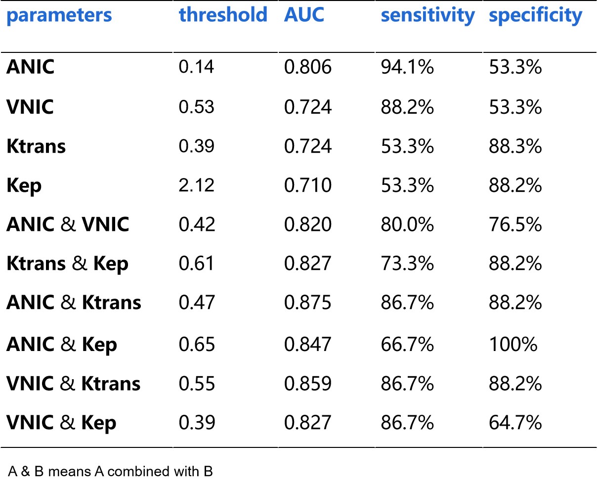
Table.2 Threshold, AUC, sensitivity and specificity of each parameter and combined parameter.
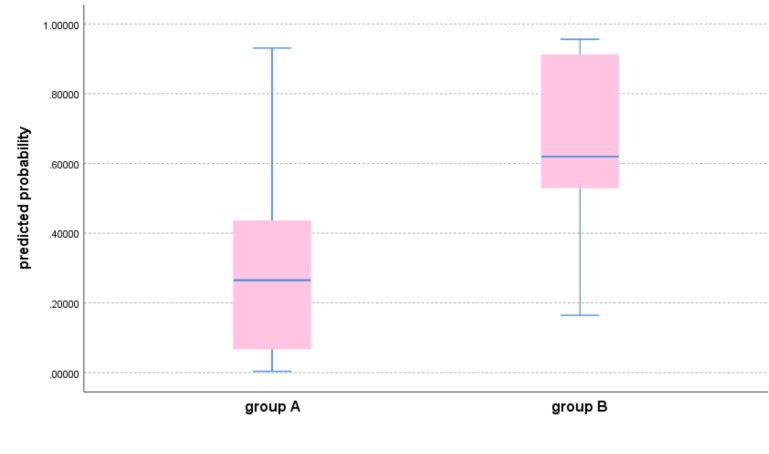
Figure.2 The predicted probability of ANIC and Ktrans in the two groups.