1084
Comparing Preservation of Ex-Vivo Lungs for Transplantation, Using 31P MRS: Extended Ex-Vivo Lung Perfusion and Cold Static Storage1University of Pennsylvania, Philadelphia, PA, United States
Synopsis
Due to a shortage of transplantable lungs, careful preservation of viable donor lungs is of paramount importance. Ex-Vivo Lung Perfusion (EVLP) and cold static storage are two clinical techniques used to preserve donor lungs prior to transplantation. In this study, magnetic resonance spectroscopy was used to compare the ability of un-ventilated normothermic EVLP vs. cold static storage to preserve rat lungs’ ATP energy status over a 5-hour ischemic period. The EVLP model was slightly better than cold static storage at sustaining the lungs’ ATP.
Introduction
Lung transplantation is a curative treatment for many with end-stage lung disease, but a shortage of transplantable donor lungs means that only slightly more than 30% of waiting list patients receive transplant, and many die before organs become available. To address this critical issue, numerous efforts aimed at expanding the organ pool by utilizing lungs from donors who are traditionally disqualified—e.g., age>45, smoking history, brain dead, etc.—are currently underway1. Ex-Vivo Lung Perfusion (EVLP) can be used to preserve both ideal and extended criteria donor lungs by delivering a flowing perfusate through the organ, thereby providing a mechanism for therapeutic intervention, oxygenation, nutrient delivery, and assessment that has the potential to improve transplant outcomes2. EVLP differs from the current gold standard method of donor lung maintenance, which involves flushing and statically storing the excised lung in a bag of preservation solution, this bag is then placed in two bags of saline in a 4-8oC icebox3. Evaluating metabolic biomarkers in donor lungs undergoing preservation may provide an early signal of lung viability. In this study in a rat model, magnetic resonance spectroscopy was used to compare preservation using un-ventilated normothermic EVLP and the gold standard (GS) of cold static storage over a period of extended (5-hour) ischemia by monitoring lungs’ energy status.Method
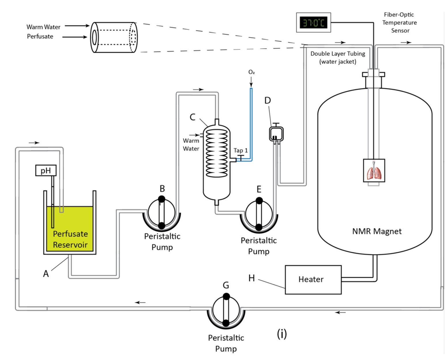
Sprague Dawley (n=8) male rats were mechanically ventilated with oxygen for 10 minutes to remove gaseous nitrogen from the lungs (VT=3.0 ml, f=38 min-1, FiO2=1.0, PEEP=3 cmH2O, 10% sigh, 33:67 I:E). Lungs were perfused through the pulmonary artery at a 10 ml/min flow rate with 500 ml of an extracellular type low potassium perfusate: modified Krebs-Henseleit buffer (119 mM NaCl, 25 mM NaHCO3, 1.3 mM CaCl2, 1.2 mM MgSO4, 4.7 mM KCl, 10 mM glucose, 2 mM lactate, 0.2 mM pyruvate, and 3% (w/v) fatty-acid-free bovine serum albumin). The trachea was sutured upon expiration, and lungs were excised and placed in a 20 mm NMR tube before being transferred to the MRI machine (AVANCE III 9.4T, Bruker Inc.). This setup is shown in Figure 1. The perfusate was passed through an oxygenating column under a constant flow of oxygen and warmed via passage through heated water-jacketed tubing to maintain a temperature of ~37oC in the NMR tube. pH was periodically adjusted to ~7.4 by adding 1.2 N HCl or NaOH to the perfusate reservoir. For the gold standard cold storage model (GS), six lungs were removed from the magnet after 1 hour of perfusion, flushed and statically stored in 20 ml of 4oC Perfadex+R (XVIVO Perfusion) for 3 hours, then gradually rewarmed, perfused at 37oC, and returned to the magnet for 1 hour. The remaining lungs (n=2) remained in the spectrometer throughout the entire 5-hour study (EVLP model). 31P spectra were obtained using a 25 mm dual-tuned (1H/31P) coil (Bruker Inc.) with the following acquisition parameters: TR=1 s, FA=60o, SW=8 kHz, NP=2048, NA=1024, with a total acquisition time of ~17 minutes. Data were processed using custom routines in MATLAB2018 and RStudio 1.2.13.Results and Discussion
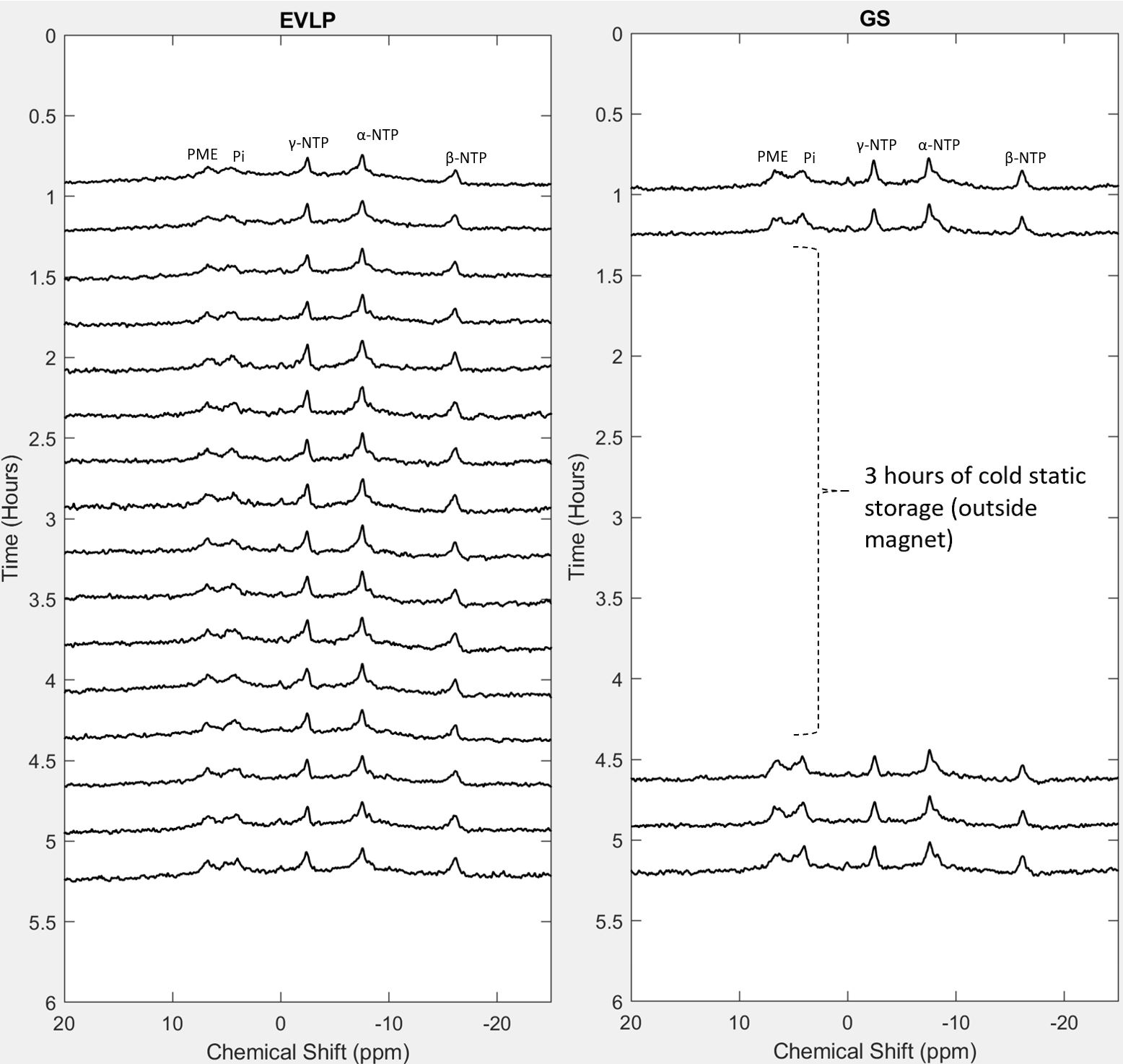
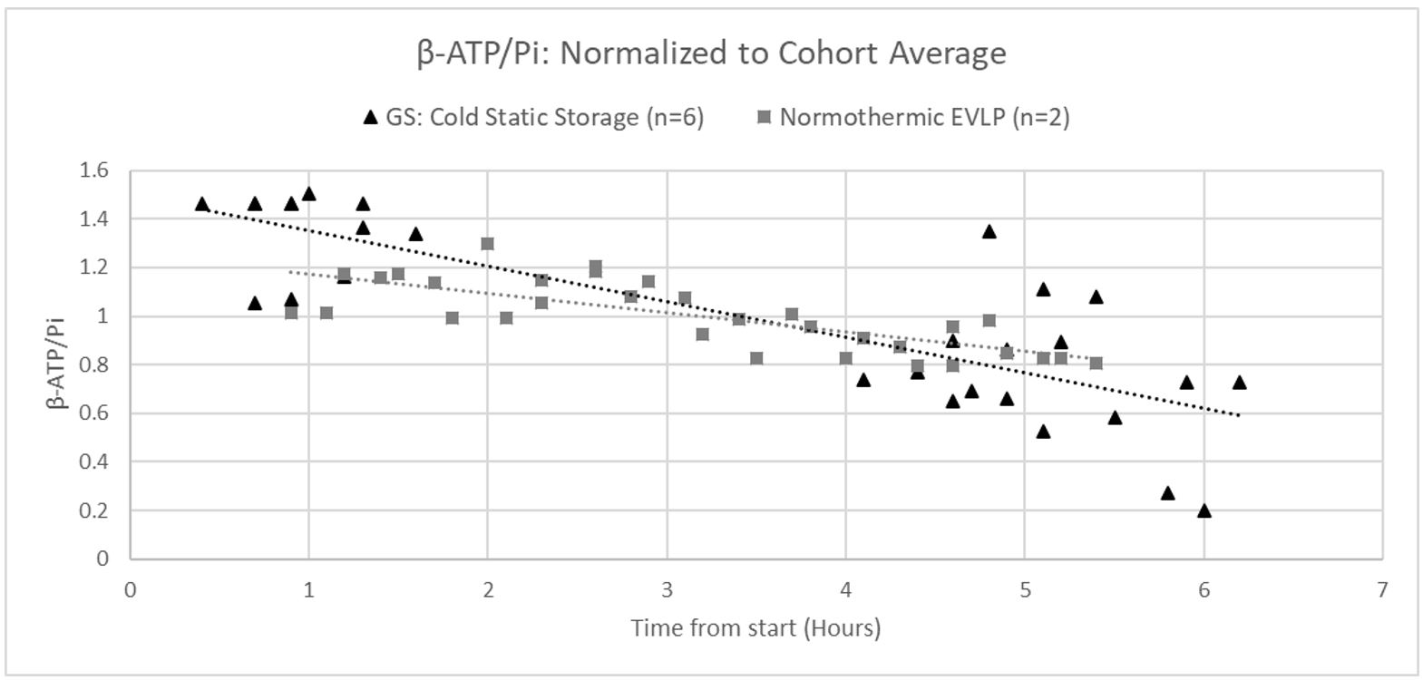
Figure 2 displays representative 31P spectra for both EVLP and GS models. The ex-vivo lung’s energy status and mitochondrial function declined over time, as can be seen spectrographically from the decline in β-ATP as well as the rise in Pi and phosphomonoester signals. Figure 3 displays a normalized time series of the ratio of β-ATP/Pi, quantifying this shift from high to low energy phosphates. In the admittedly small number of studies reported here, the EVLP model exhibited a slower rate of decline in β-ATP/Pi than the GS model, suggesting that EVLP was better at supporting lung ATP. However, substantial variability was observed in the GS results, complicating their interpretation. Specifically, two of the GS experiments exceeded the 5-hour timeframe by well over 30 mins, which may account for the low β-ATP/Pi ratios observed near their endpoints. Further highlighting the potential impact of extended ischemic time, a study by Shaghaghi, et al. found that the rate of decline in ex-vivo lung β-ATP peak intensity accelerated once β-ATP fell below 50% of its baseline value4. The GS model also involved more experimental steps, such as removing the lungs from the magnet, restarting pumps, etc., which may have contributed to lung insult or injury. While EVLP was better at preserving β-ATP/Pi, this metric’s connection to clinical lung transplant viability remains unclear—once donor lungs have been screened, approved, and transported to transplant centers, surgeons review the organ’s history and inspect the lungs, but do not assess 31P biomarkers.Conclusion
Analysis of 31P spectra and β-ATP/Pi time series suggests that normothermic EVLP may improve upon cold static storage in preserving donor lungs’ ATP energy status prior to transplantation. However, this finding is complicated both by our study’s small sample size as well substantial variability in β-ATP/Pi values due to some GS experiments exceeding 5 hours in length. To further compare EVLP with cold static storage and assess β-ATP/Pi’s value as a metric for clinical donor lung evaluation, future testing will focus on re-ventilating the collapsed lung after 5-hour preservation, transplanting lungs at various β-ATP/Pi values, and attenuating the observed β-ATP/Pi variability due to experiment length.Acknowledgements
No acknowledgement found.References
1. Chaney J, et al. Lung Donor Selection Criteria. J Thorac Dis. 2014;6(8):1032-1038
2. Cypel M, et al. Normothermic Ex Vivo Perfusion Prevents Lung Injury Compared to Extended Cold Preservation for Transplantation. Am J Transplant. 2009 Oct;9(10):2262-9.
3. Michel SG, et al. Innovative Cold Storage of Donor Organs Using the Paragonix Sherpa PakTM Devices. Heart Lung Vessel. 2015;7(3):246-255.
4. Shaghaghi H, et al. Ascorbic Acid Prolongs the Viability and Stability of Isolated Perfused Lungs: A Mechanistic Study Using 31P and Hyperpolarized 13C Nuclear Magnetic Resonance. Free Radical Biology and Medicine. 2015; 89: 62-71
Figures