1066
Altered structural asymmetries of mild white matter injury in preterm infants with different cognitive outcomes1Department of Diagnostic Radiology, the First Affiliated Hospital of Xi’an Jiaotong University, Xi‘an, China, 2MR Research China, GE Healthcare, Beijing, China
Synopsis
Punctate white matter lesions (PWMLs) are common in the preterm. The mild PWMLs may result in cognitive impairments and lesion location is closely associated with neurodevelopmental outcomes. Cognition is a high-order function with multiregional information integration and functional hemispherical lateralization. Therefore, this study aims to investigate the white matter (WM) asymmetries of mild PWMLs in preterm infants with different cognitive outcomes at a corrected age of 6 months by diffusion tensor imaging. Left lateralization of WM development is obviously reduced in mild PWMLs with cognitive delay, while WM asymmetries of PWMLs with normal cognition score are similar with controls.
Introduction
White matter (WM) injury is common in preterm neonates, with the most common punctate white matter lesions (PWMLs) can be detected on conventional MRI as hyperintensity on T1WI and hypointenity on T2WI[1]. It is important to note that the lesions can gradually disappear along with time and are easily missed diagnosis, especially the mild subgroup. Since development of brain structures is the fundamental of function, early quantitative assessment of the alterations has a great of importance for the prognosis[2]. Previous studies have reported that mild PWMLs may mainly result in cognitive impairments and preliminary found lesion location is closely associated with the neurodevelopmental outcomes[3, 4]. However, cognition is a high-order function with multiregional information integration and functional hemispherical lateralization[2]. The structural asymmetries in PWMLs are not as well characterized. Therefore, this study aims to investigate the WM asymmetries of mild PWMLs in preterm infants with different cognitive outcomes at a corrected age (CA) of 6 months by diffusion tensor imaging.Methods
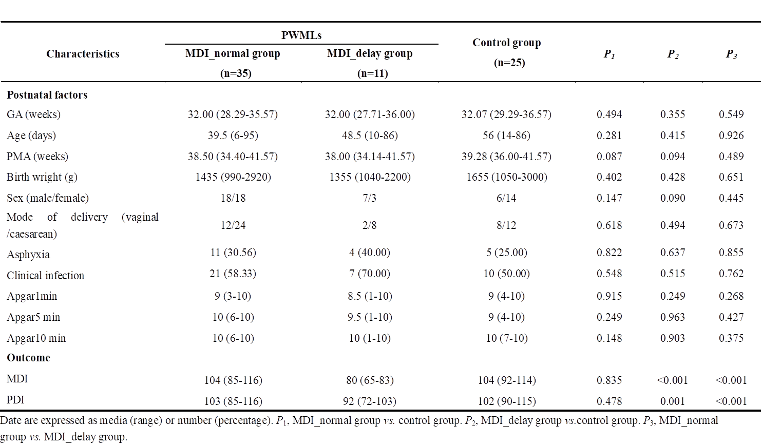
The local institutional review board approved this study and all the written informed consents were obtained from parents of the infants. Subjects Preterm infants (gestational age<37 weeks and postmenstrual age≤45 weeks) with evidence of mild PWML (diagnosed by conventional MRI: ≤10 lesions or 1 large lesion [diameter≥5mm]) and controls were included. Bayley Scales of Infant Development II was used to evaluate the developmental outcomes at a CA of 6 months. According to the mental developmental index (MDI), the subjects with mild PWMLs were classified into normal group (MDI≥85) and delayed group (MDI<85). MRI Protocols All MR examinations were performed using a 3T scanner (Signa HDxt, GE Healthcare, Milwaukee, Wisconsin) with an 8-channel head coil. The scanning parameters of DTI sequence were as follows: 30 gradient directions; the number of excitations=1; b values=0 and 600s/mm2; TR/TE=11000/69.5ms; matrix size=128×128; slice thickness=2.5mm with no gap and FOV=180mm. Data and statistical analysis Tractography atlas for infants was obtained by using the registration between local infants and the JHU template. After manual segmentation of the PWMLs on T1WI, the lesion volumes on 20 main fiber were calculated on individual level[5].. The voxel-based differences in FA values of the bilateral hemispheres were calculated by TBSS analysis. Finally, the WM asymmetry index (AI) was calculated by the following equation: AI= (Left - Right)/0.5* (Left + Right)[6]. Wilcoxon signed-rank tests were used to assess between group differences in demographics, lesion volume, AI and neurodevelopmental outcomes. The classification variables of the information were evaluated using Chi-square tests. Tests were considered significant at P<0.05. In multiple comparisons, P<0.017 (0.05/3) was considered statistically significant after the Bonferroni correction.Results
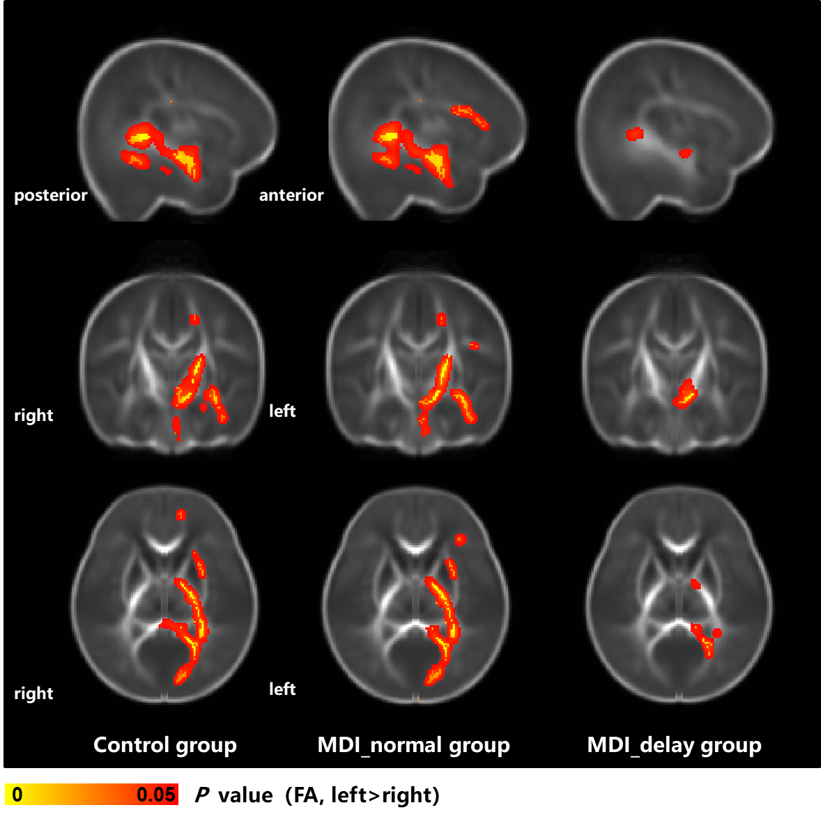
Forty-six preterm infants with mild PWMLs and 25 controls were enrolled. Thirty-five and 11 subjects were classified into MDI_normal and MDI_delayed group, respectively. No significant differences in demographics were found among groups, and the MDI of delay group was significantly lower than other two groups (Table 1). Compared with normal group, the lesion volume on SCC, IFOF_L, UF_L and AF_L were significantly larger in MDI_delayed group than those in MDI_normal group (P<0.05, Figure 1). Higher FA regions of control group were detected in left CST (from cerebral peduncle to internal capsule), SCC, IFOF/ILF, UF and AF. The MDI_normal group performed the consistency results with controls and part of left anterior corona radiata also showed the increased FA. For MDI_delayed group, the differences in FA between hemispheres were obviously decreased, and the higher FA regions were only found in part of left CST, SCC and IFOF/ILF (Figure 2). The AI alterations were different between the 2 PWMLs groups. No significant difference was found between control and MDI_normal groups (P>0.017). AI of AF in MDI_delayed group was significantly lower than those in other two groups (P<0.017, Figure 3).Discussion
This study found that lesions of preterm PWMLs with delayed cognition were mainly distributed in the left long association fibers, and further reduce the left asymmetries of WM development. The PWMLs main involvement of the left hemisphere may be associated with its less maturity compare to the contralateral[[7]]. Previous study have found the left lateralization of WM can be detected at fetal and postnatal period and which is mainly participated in the infant sensorimotor stage[8]. The left lateralization pattern similar to that of controls may be the structural basis for the good prognosis of MDI_normal group. AF is the key fiber connecting the temp-parietal lobes, and it is also the area prone to damage. Reduced left AI of AF in MDI_delayed group may be related to altered local connections in left hemisphere, and further affected the information transmission and integration integration to front-parietal lobes, and ultimately leading to cognitive delay[9].Conclusion
WM asymmetries variations are different across mild PWMLs preterm infants with different cognitive outcomes. Left lateralization of WM development is obviously reduced in mild PWMLs with cognitive delay, while the WM asymmetries of PWMLs with normal cognition score are similar with controls.Acknowledgements
This study was supported by the National Natural Science Foundation of China (No. 8197071015, 81971581, 81771810, 81471631 and 51706178), the 2011 New Century Excellent Talent Support Plan of the Ministry of Education, China (NCET-11-0438) and the Clinical Research Award of the First Affiliated Hospital of Xi’an Jiaotong University (No. XJTU1AF-CRF-2020-005).References
[1] Wagenaar N, Chau V, Groenendaal F, et al. Clinical Risk Factors for Punctate White Matter Lesions on Early Magnetic Resonance Imaging in Preterm Newborns [J]. The Journal of pediatrics, 2017, 182(34-40.e1.
[2] Ocklenburg S, Friedrich P, Güntürkün O, et al. Intrahemispheric white matter asymmetries: the missing link between brain structure and functional lateralization? [J]. Rev Neurosci, 2016, 27(5): 465-80.
[3] Guo T, Duerden EG, Adams E, et al. Quantitative assessment of white matter injury in preterm neonates: Association with outcomes [J]. Neurology, 2017, 88(7): 614-22.
[4] Cayam-Rand D, Guo T, Grunau RE, et al. Predicting developmental outcomes in preterm infants: A simple white matter injury imaging rule [J]. Neurology, 2019, 93(13): e1231-e40.
[5] Miaomiao Wang XL, Congcong Liu, Jian Yang. Reduced structural asymmetries in infants with mild white matter injury with cognitive development delay [M]. ISMRM, 2020.
[6] Toga AW, Thompson PM. Mapping brain asymmetry [J]. Nature reviews Neuroscience, 2003, 4(1): 37-48.
[7] Vasung L, Rollins CK, Yun HJ, et al. Quantitative In vivo MRI Assessment of Structural Asymmetries and Sexual Dimorphism of Transient Fetal Compartments in the Human Brain [J]. Cerebral cortex (New York, NY : 1991), 2019, bhz200.
[8] Taylor DC. Differential rates of cerebral maturation between sexes and between hemispheres. Evidence from epilepsy [J]. Lancet (London, England), 1969, 2(7612): 140-2.
[9] Gotts SJ, Jo HJ, Wallace GL, et al. Two distinct forms of functional lateralization in the human brain [J]. Proceedings of the National Academy of Sciences of the United States of America, 2013, 110(36): E3435-E44
Figures