1065
An application of BOLD-fMRI on functional changes in shenmen and neiguan electroacpunctured population with chronic partial sleep-deprivation
Wang Chunyan1, Qiu Ganbin1, Weiyin Vivian Liu2, and Ma Liheng1
1Medical Imaging, the first affiliated hospital of Guangdong pharmaceutical university, Guangzhou, China, 2MR Research, GE Healthcare, Beijing, Beijing, China
1Medical Imaging, the first affiliated hospital of Guangdong pharmaceutical university, Guangzhou, China, 2MR Research, GE Healthcare, Beijing, Beijing, China
Synopsis
An application of BOLD-fMRI on functional changes in shenmen and neiguan electroacpunctured population with chronic partial sleep-deprivation
synopsis
Quality of sleep affects health status. Individual performance and alertness decreased when sleep deprivation (SD) occurred. Acupuncture therapy have been used to treat patients with chronic pain, depression and even drug addiction. In our study, we found electroacupuncturing at Shenmen and Neiguan points could cause wide-ranged changes in the resting brain activities and functional connection in both controls and chronic partial sleep deprivation (CPSD) group, showing improved sleep states after specific brain regions were acupunctured. Two methods showed inconsistent results, suggesting that not only one analysis method should be utilized in order to comprehensively explain internal neural mechanism of SD.INTRODUCTION
Quality of sleep affects health status. Individual performance and alertness decreased when sleep deprivation (SD) occurred. Long-term SD decreases the health condition. So far, there is no final conclusion on changes of brain activities in sleep-deprived patients. Acupuncture therapy have been used to treat patients with chronic pain, depression and even drug addiction. However, no research on the acupuncture-sleep relationship has been reported. In this study, resting-state functional magnetic resonance (rs-fMRI) BOLLD technique with different analytical methods were used to evaluate the changes in the brain of the volunteers with SD and to explore the effect of the acupuncture.Methods
43 patients with chronic partial sleep deprivation (CPSD) (24 male, 19 female) and 48 healthy control (HC) (25 male, 23 female) underwent examinations on 3.0T MR scanner (Discovery 750w, GE Healthcare). The first rs-fMRI BOLD scan and cognition behavioral scale test (ANT tests) were done before acupunctured. Then, all subjects were electro-acupunctured at the bilateral Shenmen with Neiguan points and stimulated for 30 minutes after "De Qi" and underwent the second rs-fMRI scan after 20-minute rest. ALFF and ReHo were analyzed in SPM10 software. The group differences of ALFF and ReHo were compared with two independent sample T-tests and pre- and post-acupuncture effect was tested by paired T test. Pearson correlation was used to compute the correlation between the behavior and differential brain regions in the CPSD group. First ten representative differential brain regions were selected as seeds to establish the whole brain functional connectivity map.Results
The ANT experiments suggested that the response time prolonged, the leakage rate increased and the correct rate decreased in CPSD group. The Pearson correlation analysis suggested that the β value of ALFF in the hippocampus was positively correlated with the correct rate (r = 0.637, P = 0.001) and negatively correlated with the leakage rate (r = -0.427, P = 0.047). The ReHo value of left superior frontal gryus was negatively correlated with the reaction time (r =-0.514, P = 0.014). Significant differences of ALFF, ReHo and functional connectivity were found between HC group and CPSD group (Figure 1) . The significant pre- and post-electroacupuncture difference of ALFF, ReHo and functional connectivity were found in and between the HC and CPSD group (Figure 2-4).Discussion and conclusions
The present study showed that the altered activities of brain regionsafter acupuncture were mainly involved in default mode network (DMN), attention network and limbic system. The abovementioned acupoints were related to the brain regions involved in cognition and executive control, which may improve the high cognition functions especially emotion and memory, providing a new vision for exploration of the combined Shenmen and Neigang for sleep disorders.In normal conditions, after Shenmen and Neiguan points were acupunctured, the thalamus was positively activated and its functional connection with the left amygdala was also significantly enhanced, indicating that the internal mechanism of continuous arousal maintenance was related to the functional change of the thalamus, but the specific mechanism remains to be further studied.This study suggests that the strength of the functional network connection mainly responsible for execution, attention and memory decreased while that maintains arousal increased. This indicates that long-term exposure to CPSD will damage the brain regions responsible for cognitive functions such as attention and memory. In order to maintain basic survival needs, the body will allocate more energy to the brain regions that maintain the arousal state in order to maintain the alert state of the brain, thus enhancing the functional connectivity of corresponding brain regions.In this study, CPSD population could cause ALFF and ReHo changes in multiple brain regions. However, the results analyzed by the two methods were not completely consistent, suggesting that a single analysis method may not be able to fully explain its internal neural mechanism.KEY WORDS
electroacupuncture; Shenmen point; Neiguan point;sleep deprivation; functional magnetic resonance imaging; regional homogeneity;amplitude of low frequency fluctuationAcknowledgements
We would like to thank the first Affiliated Hospital of Guangdong Pharmaceutical University for the cooperation of the machines and personnelReferences
[1] Wang X S, Armstrong M E, Cairns B J, et al. Shift work and chronic disease: the epidemiological evidence.[J]. Occup Med, 2011, 61(2):443-444.[2] Avinun R, Nevo A, Knodt A R, et al. Reward-Related Ventral Striatum Activity Buffers Against the Experience of Depressive Symptoms Associated with Sleep Disturbances.[J]. Journal of Neuroscience, 2017, 37(40):9724.[3] Angel J D, Cortez J, Juárez D, et al. Effects of sleep reduction on the phonological and visuospatial components of working memory[J]. Sleep Sci, 2015, 8(2):68-74.[4] Taheri M, Arabameri E. The effect of sleep deprivation on choice reaction time and anaerobic power of college student athletes [J]. Asian J Sports Med, 2012, 3(1): 15-20.[5] Krause A J, Simon E B, Mander B A, et al. The sleep-deprived human brain.[J]. Nature Reviews Neuroscience, 2017, 18(7):404.[6] Barbeau E B, Lewis J D, Doyon J, et al. A greater involvement of posterior brain areas in interhemispheric transfer in autism: fMRI, DWI and behavioral evidences.[J]. Neuroimage Clin, 2015, 8:267-280.[7] Gruber S A, Dahlgren M K, Sagar K A, et al. Decreased Cingulate Cortex activation during cognitive control processing in bipolar disorder[J]. Journal of Affective Disorders, 2017, 213:86-95.[8] Terry D P, Sabatinelli D, Puente A N, et al. A Meta-Analysis of fMRI Activation Differences during Episodic Memory in Alzheimer's Disease and Mild Cognitive Impairment[J]. Journal of Neuroimaging, 2015, 25(6):849-860.[9] Zang Y, Jiang T, Lu Y, et al. Regional homogeneity approach to fMRI data analysis.[J]. Neuroimage, 2004, 22(1):394-400.[10] Peng D C, Dai X J, Gong H H, et al. Altered intrinsic regional brain activity in male patients with severe obstructive sleep apnea: a resting-state functional magnetic resonance imaging study[J]. Neuropsychiatric Disease & Treatment, 2014, 10(default):1819.[11] Guadagni V, Burles F, Ferrara M, et al. The effects of sleep deprivation on emotional empathy.[J]. Journal of Sleep Research, 2014, 23(6):657.[12] Raichle M E, MacLeod A M, Snyder A Z, et al. A default mode of brain function [J]. Proceedings of the National Academy of Sciences, 2001,98(2): 676-682.[13] Wang Z , Liang P , Zhao Z , et al. Acupuncture Modulates Resting State Hippocampal Functional Connectivity in Alzheimer Disease[J]. PLoS ONE, 2014, 9(3):e91160.[14] Yeo S , Choe I H , Noort M V D , et al. Acupuncture on GB34 activates the precentral gyrus and prefrontal cortex in Parkinson’s disease[J]. BMC Complementary and Alternative Medicine, 2014, 14(1):336.[15] Zhenyu L, Wenjuan W , Lijun B , et al. Exploring the Patterns of Acupuncture on Mild Cognitive Impairment Patients Using Regional Homogeneity[J]. PLoS ONE, 2014, 9(6):e99335-.Figures

Correlation between scores of the ANT task and ALFF and ReHo of CPSD group before acupuncture. (A) The ALFF value of the right parahippoclial gyrus was positively correlated with the accuracy of ANT task (r=0.637, P= 0.001) and negatively correlated with the omission rate (r=-0.427, P=0.047). (B) The ReHo value of the right superior frontal gyrus was negatively correlated with the reaction time in ANT task (r= -0.514P =0.014).
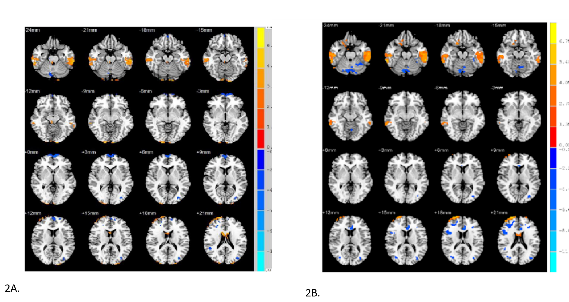
Significant differences of pre- electroacupunctured (A) ALFF and (B) ReHo values between HC group and CPSD group. Warm colors represented increased ReHo values in brain regions, while cool colors decreased ReHo values in brain regions. Warm and cool colors represented increased and decrease d ALFF values, respectively.

Significant pre- and post-electroacupunctured differences of ALFF in both groups. In HC group, there were significant differences of (A) ALFF and (B) ReHo values before and after acupuncture. (C) In CPSD group, there were significant differences in (C) ALFF and (D) ReHo values before and after acupuncture. Warm and cool colors represented increased and decreased ALFF values, respectively.

(A)Pre-acupunctured differences of function connectivity between CPSD and HC groups. (B)The Pre- and post-acupunctured difference of functional connectivity in HC group. (C)The Pre- and post-acupunctured difference of functional connectivity in CPSD group. The red globule is the seed point and the blue globule is the region in functional connection to the seed point. The solid red line represents the enhanced connection, and the dotted red line represents the reduced connection strengt