1063
Parahippocampal gyrus neuroanatomical and functional connectivity alterations in mTBI at the acute stage:a VBM and rsfMRI study1Second Clinical School, Lanzhou University, Lanzhou, China, 2Lanzhou University Second Hospital, Lanzhou, China, 3Siemens Healthineers, Shanghai, China
Synopsis
mTBI is not a static event, but a progressive injury. The neuroanatomical and functional alterations of mild traumatic brain injury (mTBI) at the acute stage can be an initial step of damages leading to cognitive and emotional deficit which can be developed in future in long-term period of injury. We analyzed early alterations in the gray matter volume (GMV) and functional connectivity (FC) alterations of mTBI patients within 7 days after injury, and found that early disruptions in left parahippocampal gyrus may appear in mTBI, which might be potentially related to the cognitive and emotional impairments.
Introduction
Cognitive and emotional impairments is frequent among patients with mild traumatic brain injury (mTBI), which may reflect alterations of brain structural and functional properties. The relationship between brain microscopic changes in the acute phase and cognitive and emotional deficit remain unclear. Just a single concussive episode induces measurable changes in brain structure, manifesting as diffuse and local patterns of altered neuromorphometry.1 Voxel based morphometry (VBM) analysis have enabled the quantification of regional gray matter volume (GMV) throughout the cortex. Resting-state fMRI and seed based region of interest analysis have been used to identify changes in functional connectivity (FC) in mTBI. The parahippocampal gyrus serves as the main cortical input of the hippocampus and has an important relationship with cognition and emotion.2 The aim of this study is to examine parahippocampal gyrus structure disruption and functional connectivity alterations in acute period of mTBI using VBM and rsfMRI.Methods
Forty-three mTBI patients within 7 days after injury and thirty-seven age-, sex-, and educational -matched healthy controls were recruited in this study. All subjects underwent the same neuropsychological testing (Mini Mental State Examination (MMSE) and Self-rating Depression Scale (SDS)) and neuroimaging protocol on a 3T MR scanner (Magnetom Verio, Siemens Healthcare, Erlangen, Germany). Sagittal three-dimensional turbo spin-echo (3D-TSE) T1WI (TI = 900 ms and flip angle = 12°) were acquired with 256-mm FOV and 192 contiguous partitions (1.0 mm) at 256 × 256 matrix. The resting-state fMRI were acquired with axially using a gradient-echo echo-planar imaging (GE-EPI) sequence, with TE = 30 ms, TR =2000 ms, 90 flip°, 128 × 128 acquisition matrix, 220 mm2 FOV, 36 slices and 3.0 mm thickness. The structural and functional images were preprocessed using SPM12 running on MATLAB (R 2013b). The GMV change between two groups was analysed by a two-sample t test. Seed-based analysis was performed in order to reveal disturbances in functional connectivity. Spearman’s correlation analysis was calculated between the MMSE/SDS scores and the GMV.Results
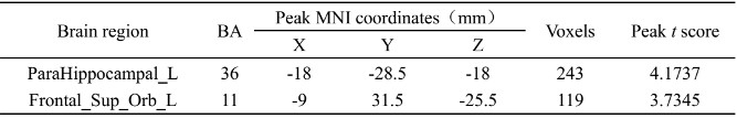
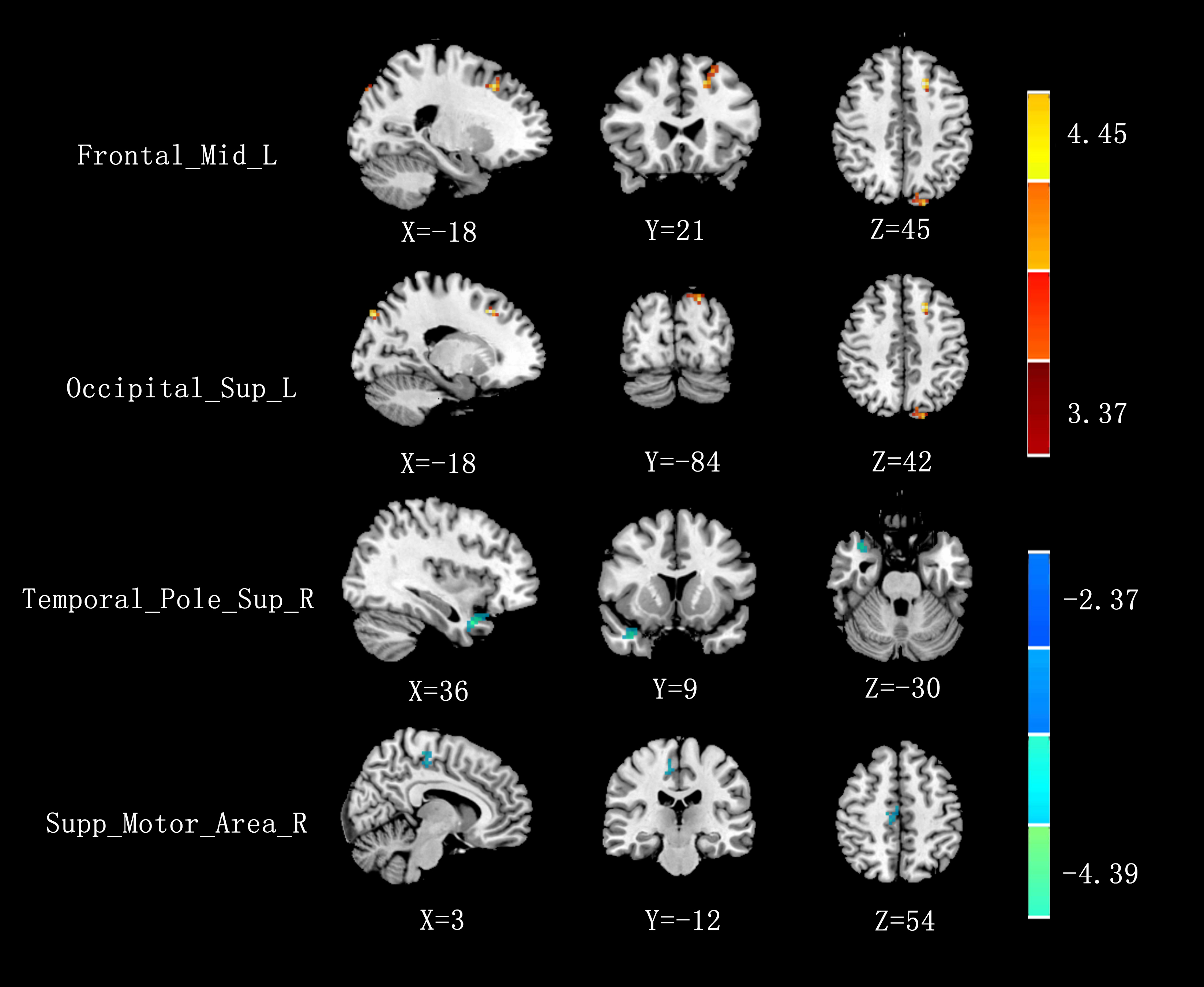
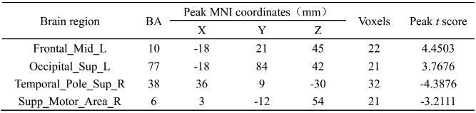
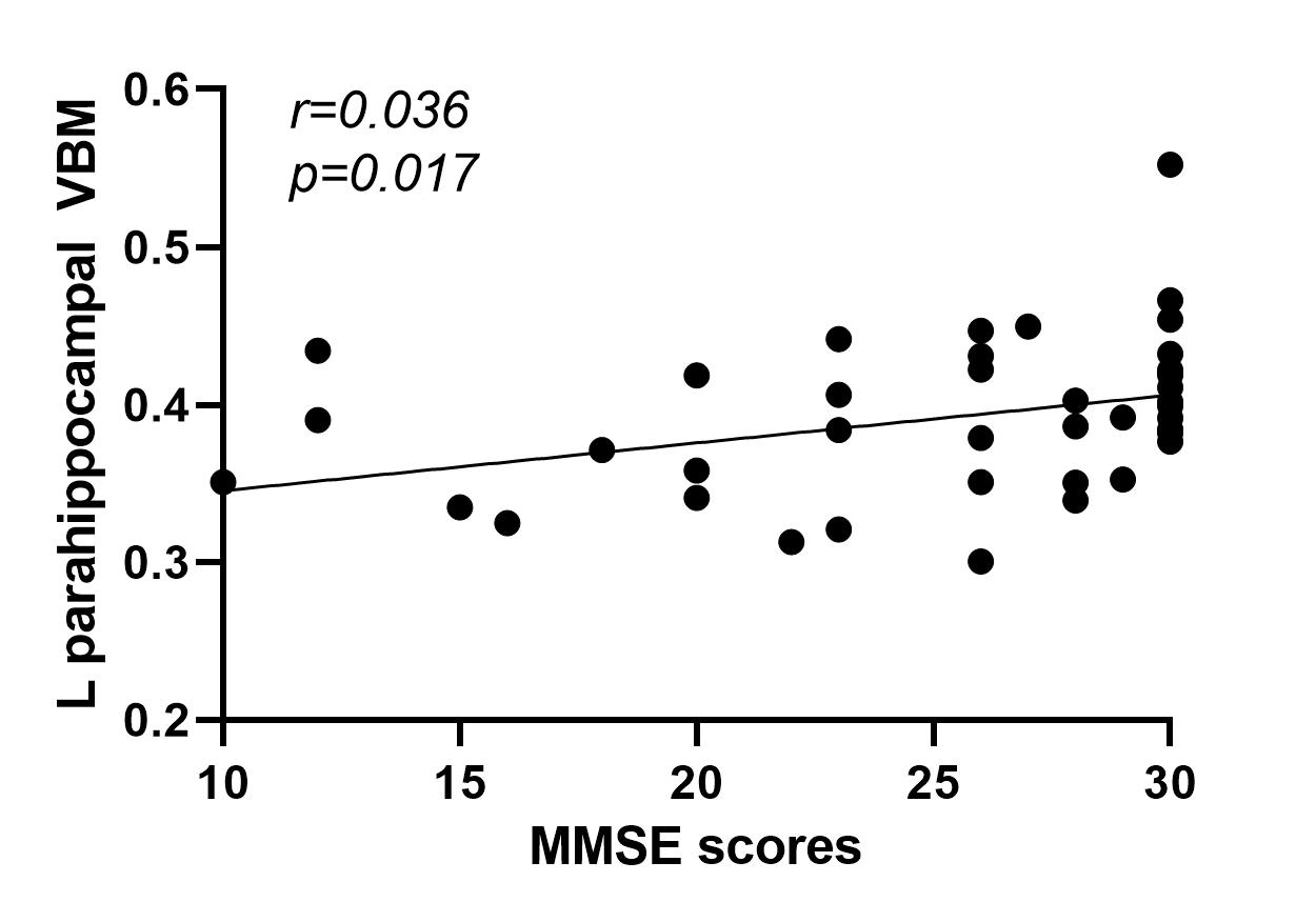
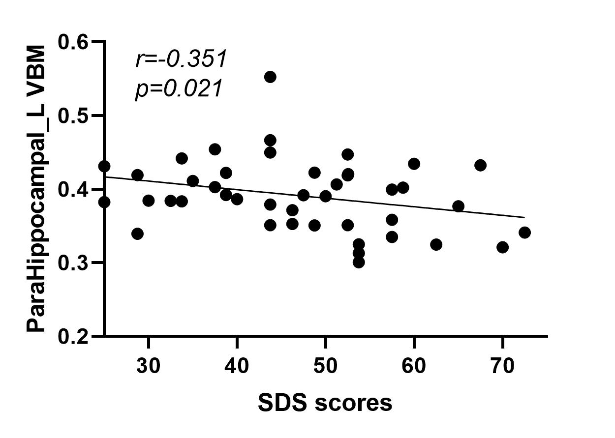
A whole-brain comparison showed GMV increase in the left parahippocampal gyrus and left orbital part of superior frontal gyrus in patients with mTBI at the acute stage compared with healthy controls (Table 1). Relative to controls, mTBI patients showed increased connectivity between the seed region of left parahippocampal gyrus and left middle frontal gyrus and left superior occipital gyrus, while the connectivity with right superior temporal gyrus and right supplementary motor area was weakened (Fig. 1 and Table 2). The Spearman analysis showed that there was positive correlation between the MMSE scores and the increased GMV in the left parahippocampal gyrus in patients with mTBI at the acute stage compared to healthy controls (r = 0.363, p = 0.017) (Fig. 2). In addition, a weak negative correlation between SDS and GMV alteration in the left parahippocampal gyrus (r = -0.351, p = 0.021) (Fig. 3).Discussion
Approximately 20%–30% of all patients experience some degree of neurologic and psychologic problem following mTBI, especially impaired cognitive and emotion function.3 As a part of the hippocampal circuit, the parahippocampal gyrus is associated with higher neural functions such as emotion, learning and memory. The GMV was significantly greater suggest that the left parahippocampal gyrus displays compensatory remodeling involved in cognitive regulation.4 In addition, the parahippocampus plays an important role in the neuropathology of depression and may be associated with increased depressive symptoms in mTBI patients. Interestingly, our study demonstrated significant FC increased/reduction between the left parahippocampal gyrus and ipsilateral/contralateral areas. This asymmetry in connectivity during the resting state is small but consistent.5Acknowledgements
This article is supported by the National Natural Science Found (No. 8196070049)References
1. Sussman D, da Costa L, Chakravarty MM, et al. Concussion induces focal and widespread neuromorphological changes. Neurosci Lett. 2017; 650: 52-59.
2. Li F, Lu L, Chen H, et al. Neuroanatomical and functional alterations of insula in mild traumatic brain injury patients at the acute stage. Brain Imaging Behav. 2020; 14(3): 907-916.
3. Cassidy JD, Carroll LJ, Peloso PM, et al. Incidence, risk factors and prevention of mild traumatic brain injury: results of the WHO Collaborating Centre Task Force on Mild Traumatic Brain Injury. J Rehabil Med. 2004; (43 Suppl): 28-60.
4. Killgore Wds, Singh P, Kipman M, et al. Gray matter volume and executive functioning correlate with time since injury following mild traumatic brain injury. Neurosci Lett. 2016; 612: 238-244.
5. Raemaekers M, Schellekens W, Petridou N, Ramsey NF. Knowing left from right: asymmetric functional connectivity during resting state. Brain Struct Funct. 2018; 223(4): 1909-1922.
Figures