0996
Assessment of Cardiovascular Characterization in Rat Model of non-Targeted Heart Radiation Using MRI1Medical College of Wisconsin, Milwaukee, WI, United States
Synopsis
The effect of exposure outside of the geometric radiation field on non-targeted organs as it results in cardiac disease is uncertain. Characterization of myocardial tissue abnormalities and cardiac dysfunction with cardiac MRI represents a valuable tool to define the presence and stage of a disorder at given timepoint. In this study, cardiac MRI was performed in an established rat model of irradiation injury to the non-targeted heart at different timepoints post irradiation. The results showed the value of myocardial strain and T1 measurements as sensitive markers of cardiovascular changes due to non-direct irradiation effect on the heart.
Introduction
Radiotherapy is a cornerstone of successful cancer treatment, with over one-half of all eligible patients receiving radiotherapy. Radiotherapy for cancers below the diaphragm involves direct irradiation of the neoplasm in the targeted organ. The effect of this exposure outside of the geometric radiation field on non-targeted organs as it results in cardiac disease is uncertain. Characterization of myocardial tissue abnormalities and cardiac dysfunction with cardiac MRI represents a valuable tool to define the presence and stage of a disorder at given timepoint, and to serially and noninvasively monitor small animal models for progression of disease. We hypothesize that radiotherapy targeted to organs below the diaphragm results in adverse events outside of the irradiated field in the non-targeted heart. In this study, cardiac MRI was performed in an established rat model of irradiation injury to the non-targeted heart at different timepoints post irradiation to evaluate non-direct irradiation effect on the heart.Methods
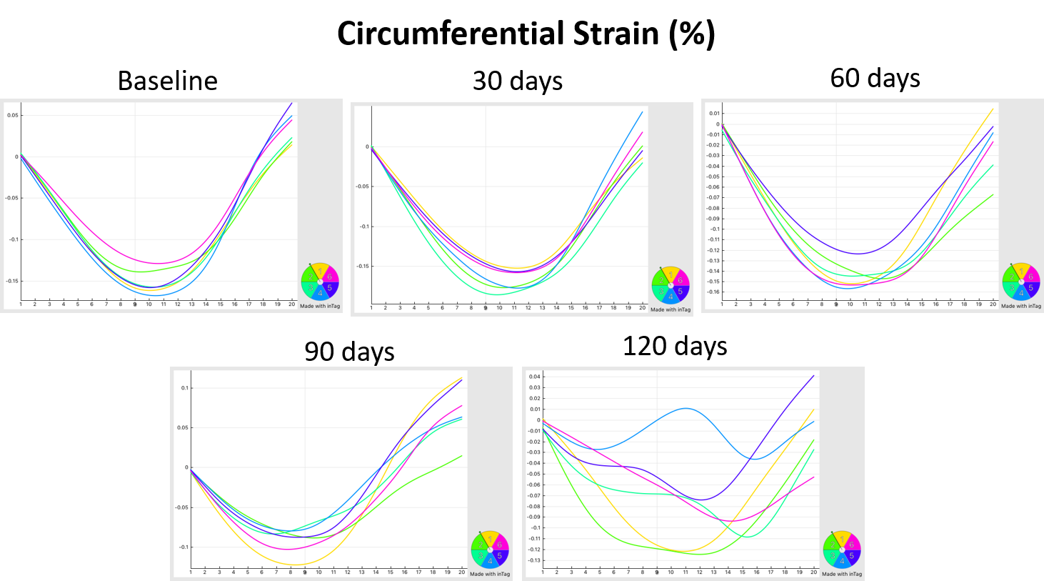
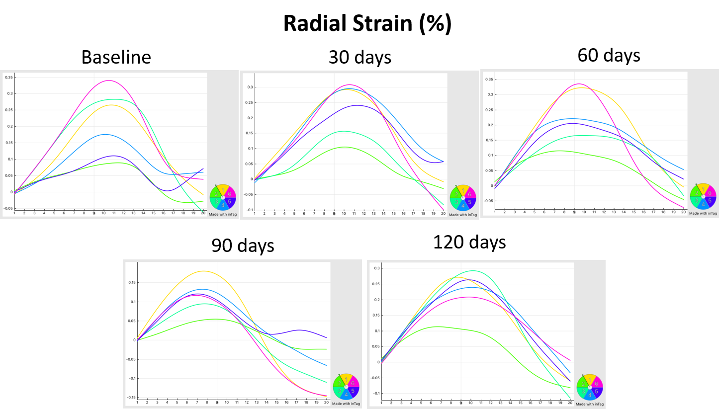
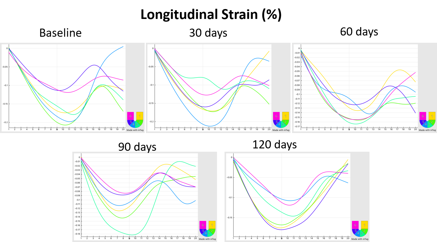
Male WAG rats at 6 weeks of age (n=4) were irradiated below the diaphragm with three 5Gy fractions of X-rays and studied over the 120-day follow up period. Cardiac MRI was conducted at baseline and 30, 60, 90, and 120 days after irradiation. The rats were imaged on a 9.4 T small-animal MRI scanner with 30-cm bore diameter (Bruker, Ettlingen, Germany) using a 4-element surface coil. The rats were scanned in the prone position, with the heart region positioned at the center of the coil. The cardiac MRI exam included cine, tagging, inversion recovery T1 imaging, and multi-echo spin-echo T2 imaging sequences to evaluate global function, regional function, and myocardial tissue characterization. Elevations in T1 and T2 values have been previously associated with increased tissue fibrosis and edema, respectively. Both long-axis and short-axis slices covering the heart were acquired. The cine, inversion recovery, and spin-echo images were analyzed using the cvi42 (Circle Cardiovascular Imaging, Calgary, Canada) software to measure ejection fraction (EF), mass, end-diastolic volume (EDV), and T1/T2 maps. The tagged images were analyzed using the SinMod technique (InTag, Lyon, France) to measure myocardial circumferential (Ecc), radial (Err), and longitudinal (Ell) strains. Values reported as mean ± SEM. Statistical t-test was used to compare measurements from experimental and control rats and at different imaging timepoints. P<0.05 was considered significant.Results
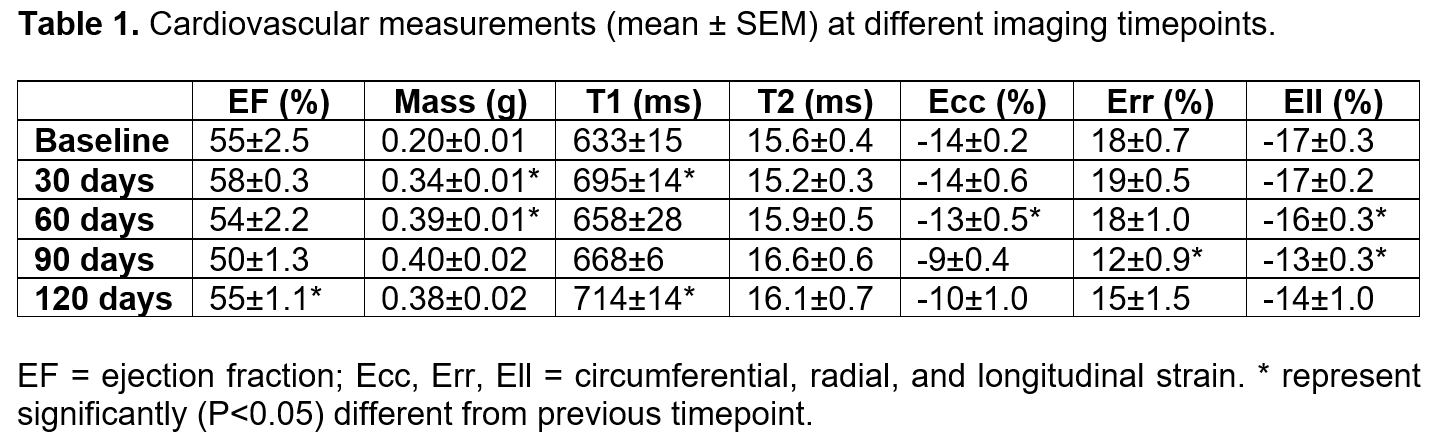
The results showed normal left-ventricular (LV) EF at baseline (55±2.5%), followed by slight increase in EF at 30-days (58±0.3%), which slightly decreased at 60-days (54±2.2%) and 90-days (50±1.3%), then increased at 120-days (55±1.1%). LV mass significantly increased post irradiation (0.34±0.01g, 0.39±0.01g, 0.40±0.02g, and 0.38±0.02g at 30-, 60-, 90-, and 120-days, respectively) compared to baseline (0.20±0.01g). There were slight differences in T2 between different imaging timepoints (15.6±0.4ms, 15.2±0.3ms, 15.9±0.5ms, 16.6±0.6ms, and 16.1±0.7ms at baseline, 30-, 60-, 90-, and 120-days respectively). Apparent T1 value showed significant increase from baseline (633±15ms) to 30-days (695±14ms), which slightly decreased at 60-days (658±28ms) and 90-days (668±6ms), although it was still larger than baseline, before significantly increase at 120-days (714±14ms). LV strain measurements were almost maintained up to 60-days, but significantly dropped after that. Ecc= -14±0.2%, -14±0.6%, -13±0.5%, -9±0.4%, and -10±1.0% at baseline, 30-, 60-, 90-, and 120-days, respectively. The corresponding values in Err were: 18±0.7%, 19±0.5%, 18±1.0%, 12±0.9%, and 15±1.5%; and the corresponding values in Ell= -17±0.3%, -17±0.2%, -16±0.3%, -13±0.3%, and -14±1.0%, respectively.Discussion and Conclusions
The overall goal of the proposed research was to utilize cardiac MRI as a tool to determine the impact of radiation below the diaphragm on the development of cardiac dysfunction and changes in myocardial tissue characteristics in the non-targeted heart. The results showed normal LV EF at all timepoints (>50%) and hypertrophy starting at 30-days. Strain measurements showed maintained values until the 60-days timepoint, after which they significantly decreased. T1 showed significant increase at 30-days, and values at 60- and 90-days were larger than baseline, mainly representing diffused fibrosis post irradiation. T2 showed slight differences between different timepoints. It should be noted that apparent T1 values were shorter than expected true T1 values, which could be due to the influence of very short T2 time or incomplete magnetization recovery; nevertheless, as T2 measurements were similar at different timepoints, the measured T1 values should still reflect changes in true T1. In patients receiving radiotherapy for testicular, mean dose received by the heart was estimated to be 0.75 Gy, with only 14% of the cardiac volume receiving over 0.9 Gy. Therefore, this dose of cardiac radiation is unlikely to account for the observed elevation in risk. Radiotherapy initiates self-perpetuating endothelial inflammation that confers adverse risk to the whole cardiovascular system rather than just the parts directly irradiated. Inadvertent irradiation of renal tissue, leading to hypertension and a consequent increase in the risk of cardiac disease, may be partly responsible for cardiotoxicity, which could explain how large volume infra-diaphragmatic radiotherapy increases risk without significant cardiac irradiation. In conclusion, the results from this study emphasize the value of MRI, especially myocardial strain and T1 measurements, as sensitive markers of changes in regional cardiac function and tissue composition due to indirect effects on the heart from radiation of organs below the diaphragm.Acknowledgements
Funding from Daniel M. Soref Charitable Trust, MCW, USA.References
1. Lenarczyk et al. FASEB BioAdvances. 2:705-719.
2. Lenarczyk et al. Radiat Res. 180:247-58
3. Aravindan et al. Cancer Gene Ther. 21:54-9.
4. Ibrahim. Heart Mechanics. Magnetic Resonance Imaging. CRC Press, Boca Raton, FL. 2017
Figures