0985
Association Between Ventricular Arrhythmias and Late Gadolinium Enhancement in Patients with Apparently Normal Hearts1The First Affiliated Hospital of Nanjing Medical University, Nanjing, China, 2Cardiovascular Medicine, The First Affiliated Hospital of Nanjing Medical University, Nanjing, China, 3MR Collaboration, Siemens Healthineers Ltd., Shanghai, China, 4radiology, The First Affiliated Hospital of Nanjing Medical University, Nanjing, China
Synopsis
Cardiac magnetic resonance (CMR) is the gold standard for evaluating myocardial fibrosis. This study investigated the association between the occurrence and morphological features of ventricular arrhythmias (VAs) and left ventricular late gadolinium enhancement (LV-LGE) in patients without structural heart diseases. Seventy-eight patients with apparently normal hearts were enrolled and underwent simultaneous electrocardiogram and CMR examinations. Polymorphic VAs and ST-segment depression indicated a higher probability of having LV-LGE on CMR. The findings can help clinicians screen patients for CMR.
introduction
Ventricular arrhythmias (VAs) are common abnormalities seen on electrocardiograms (ECGs) and include premature ventricular complexes, non-sustained ventricular tachycardia, accelerated idioventricular rhythm, sustained ventricular tachycardia, and ventricular fibrillation1. VAs are usually benign2. However, frequent and polymorphic VAs, as well as malignant VAs, can contribute to prognoses such as cardiomyopathies, high risk of heart failure, and even sudden cardiac death2.Myocardial fibrosis provides potential substrates for the initiation and perpetuation of VAs3. Cardiac magnetic resonance (CMR) with late gadolinium enhancement (LGE) is a noninvasive tool that can accurately identify and quantify ventricular myocardial fibrosis3. Because of long examination times, it is difficult to perform CMR on patients with ECG abnormalities. This study aimed to explore the association between VAs' occurrence and morphological features and left ventricular LGE (LV-LGE) characteristics in patients with apparently normal hearts to help guide clinical work.methods
Seventy-eight patients with apparently normal hearts were enrolled. The patients underwent simultaneous 24-hour ambulatory Holter ECG and CMR examinations. A 3T MR scanner (MAGNETOM Skyra, Siemens Healthcare, Erlangen, Germany) was used for the CMR scan including LGE. The presence and extent of LGE were determined and compared based on the occurrence and morphological features of VAs. The clinical characteristics were also recorded and calculated.results
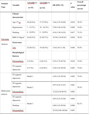
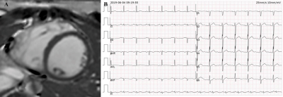
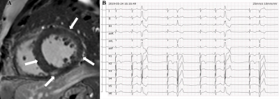
LV-LGE was observed in 19 (37.3%) and 4 (14.8%) patients with and without VAs, respectively (P=0.039). It was more frequently observed in patients with polymorphic VAs (P=0.024) and ST-segment depression (P=0.001), and its extent was greater in polymorphic VAs than monomorphic VAs, with a difference that approached significance (P=0.055) (Table 1). In multivariate analyses adjusted for other clinical variables, the presence of ST-segment depression (hazard ratio: 8.8; 95% CI: 2.2-35.5; P=0.002), drinking (hazard ratio: 6.8; 95% CI: 1.6-28.6; P=0.008), and polymorphic VAs (hazard ratio: 25.2; 95% CI: 3.9-164.1; P=0.001) were associated with greater prevalence of LV-LGE (Table 2). An example of a patient without VAs but with LGE is shown in Figure 1. Another example of a patient with polymorphic VAs and multiple LGE was present in Figure 2.discussion
This study evaluated the association between VAs' occurrence and morphological features and presence of LGE in patients with apparently normal hearts. The results demonstrated that LGE is more common in patients with VAs, especially in patients with polymorphic VAs and ST-segment depression. Specifically, polymorphic VAs and ST-segment depression were associated with 25.2-fold and 8.8-fold increases, respectively, in the occurrence of LV-LGE. The frequency and burden of Vas showed no effect on the presence of LV-LGE. LV-LGE extent tended to increase in patients with polymorphic VAs.conclusions
The presence of polymorphic VAs and ST-segment depression indicated a higher probability of having LV-LGE on CMR. Because LGE plays a vital role in risk stratification, therapies should be administered early to prevent myocardial fibrosis in patients with certain ECG characteristics and without known structural heart diseases.Acknowledgements
noneReferences
1. Marthe A J Becker, Jan H Cornel , Peter M van de Ven, et al. The prognostic value of late gadolinium-enhanced cardiac magnetic resonance imaging in nonischemic dilated cardiomyopathy: a review and meta-Analysis. JACC Cardiovasc Imaging, 2018, 11(9):1274-1284.2.
2. Luebbert J, Auberson D, Marchlinski F. Premature ventricular complexes in apparently normal hearts. Card Electrophysiol Clin, 2016, 8(3):503-514.3.
3. Shin DG, Lee HJ, Park J, et al. Pattern of late gadolinium enhancement predicts arrhythmic events in patients with non-ischemic cardiomyopathy. Int J Cardiol, 2016, 222:9-15.
Figures

Table 1. Association between left ventricular late gadolinium enhancement and ventricular arrythmias frequency and morphological features


