0973
A preliminary study on myocardial strain evaluation of ventricular septal bulge and hypertrophic cardiomyopathy in the elderly
Jingwen Dai1, Min Zhang1, Jing An2, and Min Chen1
1Beijing Hospital, National Center of Gerontology, Beijing, China, 2Siemens Shenzhen Magnetic Resonance Ltd, Shenzhen, China
1Beijing Hospital, National Center of Gerontology, Beijing, China, 2Siemens Shenzhen Magnetic Resonance Ltd, Shenzhen, China
Synopsis
To investigate the differences in basal septal hypertrophy in elderly people between ventricular septal bulge(VSB) and hypertrophic cardiomyopathy(HCM) based on myocardial strain analysis, A total of 13 VSB subjects, 9 HCM patients, and 8 healthy controls underwent cardiac MR scan. The strain parameters shows significant differences between VSB and HCM, and no significant differences between VSB and healthy controls. These findings may suggest a different underlying pathological change and provide a potential diagnostic clue in distinguishing VSB and HCM in elderly people.
Introduction
Ventricular septal bulge (VSB) is known as a localized hypertrophy of the basal ventricular septum and is often described in elderly patients. It has been considered to be an incidental finding related to older age and hypertension1. It is sometimes difficult to distinguish from hypertrophic cardiomyopathy (HCM) especially in elderly patients, and its impact on cardiac function remains uncertain. The aim of this study is to evaluate the LV strain changes in elderly people with VSB, and to investigate the diagnostic differences by strain analysis in discriminating VSB and basal septal hypertrophic HCM in the elderly.Methods
This study recruited 13 VSB subjects (9 males), 9 HCM patients (6 males), and 8 healthy controls (4 males). All the subjects were over 50 years old. HCM and VSB were diagnosed with conventional echocardiography by assessing the wall thickness and morphology of the basal septum. HCM was defined by a wall thickness ≥15 mm in one or more segments including basal septum that cannot be solely explained by abnormal loading conditions. VSB were defined as localized basal septal hypertrophy with an increased wall thickness>12mm in the absence of other cardiac diseases. CMR studies were acquired on a 3.0T MR (MAGNETOM Prisma; Siemens Healthcare, Erlangen, Germany) using an 18-channel body coil. ECG-gated, breath-hold, balanced steady-state free-precision (bSSFP) cine images were collected in the LV long-axis views of 2-, 3-, and 4-chamber views, and a short-axis stack covering the entire LV. Imaging parameters were FOV = 340mm x 340mm, slice thickness = 6mm, TR/ TE=39.4ms/1.4ms, flip angle (FA) = 60°, voxel size = 1.6mm x 1.6mm x 6.0mm. Left ventricular strain was analyzed on long- and short-axis cine images using tissue tracking software (cvi42, Circle Cardiovascular Imaging, Calgary, Canada). Data were analyzed using SPSS (version 20.0), and the Chi-squared test was used.Results
This study included 13 VSB patients (72.0±8.5 years old), 9 HCM patients (57.4±5.0 years old), and 8 healthy controls (60.8±7.2 years old) (Figure 1). Compared with HCM patients, subjects with VSB were older (p=0.013). All the subjects had a normal LV ejection fraction (LVEF). The global radial, circumferential, and longitudinal strain had no significant difference between normal controls and VSB subjects along with the basal radial, circumferential strain. The global and basal radial, circumferential, and longitudinal strain were significantly different in HCM compared with NC and VSB subjects (Figure 2).Discussion
This is a preliminary study in evaluation of hypertrophy of the basal septum in the elderly using strain analysis. VSB is more frequently described in echocardiography. However, the understanding and evaluation by cardiac magnetic resonance is rare and needs to be improved. The results between healthy people and subjects with VSB indicating that in the elderly people, the changed morphology of the basal septum does not represent abnormal deformation of myocardial motion which may suggest a more benign clinical course with VSB. In spite of hypertrophic basal septum, the significant strain differences may reveal a different underlying pathological and phenotype basis of myocardial hypertrophy in the elderly people between VSB and HCM, and also suggest a potential diagnostic clue for differentiate VSB and HCM in the elderly. However, further study with a larger sample size and more detailed analysis is needed to verify this initial results.Conclusion
Ventricular septal bulge in elderly subjects showed no abnormal myocardial strain compared with healthy people. Myocardial strain parameters differentiate between VSB and HCM which may provide a potential clue in distinguishing different patterns of hypertrophic basal septum in elderly people.Acknowledgements
No acknowledgement found.References
1. Canepa M, Malti O, David M, et al. Prevalence, Clinical Correlates, and Functional Impact of Subaortic Ventricular Septal Bulge (from the Baltimore Longitudinal Study of Aging). Am J Cardiol 2014; 114:796–802.Figures
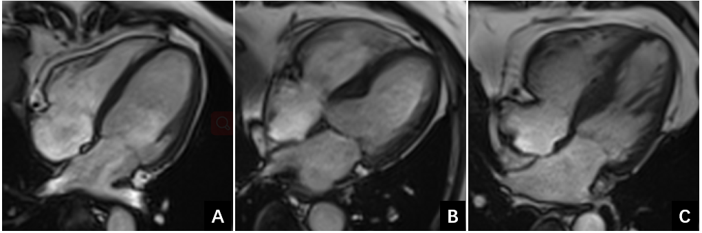
Figure 1. 4-chamber view of long axis at
end-diastolic phase
A: 60-year-old
healthy woman with a normal myocardial morphology.
B: 71-year-old
man with an isolated basal septum hypertrophy (15mm) protruding into the outflow
tract of the left ventricle was diagnosed as VSB.
C:56-year-old man with a spindle-shaped
basal septum hypertrophy (16mm) was diagnosed as HCM.
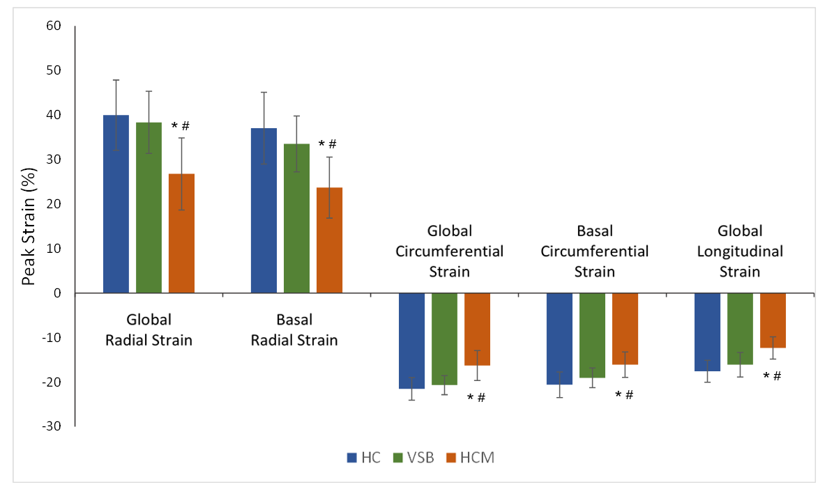
Figure 2. Radial, circumferential, and longitudinal
peak strain in three groups. All comparisons between healthy control (NC) and
hypertensive patients with basal septal hypertrophy (VSB) were nonsignificant.
* p<0.05 compared with HC. # p<0.05 compared with VSB.