0950
Differential diagnosis of Endometrial carcinoma and polyps using Amide proton transfer-weighted imaging and permeability analysis1The First Affiliated Hospital of Dalian Medical University, Dalian, China, 2Philips Healthcare, Beijin, China
Synopsis
The accurate differential diagnosis of endometrial carcinoma and endometrial polyps always met many challenges. In this study, we evaluated the capability of differentiation of endometrial carcinoma from endometrial polyps with APT signal intensity (SI) and permeability parameters. The diagnostic efficiency is the highest using APT combined with Kep. APT and permeability parameters are potentially a promising and valuable method in differentiation of endometrial carcinoma from endometrial polyps.
Introduction
The accurate differential diagnosis of endometrial carcinoma and endometrial polyps is hard to made because of not only the resemblance of these lesions to each other but also the disturbance from the background of endometritis, artifacts, and so on1. Amide proton transfer-weighted (APTw) imaging is a molecular imaging technique with the amide protons weighted contrast based predominantly on the concentration and exchange environment (pH, temperature, etc) of mobile cellular proteins and bulk water2. The volume transfer constant (Ktrans) is an indicator of capillary permeability calculated using dynamic contrast-enhanced (DCE) MR to reflect the transfer between blood plasma and Extravascular Extracellular Space (EES). The similar permeability paraments included rate between EES and blood plasma (Kep), extravascular volume fraction (ve) and plasma volume fraction (vp). DCE-MRI has been extensively used for diagnosis, prognosis and therapy monitoring of various diseases including cancer3. This study aims to evaluate the differential diagnosis capability between endometrial carcinoma and endometrial polyps using APT value and permeability indictors (Ktrans, Kep, ve, vp).Methods
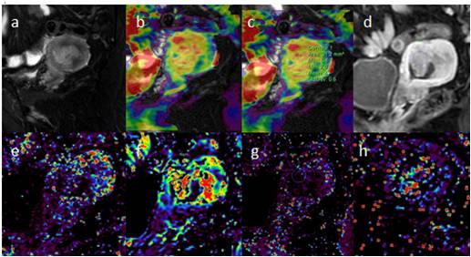
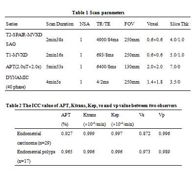
Twenty-nine patients (mean age: 52.76 ± 8.09, range: 33-67 years) with endometrial carcinoma and seventeen patients (mean age: 47.88 ± 11.88, range: 29-71 years) with endometrial polyps were recruited in this study. All of patients were confirmed by operation and pathology and were ask to sign the informed consent. MR imaging was performed on a 3.0 T MR scanner (Ingenia CX, Philips Healthcare, the Netherlands) with a 16-channel abdominal coil. Scan protocol included of T2-SPAIR-MVXD, APT weighted and dynamic enhancement (40 phase) sequence and scan parameters were shown in Table 1. The APT imaging and permeability parameters mapping were calculated on the IntelliSpace Portal 10.0 (ISP 10, Philips Healthcare, the Netherlands). Three ROIs were placed manually on each cyst (Fig. 1). The average signal intensity of APT and permeability parameters (Ktrans, Kep, ve, vp) were measured by two experienced neuroradiologists. The intra-class correlation coefficient (ICC) was used to test the consistency of the two observers. The differences between the two groups of APT and Ktrans, Kep, ve, vp values were compared using Mann-Whitney U test. Logistic regression and ROC were plotted to calculate the diagnostic efficiency of endometrial polyps. Delong test was used to compare the diagnostic efficacy.Results
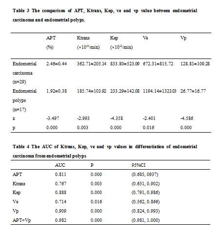
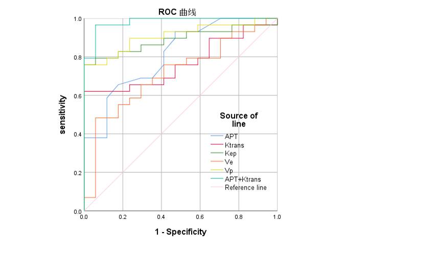
Inter-observer ICCs of APT, Ktrans, Kep and vp are more than 0.9 of the two observers in endometrial carcinoma and endometrial polyps, except ICC of ve in endometrial carcinoma of 0.872. Elevated APT SI and Ktrans, Kep and vp values in endometrial carcinoma were found with significant statistical difference (p < 0.05) (Table 2). Decreased ve in endometrial carcinoma was also detected. As for ROC curves, the AUC values of APT, Ktrans, Kep, ve, and vp value are all more than 0.7. The diagnostic efficiency of APT combining with Kep is the higher with statistic difference of those of APT, Ktrans, Kep and Ve (p = 0.0039, 0.0014, 0.0479, 0.0008, respectively).Discussion
In this study, we found that increased APT SI in endometrial carcinoma. The reason may contribute to the higher cellular density and more over-expressed proteins in endometrial carcinoma than endometrial polyps. This results is in agreement with the APTw hyperintensity based on MR spectra of perfused tumor cells4. Permeability analysis produces a minimally invasive visualization of tumor angiogenesis. Because of the disorganization and increased permeability of the tumor vessels, higher Ktrans, Kep and vp value of Endometrial carcinoma are found than that of endometrial polyps, which reflect the material transport in the intravascular and extravascular spaces. However, the vp value reflect the vascular (plasma) gap volume fraction and is higher in malignant tumor.Conclusions
APTw SI and permeability indictors based on DCE-MRI are potentially a promising and valuable non-invasive method in differentiation of endometrial carcinoma from endometrial polyps.Acknowledgements
No acknowledgement found.References
1. Silverberg SJMpaojotUS, Canadian Academy of Pathology I. Problems in the differential diagnosis of endometrial hyperplasia and carcinoma. 2000;13(3):309-327.
2. Zhou J, Payen J, Wilson D, Traystman R, van Zijl PJNm. Using the amide proton signals of intracellular proteins and peptides to detect pH effects in MRI. 2003;9(8):1085-1090.
3. Kim HJJon, science. Variability in Quantitative DCE-MRI: Sources and Solutions. 2018;4(1).4. Zhou J, Heo H, Knutsson L, van Zijl P, Jiang SJJomriJ. APT-weighted MRI: Techniques, current neuro applications, and challenging issues. 2019;50(2):347-364.
Figures