0943
Evaluating the cytokeratin 19 (CK-19) status via neural network model established by the SWI-derived radiomics features1Sun Yat-sen University Cancer Center, Guangzhou, China, 2Central Research Institute, United Imaging Healthcare, Shanghai, China
Synopsis
Susceptibility Weighted Imaging (SWI) has shown tremendous clinical significance for identifying the hepatic micro-structural abnormalities such as micro-bleeding, vascularity, nodule and so forth. Recent studies concluded that cytokeratin 19 (CK-19) is an important marker for prognostic prediction of hepatocellular carcinoma (HCC). We hypothesized that the neural network model can be established by means of extracting high throughput radiomics features from SWI images for noninvasively evaluating the CK-19 status with high accuracy. The results demonstrated that such deep learning based neural network model yielded excellent diagnostic performance for predicting the CK-19 status.
Introduction
With notoriously high incidence and mortality, HCC has been a tremendous threat to human health for the long time. Early prediction of the prognosis with high accuracy will be of great importance for guiding the clinical management of HCC. Some recent studies unveiled that the CK-19 status is closely associated with the prognosis of HCC.1-3 Therefore, CK-19 is valuable for noninvasively predicting the prognosis of HCC. Susceptibility Weighted Imaging (SWI), one functional MRI technique, has attracted innumerable attention from investigators due to the powerful capability of visualizing the micro-structural abnormalities containing haemorrhage, vascularity, nodule and so on, which are representative characteristics during the progression of hepatic diseases.4 In the past few years, Radiomics, with the aim of extracting quantitative image features with high throughput, has been widely applied for exploring the meaningful diagnostic markers and models.5 However, to the best of our knowledge, hardly has the SWI and Radiomics been integrated via deep learning based neural network for noninvasively predicting the prognostic markers of HCC. This research, hence, aims to evaluate the CK-19 status via neural network model established by the SWI-derived radiomics features.Methods
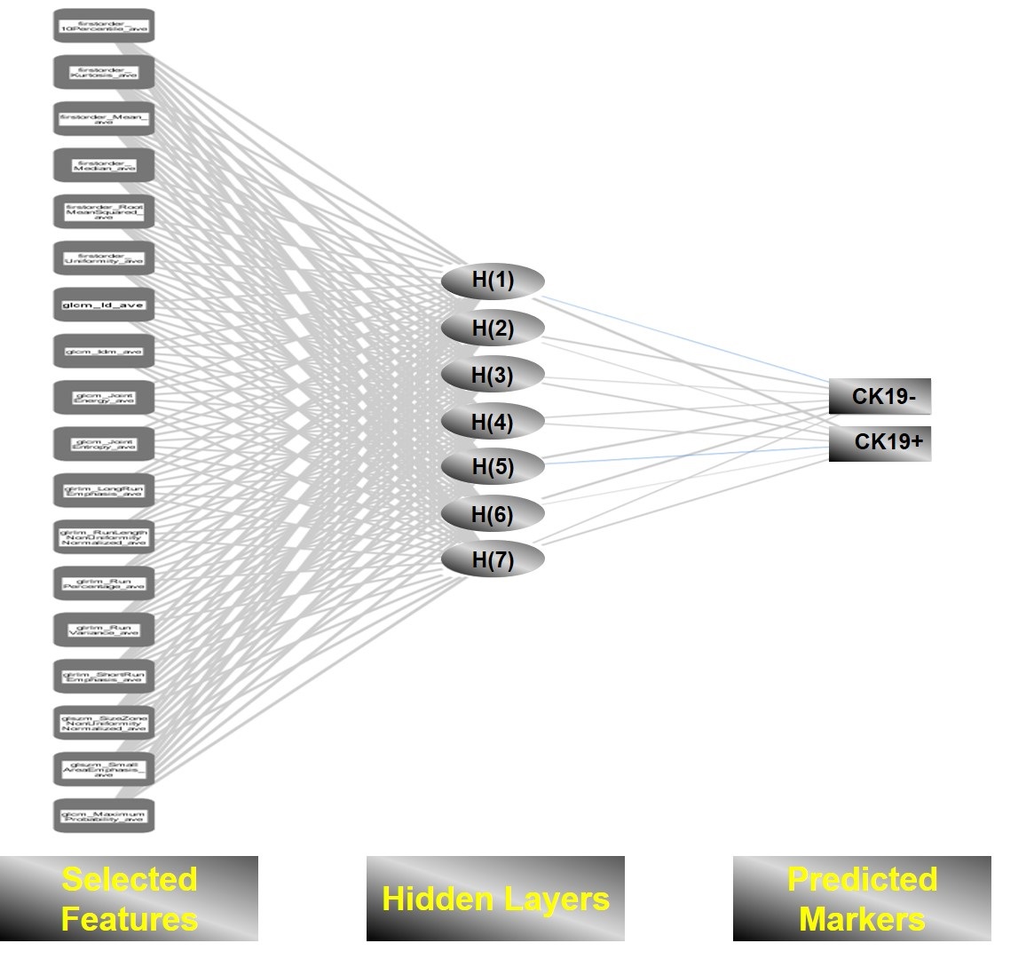
From March 2019 to October 2020, a total of 90 patients with HCC were included into this prospective study. MR examinations were conducted with a commercial 3.0 T scanner (uMR 780, United Imaging Healthcare Co Ltd). Detailed parameters of SWI sequences were listed as the following: TR/TE: 140.0 ms/10.0 ms, flip angle: 30 °, slice thickness: 5.0 mm, Matrix/ Field of View: 368×216/280 mm × 380 mm. The expression status of CK-19 (positive or negative) of each patient was obtained from our electronic medical record system. After the segmentation of the whole tumor by two experienced abdominal radiologists, the radiomics features were extracted by means of an open-sourced software named Pyradiomics (https://pyradiomics.readthedocs.io/en/latest/). Specifically, 107 radiomics features were extracted from the SWI images of each patient. Next, interobserver agreement coefficient (ICC) was used to quantify the reproducibility of features. The radiomics features with poor reproducibility (ICC < 0.8) were removed. Then, the feature selection was carried out by means of LASSO regression. Next, the selected features were utilized to establish the deep learning based neural network model, Artificial Neural Network (ANN). The diagnostic performance was evaluated by means of ROC analysis 5-fold cross validation.Results
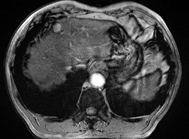
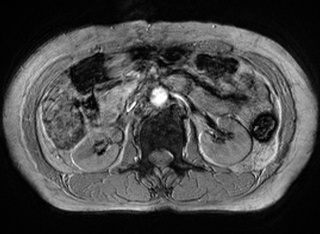
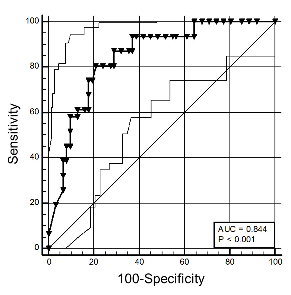
Figure 1 and Figure 2 respectively display the SWI images of one patient with negative CK-19 expression and one patient with the positive CK-19 expression. MR images show that micro-haemorrhage and neovascularization are more common in patients with positive CK-19 expression. Moreover, the results indicated that 90 features were with high reproducibility and stability (ICC > 0.8). Moreover, a total of 17 features were finally selected by means of LASSO Regression. The structure of ANN model was exhibited in Figure 3. Finally, the ANN model yielded an excellent diagnostic performance with AUC of 0.844 for identifying the CK-19 status of HCC.Discussion
Recently, CK-19, a histopathologic index, has drawn the attention of plenty of investigators. Their results indicated that CK-19 is of great clinical value for prognostic prediction of HCC. Specifically, recurrence, poor prognosis and lymph node metastasis are much more common in the patients with positive expression of CK-19. Thus, CK-19 has been widely regarded as a valuable prognostic marker of HCC. The study revealed that the ANN model established via the SWI-derived radiomics features was able to yield fascinating diagnostic performance. The potential causes were speculated as the followings: 1) Previous studies have concluded that SWI is able to visualize some pathological changes during the progress of HCC such as nodular dysplasia, hyper-vascularity and hemorrhage.6-8 Theses pathological changes may be associated with the expression of CK-19, which results in the indirect association between the SWI image manifestations and the expression of CK-19. 2) The radiomics technology is able to provide hundreds of quantitative diagnostic markers. 3) Deep learning based neural network is robust for classifying the different subgroups (positive CK-19 vs negative CK-19) with high accuracy.Conclusion
Deep learning based neural network established by SWI-derived radiomics features holds great potential in identifying the CK-19 status with high accuracy.Acknowledgements
NoneReferences
1. Zhuang PY, Zhang JB, Zhu XD, et al. Two pathologic types of hepatocellular carcinoma with lymph node metastasis with distinct prognosis on the basis of CK19 expression in tumor. Cancer: Interdisciplinary International Journal of the American Cancer Society 2008;112(12):2740-2748.
2. Zhuo J-Y, Lu D, Tan W-Y, Zheng S-S, Shen Y-Q, Xu X. CK19-positive Hepatocellular Carcinoma is a Characteristic Subtype. Journal of Cancer 2020;11(17):5069.
3. Choi S-Y, Kim SH, Park CK, et al. Imaging Features of Gadoxetic Acid–enhanced and Diffusion-weighted MR Imaging for Identifying Cytokeratin 19-positive Hepatocellular Carcinoma: A Retrospective Observational Study. Radiology 2018;286(3):897-908.
4. Haacke EM, Xu Y, Cheng YCN, Reichenbach JR. Susceptibility weighted imaging (SWI). Magnetic Resonance in Medicine: An Official Journal of the International Society for Magnetic Resonance in Medicine 2004;52(3):612-618.
5. Gillies RJ, Kinahan PE, Hricak H. Radiomics: images are more than pictures, they are data. Radiology 2016;278(2):563-577.
6. Li Rk, Zeng Ms, Rao Sx, et al. Using a 2D multibreath‐hold susceptibility‐weighted imaging to visualize intratumoral hemorrhage of hepatocellular carcinoma at 3T MRI: Correlation with pathology. Journal of Magnetic Resonance Imaging 2012;36(4):900-906.
7. Chang S-X, Li G-W, Chen Y, et al. Characterizing venous vasculatures of hepatocellular carcinoma using a multi-breath-hold two-dimensional susceptibility weighted imaging. PloS one 2013;8(6):e65895.
8. Chen W, DelProposto Z, Liu W, et al. Susceptibility-weighted imaging for the noncontrast evaluation of hepatocellular carcinoma: a prospective study with histopathologic correlation. PLoS One 2014;9(5):e98303.
Figures