0924
3D Free-breathing Multitasking T1-T2 Mapping in Small Animals on a 3-Tesla System: A Preliminary Study on a Murine Model with Liver Metastasis1Biomedical Imaging Research Institute, Cedars-Sinai Medical Center, Los Angeles, CA, United States, 2Chemistry Department, Georgia State University, Atlanta, GA, United States, 3Department of Medicine, Cedars-Sinai Medical Center, Los Angeles, CA, United States, 4Bioengineering, University of California, Los Angeles, Los Angeles, CA, United States, 5Division of Digestive and Liver Diseases, Cedars-Sinai Medical Center, Los Angeles, CA, United States
Synopsis
MRI is a promising tool for the non-invasive study of animal model, but continues to face technical challenges. In this work, a Multitasking T1-T2 mapping technique was proposed for mouse abdominal imaging on 3T, which achieved 3D coverage, motion-resolved acquisition, and simultaneous T1 T2 mapping within 10 minutes. The study was performed on a murine model with liver metastasis of colorectal cancer. Data at multiple time points after tumor injection were acquired with different contrast agent, Eovist and ProCA.collagen1. The results demonstrated that 10-min Multitasking technique produced improved images with clear tumor delineation compared to conventional MRI series (50 minutes).
Introduction
Studies on living animals are critical to oncology research1-3. MRI has been an emerging tool to noninvasively monitor tumor progression/regression in small animals, such as murine models of cancer4. However, there are still demanding technical challenges that hamper the application of advanced MRI techniques to small animal studies. First, to achieve adequate spatial resolution for small animal anatomy, images are usually performed at high field systems, such as 4.7T – 11.7T, at the expense of additional field inhomogeneity and less translatability to human studies5. Second, imaging of the abdomen, which is essential for multiple type of cancer studies, is susceptible to respiratory motion-related artifacts. Respiratory triggering can improve image quality, but at the cost of significantly lengthened scan time and experimental complexity. To address these limitations, and to develop an accelerated, motion-resolved imaging tool for mouse studies, we proposed a Multitasking technique on 3.0T scanner6, which achieved 3D-coverage, free-breathing, and simultaneous T1-T2 mapping in a single 10-min scan. The feasibility of the Multitasking technique was tested on a murine model with liver metastasis of colorectal cancer.Methods
Animal Model: 8- to 12-week-old female mice were fed with a low-fat diet (LFD; 12% fat calories) or high-fat diet (HFD; 60% fat calories) for 6 weeks. After a laparotomy, MC38 cells (200000 cells; C57BL/6 background CRC cell line) were injected through the spleen. Two weeks following injection (8 total weeks of LFD/HFD feeding), tumors were harvested and analyzed.Contrast Agent: A novel protein-based MRI contrast agent ProCA32.collagen1 with collagen type I targeting capability was used in this work7. The R1 and R2 relaxivity of ProCA32.collagen1 are 14 to 20-fold higher than clinically used contrast agents. The uniqueness of both high relaxivity and collagen specificity enable improved detection of colorectal cancer metastasis.
Sequence Design: As shown in Figure 1, continuous acquisition with prototype FLASH following non-selective varied-duration T2-IR preparations was used to generate T1 and T2 contrasts8. Cartesian acquisition with randomized reordering in ky and kz directions was implemented. The center k-space line was collected every 4 readouts as training data8.
Image Reconstruction: The 6D images were formed as a 4-way tensor with voxel location index (by collapse three spatial dimensions into one), inversion recovery, T2 preps, and respiratory dimensions. The high correlation along and across multiple image dimensions induces $$$\mathcal{A}$$$ to be low-rank and thus can be decomposed into the product of the basis matrices of dimensions6:$$\mathcal{A}=\mathcal{G} \times_{1}\mathbf{U}\times_{2}\mathbf{V}\times_{3}\mathbf{W}\times_{4}\mathbf{Q}$$where the columns of $$$\mathbf{U}$$$, $$$\mathbf{V}$$$, $$$\mathbf{W}$$$ and $$$\mathbf{Q}$$$ contain the basis functions for spatial, IR, T2 preps, and respiration, respectively, and $$$\mathcal{G}$$$ denotes the core tensor. The core tensor and the temporal basis matrices $$$\mathbf{V}$$$, $$$\mathbf{W}$$$ and $$$\mathbf{Q}$$$ were first recovered from the training data containing center k-space information, while the spatial basis matrices $$$\mathbf{U}$$$ were then recovered by fitting the temporal bases to the imaging data.
Simultaneous T1-T2 Mapping: The signal evolution of each voxel can be formulated as:$$s\left(A,B,T_{1},T_{2}\right)=A\frac{1-e^{-\frac{T_{\mathrm{R}}}{T_{1}}}}{1-e^{-\frac{T_{\mathrm{R}}}{T_{1}}}\cos\alpha}\left[1+\left(B e^{-\frac{T_{\mathrm{E}, \mathrm{prep}}}{T_{2}}}-1\right)\left(e^{-\frac{T_{\mathrm{R}}}{T_{1}}} \cos\alpha\right)^{n}\right] \sin\alpha$$with amplitude factor $$$A$$$, IR pulse efficiency $$$B$$$, FLASH readout interval $$$T_{\mathrm{R}}$$$, flip angle $$$\alpha$$$, recovery time point $$$n=1,2,\cdots,N$$$ ($$$N$$$ is the number of TIs in an IR period), and T2 prep durations $$$T_{\mathrm{E}, \mathrm{prep}}$$$. A four-parameter voxelwise fit for $$$A$$$, $$$B$$$, $$$T_{1}$$$, $$$T_{2}$$$ were estimated accordingly.
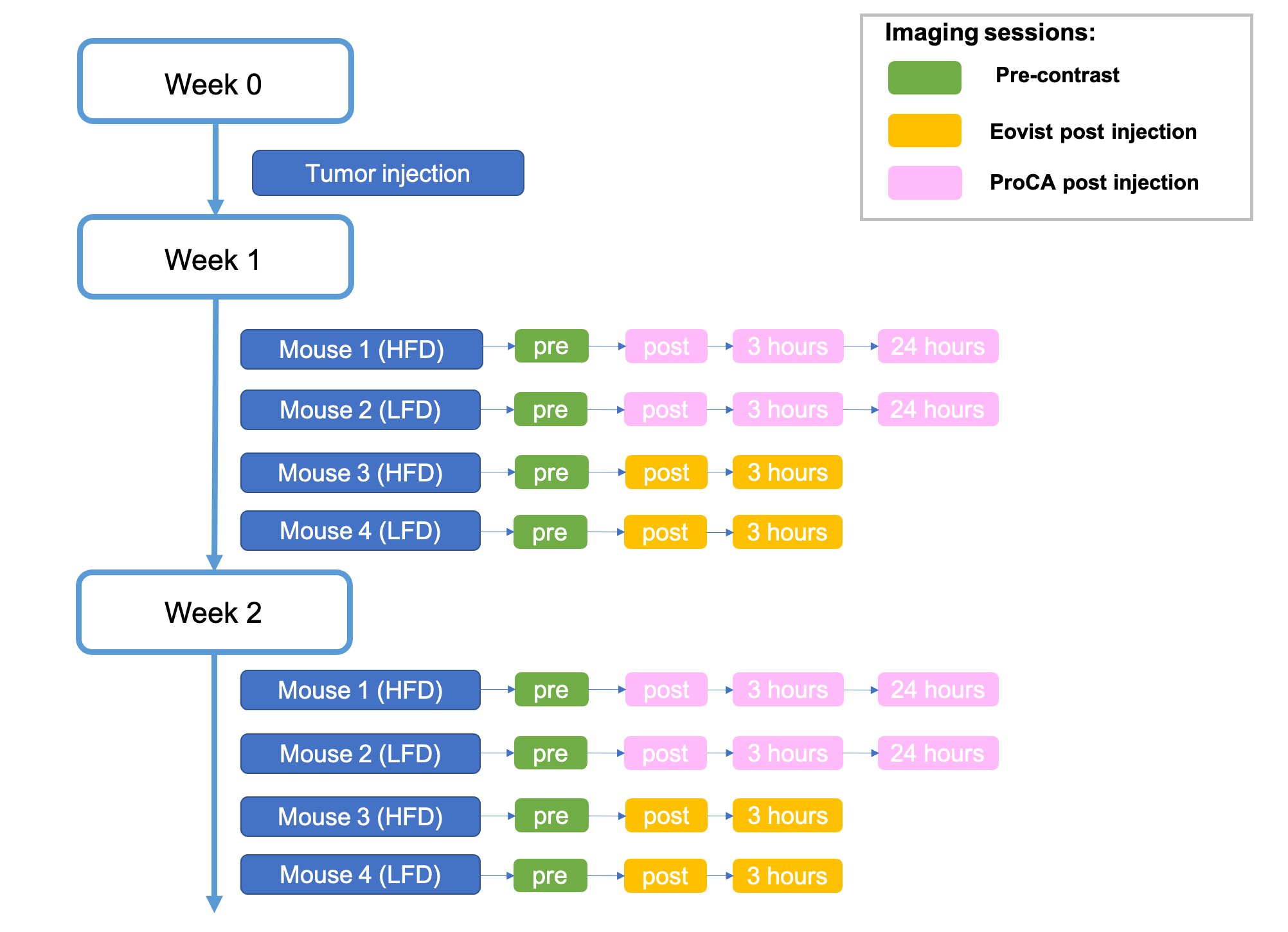
Imaging Experiments: The mice study was performed on a 3T scanner (MAGNETOM Vida, Siemens Healthcare, Germany) with a dedicated small animal coil. Four mice were scanned in this study, including 2 with HFD and the other 2 with LFD. All the mice were scanned 1 week and 2 weeks after tumor injection. Each week, each mouse was studied with Eovist or ProCA. collagen1 at multiple time points, as shown in Figure 2. In each imaging session, following images were acquired: conventional 3D T1-weighted VIBE, T2-weighted TSE, IR-TSE with multiple TIs for T1 mapping, multi-echo spin echo (MESE) for T2 mapping, and Multitasking T1-T2 mapping. All the images were acquired with free breathing. Detailed parameters were listed in Table 1.
Image Analysis: T1 and T2 mapping were performed on conventional and Multitasking images. Image quality were evaluated. The SNR of all weighted images were measured.
Results
Figure 3 illustrates the T1-T2-weighted images and T1 T2 maps from conventional methods and from Multitasking acquired on an LFD mouse at one week after tumor injection. Conventional image series took about 50 minutes. In contrast, Multitasking took 10 minutes with vastly reduced motion artifacts and sharper anatomical delineation. Figure 4 shows a set of images acquired on an HFD mouse at two weeks after tumor injection with visible tumors forming. Multitasking consistently showed better image quality with reduced artifacts. The mean and standard deviation of SNR for conventional T1- and T2-weighted images were 44±11 and 35±15, respectively; for Multitasking T1- and T2-weighted images were 52±8 and 43±12, respectively.Discussion and Conclusion
This study was an initial demonstration of the feasibility of Multitasking T1-T2 mapping for mouse abdominal imaging on 3T, which achieved 3D coverage, motion-resolved acquisition, and simultaneous T1 T2 mapping within 10 minutes. It produced better images with vastly reduced scan time (10 minutes) compared to conventional imaging series (50 minutes). Tumor were clearly depicted with the proposed method. Further analysis with histological validation will be performed.Acknowledgements
No acknowledgement found.References
1. Puaux AL, Ong LC, Jin Y, et al. A comparison of imaging techniques to monitor tumor growth and cancer progression in living animals. Int J Mol Imaging. 2011;2011:321538.
2. Bruns CJ, Liu WB, Davis DW, et al. Vascular endothelial growth factor is an in vivo survival factor for tumor endothelium in a murine model of colorectal carcinoma liver metastases. Cancer. Aug 1 2000;89(3):488-499.
3. VanSaun MN, Lee IK, Washington MK, Matrisian L, Gorden DL. High Fat Diet Induced Hepatic Steatosis Establishes a Permissive Microenvironment for Colorectal Metastases and Promotes Primary Dysplasia in a Murine Model. Am J Pathol. Jul 2009;175(1):355-364.
4. Dwek M, Brooks SA, Schumacher U. Metastasis research protocols. Springer; 2012.
5. Marzola P, Osculati F, Sbarbati A. High field MRI in preclinical research. European Journal of Radiology. Nov 2003;48(2):165-170.
6. Christodoulou AG, Shaw JL, Nguyen C, et al. Magnetic resonance multitasking for motion-resolved quantitative cardiovascular imaging. Nat Biomed Eng. 2018;2(4):215.
7. Salarian M, Yang H, Turaga RC, et al. Precision detection of liver metastasis by collagen-targeted protein MRI contrast agent. Biomaterials. Dec 2019;224:119478.
8. Xie Y, Christodoulou AG, Wang N, Li D. Quantitative Multi-Contrast Atherosclerosis Characterization (qMATCH): Comprehensive Quantitative Evaluation of Atherosclerosis in a Single-Scan. In Proceedings of the 25th Annual Meeting of ISMRM, Honolulu, HI, USA, 2017. Abstract 5148.
Figures