0902
Susceptibility artifact-insensitive ultrafast 3D gradient-echo imaging by combination of head-tilting and ERASE acquisition1Department of Biomedical Engineering, Sungkyunkwan University, Suwon, Korea, Republic of, 2Department of Intelligent Precision Healthcare Convergence, Sungkyunkwan University, Suwon, Korea, Republic of, 3Biomedical Institute for Convergence at SKKU, Sungkyunkwan University, Suwon, Korea, Republic of, 4Center for Neuroscience Imaging Research, Institute for Basic Science (IBS), Suwon, Korea, Republic of
Synopsis
Equal-TE Rapid Acquisition with Sequential Excitation (ERASE) is a 3D ultrafast gradient-echo-based Spatiotemporal Encoding (SPEN) imaging technique with a constant TE and high tolerance to main magnetic field () inhomogeneity. However, misregistration of spin isochromat can occur due to large magnetic susceptibility-induced gradients in the prefrontal brain region. Here, we propose that shim improvement by chin-up head tilting can effectively mitigate misregistration in ERASE to realize fast prefrontal brain imaging with much higher image quality than conventional EPI.
Introduction
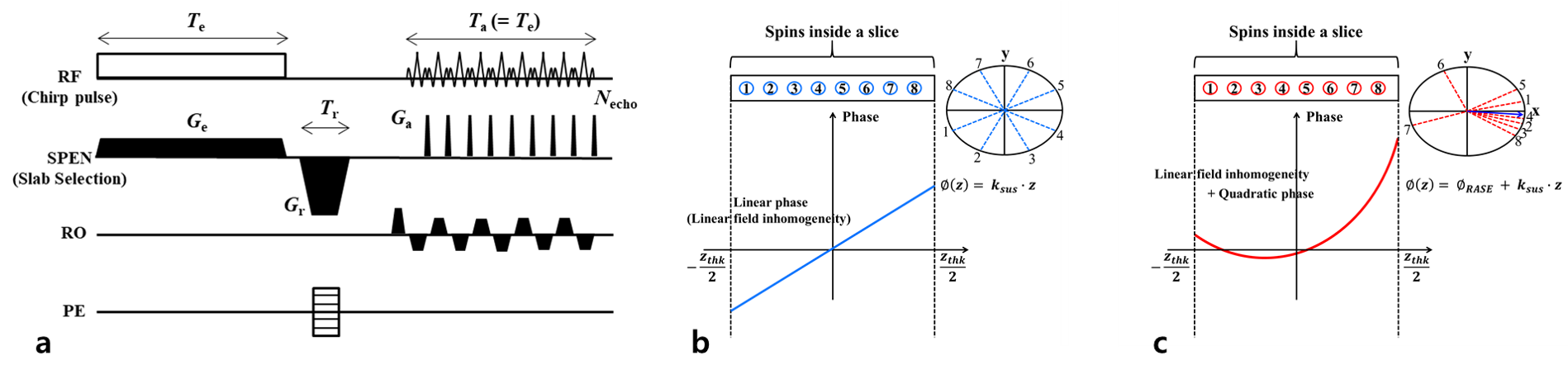
We recently developed a 3D ultrafast gradient-echo Spatiotemporal Encoding (SPEN) imaging technique, referred to as ERASE (Equal-TE Rapid Acquisition with Sequential Excitation)1 (Fig.1a). In ERASE acquisition scheme, a frequency-swept chirp pulse is used for spin excitation producing a quadratic phase that sequentially localizes a signal in both time and space.2 Due to larger gradient amplitudes and the quadratic-phase distribution (Figs.1b,c), ERASE is less sensitive to inhomogeneity than conventional gradient-echo EPI (GRE-EPI).1,2In the presence of strong magnetic susceptibility-induced gradients (Gsus) as in the prefrontal lobe near the sinus cavity, the quadratic-phase shift can lead to misregistration of spin positions and thus image distortions. Such effects can be mitigated by the recently proposed tilted-head scan method3, which effectively reduces near the sinus cavity. In this study, we show that, by combining ERASE with tilted-head brain scan, substantially improved image quality can be achieved in the prefrontal region compared to GRE-EPI. This is demonstrated on a healthy volunteer at 3T and 7T. Our method can be a promising technique to overcome image degradation in the prefrontal region in GRE-based high-field functional MRI studies.
Methods
Possible misregistration in ERASE: A quadratic-phase distribution created by a chirp pulse shifts with the application of a rephasing gradient (Gr) and blipped gradients (Ga) along the SPEN direction. The phase accrued at time ta is expressed as Eq.[1], where γ is the gyromagnetic ratio, L is the FOV in the SPEN direction, Te is the pulse duration, and z 0 is the position of on-resonance isochromat. Assuming that Gsus continues to exist during the echo time, spins that experience Gsus will have an additional phase (Eq.[2]). The vertex position of quadratic-phase shift at ta includes an additional shift (Eq.[3]). Therefore, the misregistration of spin positions can occur when the shift by Gsus exceeds the intrinsic resolution of SPEN, L/\sqrt{R} (R: pulse time-bandwidth product)1,2. To avoid misregistration, the echo time, R-value and the FOV of SPEN direction should be controlled.\phi_{ERASE}=\phi_{e}+\phi_{r}+\phi_{a}=\frac{\gamma G_{e} T_{e}}{2L}(z-\frac{2z_{0}-L}{2})^{2}-\gamma G_{r} T_{r}z+\gamma G_{a} t_{a}z Eq.[1]
\phi_{ERASE}=\frac{\gamma G_{e} T_{e}}{2L}(z-\frac{2z_{0}-L}{2})^{2}-\gamma G_{r} T_{r}z+\gamma G_{a} t_{a}z+\gamma G_{sus} T_{E}z Eq.[2]
z=z_{0}+\frac{L}{2}-\frac{t_{a}}{T_{e}}L-\frac{G_{sus}T_{E}}{G_{e}T_{e}}L Eq.[3]
Tilted-head scan: A previous study proposed that tilted-head scans for healthy subjects can reduce susceptibility-induced signal dropout and improve the image quality in the prefrontal region.3 The tilted-head brain scan has the effect of steering the region of intense B0 inhomogeneity away from its usual location in the inferior frontal lobe directly above the nasal cavity, and it can effectively achieve local B0 shimming without use of additional hardware.
Experiments: All scans followed a human study protocol approved by the Institutional Review Board (IRB) of Sungkyunkwan University. A healthy subject (male; age= 24) was scanned with normal and tilted head orientations in both at 3T (Siemens Prisma) with a 64-channel head-and-neck coil and at 7T (Siemens Terra) with a 32-channel head coil. 2D GRE-EPI and ERASE scans were performed to compare the signal dropout and recovery in the prefrontal region. Scan parameters are listed in Table 1. For tilted-orientation scans taken at 7T, custom-made dielectric pads were used to compensate for signal loss in the cerebellar region. The tilt angles relative to the normal orientations at 3T and 7T were 27.7° and 27.5°, respectively. B0 maps were obtained from the double-echo GRE phase images to verify the improvement of homogeneity. To compare image distortion, we manually drew masks with a reference to GRE images and overlaid them on ERASE and GRE-EPI images.
Results
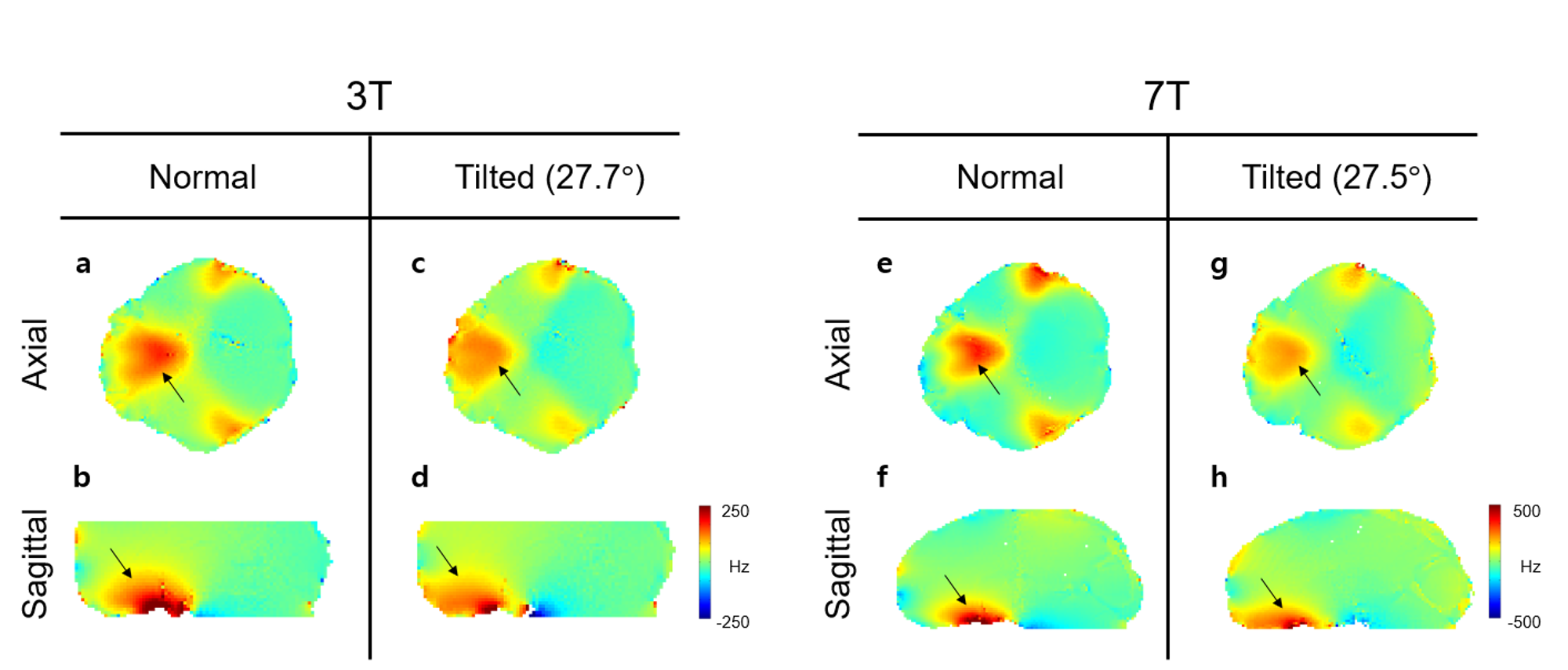
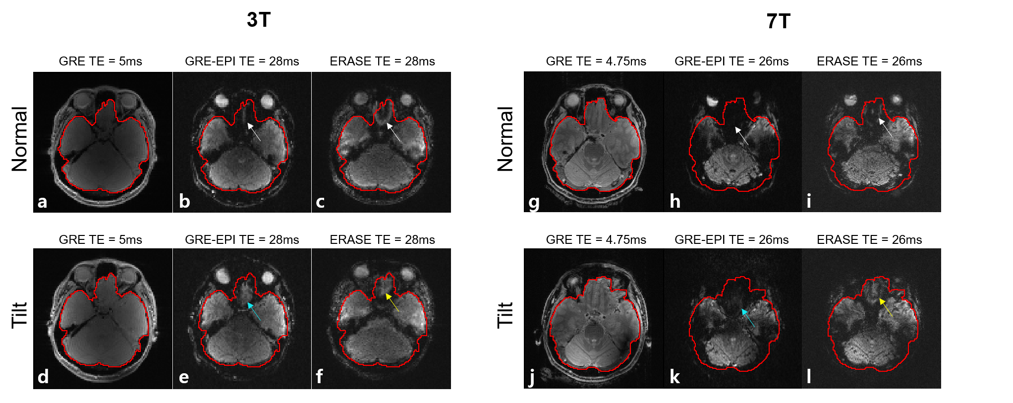
Figure 2 illustrates representative axial and sagittal-plane B0 maps. The black arrows in Fig.2 indicate B0 inhomogeneity near the prefrontal region due to tissue-air susceptibility difference between the sinus and nasal cavity. In the tilted scan (Figs.2c,d, Figs.2g,h), the local field of prefrontal region is significantly improved compared to the normal orientation scans (Figs.2a,b, Figs.2e,f). These indicate that the local B0 gradient in the head-tilted scans is indeed smaller than in the normal scans.Figure 3 shows the axial images of GRE for reference, GRE-EPI, and ERASE for both normal and head-tilted scans at 3T and 7T. This illustrates that the head-tilted ERASE scan (Figs. 3f,l) provides dramatically improved image quality in the prefrontal region compared to conventional, normal-orientation GRE-EPI (Fig. 3b,h), with the effect especially standing out in 7T images. The solid red lines indicate the manually drawn masks on the reference GRE magnitude images for better comparison of GRE-EPI and ERASE.
Discussion & Conclusion
We demonstrated that the head-tilted scan combined with ERASE sequence achieves significantly improved image quality with lesser signal dropout than GRE-EPI, especially in the prefrontal region. Reduced magnetic susceptibility-induced gradients by tilted-head scan in the prefrontal region could potentially allow more flexibility in the range of scan parameters for ERASE such as the FOV in the SPEN direction, which is currently limited by gradient-induced spin misregistration to some extent. The benefit of the proposed method particularly stands out at ultrahigh field, e.g., 7T. In future studies wed plan to apply the proposed method to high-field functional MRI with improved sensitivity and spatiotemporal resolution.Acknowledgements
This work was supported by NRF-2019M3C7A1031993 and IBS-R015-D1.References
[1] Ryu, J. K., Jung, W. B., Yu, J., Son, J. P., Lee, S. K., Kim, S. G., Park, J. Y., 2020. An equal-TE ultrafast 3D gradient-echo imaging method with high tolerance to magnetic susceptibility artifacts: Application to BOLD functional MRI. Magn. Reson. Med. DOI: 10.1002/mrm.28564.
[2] Ryu, J. K., Han, S., Oh, S. H., Lee, J., Kim, S. G., Park, J. Y., 2019. A new ultrafast 3D gradient echo-based imaging method using quadratic-phase encoding. Magn. Reson. Med. 82, 237–250.
[3] Yoo, S., Song, H., Kim, S. G., Shim, W. M., & Lee, S. K., 2020. Feasibility of head-tilted brain scan to reduce susceptibility-induced signal loss in the prefrontal cortex in gradient echo-based imaging. NeuroImage. 223, 117265.
Figures