0898
Rapid and robust DTI using a turboPROP technique with blade sharing and whole-blade acquisition
Zhiqiang Li1 and John P Karis1
1Neuroradiology, Barrow Neurological Institute, Phoenix, AZ, United States
1Neuroradiology, Barrow Neurological Institute, Phoenix, AZ, United States
Synopsis
DTI is a vital tool in many neurological applications. ssEPI is the method of choice in clinical DTI but suffers from strong geometric distortions. FSE and PROPELLER-based techniques are free from geometric distortions but have low scan efficiency. Recently, a split-blade turboPROP technique has been enhanced for brain DWI with improved image quality. In this work we develop a whole-blade acquisition mode and a blade sharing strategy with turboPROP for DTI at a speed comparable to an ssEPI-based clinical protocol. In vivo results demonstrate good image quality.
Introduction
Diffusion-tensor imaging (DTI) is a vital tool in many neurological studies. Single-shot echo-planar imaging (ssEPI) is the method of choice in clinical DTI due to its fast speed. However, ssEPI suffers geometric distortion artifacts caused by phase errors accumulated along the echo train from field inhomogeneities and tissue susceptibilities. Multi-shot EPI (msEPI)1-4 reduces the artifacts by acquiring multiple shorter echo trains that lead to less phase errors. However, the improvement is limited in areas with strong field variations, and prone to motion artifacts. Furthermore, msEPI increases the scan time by a factor approximately equal to the number of shots. Turbo-spin-echo acquisitions are free from geometric distortion artifacts, but typically have low scan efficiency. A more efficient technique, turboPROP5,6, has recently been enhanced for improved image quality by using a quadratic phase modulation scheme and a two-stage phase correction strategy7. Despite these improvements, the scan time with turboPROP is still too long for routine clinical DTI applications. In this work, we introduce a whole-blade mode and a blade sharing strategy to turboPROP to achieve fast and robust DTI with a clinically acceptable speed comparable to an ssEPI-based protocol.Methods
In the recently improved turboPROP7, a split-blade acquisition mode is used, where the odd and even echoes are placed in separate blades because odd and even echoes have different phases. In the split-blade mode, these separate blades from odd or even echoes are collected at the same blade angles, i.e., the same k-space is collected twice, leading to slow speed that is impractical for DTI. To develop a turboPROP-based technique for DTI, we propose two new features.First, a whole-blade mode is introduced to achieve a 2X acceleration compared to the split-blade mode. To avoid interference from phase differences between odd and even echoes, spin-echo (SE) formed sub-blades are collected at the center of k-space (of each blade) to simultaneously serve as both imaging and calibration data, while gradient-echo (GRE) formed sub-blades are collected in the peripheral region in k-space, as shown Fig. 1a. The reconstruction algorithm correcting for these phase differences is illustrated in Fig. 1b and 1c. After correction for the phase differences between GRE and SE (Fig. 1b), the SE images are utilized to extract information about the phase difference between odd and even echoes. This information is then subtracted from odd GRE and SE to enforce phase consistency (Fig. 1c). All echoes (odd and even, GRE and SE) from each shot are then combined to form a final blade, which is ~2 times wider than that from split-blade mode and therefore allows for a 2X acceleration.
To further accelerate the scan, another strategy, called blade sharing, is developed. In DTI scans, typically 32 or more diffusion directions are desired for accurate quantitative measurement8. With 32 or more directions, data of close diffusion directions have similar signal weighting. To utilize this similarity, we propose a blade sharing technique (Fig. 2), which keeps blade-wise undersampled data of target direction in the center of k-space, while adding data from nearby direction in the outer k-space to satisfy the Nyquist criterion. This allows for an acceleration factor that equals the number of tiers. For the 32-direction DTI scheme used in this work, the range of angle between optimized pairs of diffusion directions is 13°~16° for a 2-tier sharing scheme. Therefore, by combining the 2-tier sharing scheme and the whole-blade mode, an acceleration factor of ~4 can be achieved compared to previous split-blade turboPROP.
The sequence was implemented on a Philips 3T Ingenia scanner. Volunteer data were scanned with whole-blade mode and blade sharing using the following parameters: FOV=230x230 mm2, resolution=2.25x2.25 mm2, slice thickness=2 mm, gap=0 mm, 60 slices, ETL=12, GRASE factor=5, one b=0 and 32 directions with b=1000 s/mm2, scan time=~7:20. Data were also acquired without blade sharing to examine its performance. In this experiment, data with whole-blade and blade sharing were acquired with 2X oversampling to match the scan time of data without blade sharing for a fair comparison. Reference data were acquired with an ssEPI-based clinical protocol, with scan time about 6:40. A resolution of 2x2 mm2 was used in ssEPI considering T2*-decay-induced blurring in the phase encoding direction.
Results and Discussion
Figure 3 confirms the image quality of data acquired with blade sharing. Compared to the trace images without blade sharing, the quality of images with blade sharing are comparable, demonstrating its feasibility. The data with blade sharing in this comparison were acquired with 2X oversampling (to match scan time) and therefore can be acquired twice faster than data without sharing.The minimization of distortion artifacts with turboPROP is illustrated and compared with ssEPI in Figure 4. As expected, the distortion artifacts observed in images from ssEPI are not present in turboPROP.
Fig. 5 demonstrates the quality of quantitative measurement. The fractional anisotropic map calculated from turboPROP data is in general comparable to that from ssEPI, indicating good accuracy.
Conclusion
In this project we proposed a turboPROP technique with whole-blade acquisition and blade sharing for fast and robust DTI, with a scan time comparable to a clinical ssEPI-based protocol, and minimized distortion artifacts. Therefore, it provides a potential alternative to ssEPI for clinical DTI.Acknowledgements
No acknowledgement found.References
- Holdsworth SJ, Skare S, Newbould RD, Guzmann R, Blevins NH, Bammer R. Readout-segmented EPI for rapid high resolution diffusion imaging at 3T. Eur J Radiol. 2008;65:36-46. PMCID: PMC3360876
- Porter DA, Heidemann RM. High resolution diffusion-weighted imaging using readout-segmented echo-planar imaging, parallel imaging and a two-dimensional navigator-based reacquisition. Magn Reson Med. 2009;62:468-475.
- Chen N-k, Guidon A, Chang H-C, Song AW. A robust multi-shot scan strategy for high-resolution diffusion weighted MRI enabled by multiplexed sensitivity-encoding (MUSE). Neuroimage. 2013;72:41-47. PMCID: PMC3602151
- Jeong H-K, Gore JC, Anderson AW. High-resolution human diffusion tensor imaging using 2-D navigated multishot SENSE EPI at 7 T. Magn Reson Med. 2013;69:793-802. PMCID: PMC3424313
- Pipe JG, Zwart N. Turboprop: Improved PROPELLER imaging. Magn Reson Med. 2006;55:380-385.
- Lee C-Y, Li Z, Pipe JG, Debbins JP. Turboprop+: Enhanced turboprop diffusion-weighted imaging with a new phase correction. Magn Reson Med. 2013;70:497-503.
- Li Z, Ooi MB, Karis, JP. An enhanced turboPROP+ technique for diffusion weighted imaging. In Proceedings of the ISMRM Virtual Conference and Exhibition, 2020:p966.
- Jones DK. The effect of gradient sampling schemes on measures derived from diffusion tensor MRI: A Monte Carlo study. Magn Reson Med. 2004;51:807-815.
Figures
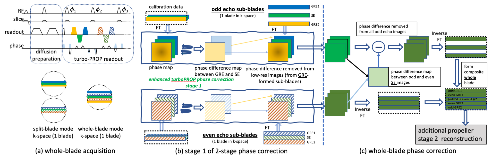
Fig. 1. (a) shows the whole-blade turboPROP sequence. Unlike the split-blade mode, the whole-blade mode places all echoes in the same k-space, increasing the blade width by a factor of ~2. Odd/even SE-formed sub-blades are collected at the center of k-space, also serving as calibration data. Odd/even GREs are placed in outer k-space. After correction for phase differences between GRE and SE (b), phase differences between odd/even SE images are computed and used to enforce phase consistency between all odd/even echoes (c). The final combined blades undergo regular PROPELLER recon.
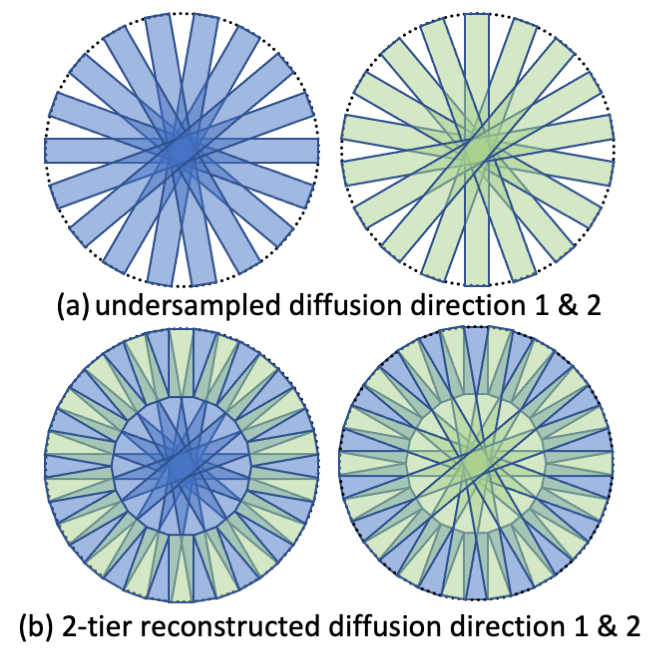
Fig. 2. Blade sharing for turboPROP. (a) shows two diffusion directions with blade-wise undersampled but supplemental k-space. (b) shows the composite full k-space for each diffusion direction by sharing data in outer k-space between these two directions.
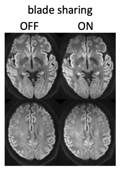
Fig. 3. Comparison of whole-blade turboPROP data without and with blade sharing, which demonstrate comparable image quality.
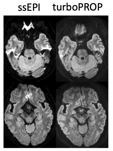
Fig. 4. Demonstration of of geometric distortion artifacts, which are observed in the trace images from ssEPI but minimized in turboPROP.
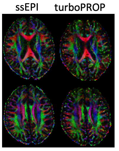
Fig. 5. Comparison of quantitative measurement (FA maps) between ssEPI and turboPROP.