0896
Strategically Acquired Gradient Echo imaging with Compressed Sensing: Comparison of quantitative images with different acceleration factors
bingbing gao1, Yuhan Jiang1, Nan Zhang1, Yanwei Miao1, Qingwei Song1, Ailian Liu1, and Peng Sun2
1the First Affiliated hospital of Dalian Medical university, Dalian, China, 2Philips Healthcare, BeiJing, China
1the First Affiliated hospital of Dalian Medical university, Dalian, China, 2Philips Healthcare, BeiJing, China
Synopsis
Strategically Acquired Gradient Echo (STAGE) imaging is a novel and useful MR sequence which can provide seven different key modalities at the same time, T1w, PDw, SWI, T1 mapping, PD mapping, T2* mapping, and SWIM. However, the acquisition time is relatively long (about 8 minutes). The compressed sensing (CS) technique is now wildly applied for accelerating MR acquisition. This study aimed to evaluate the feasibility of CS accelerated STAGE technique, and find an optimal acceleration factor (AF). According to our preliminary results, the AF of 4 was recommended.
Introduction
Multimodality MRI, a combination of imaging modalities may provide a better solution to improve the diagnosis of diseases. Strategically Acquired Gradient Echo (STAGE) imaging can provide seven different key modalities at the same time, including T1w, PDw, SWI, T1 mapping, PD mapping, T2*mapping, and SWIM. It takes a relatively long time to collect the data because a second echo must be acquired to maintain flow compensation under a separate scan. CS has been wildly applied to accelerate 3D MRI acquisitions. Here, we demonstrate the feasibility of the CS accelerated STAGE technique, and evaluate the impact of AF on the images by comparing the CS accelerated images with different AF and images without CS.Methods
The study was prospective. Fifteen volunteers (25.78±7.79, range: 22-36years, 6 men) were enrolled, who underwent a STAGE protocol (MR parameters were shown in Table 1, scan protocol was shown in Table 2) on a 3.0 Tesla Philips MR (Ingenia CX, Philips Healthcare, Best, the Netherlands) using a 32-channel head coil. Three CS STAGE with AF of 2, 3, 4 were scanned, and the routine STAGE accelerated 2 times by SENSE was used as a reference. All DICOM raw data were processed by using SPIN and STAGE software by one experienced neuroradiologist. Quantitative value-T1 value and T2* value, signal-to-noise ratio (SNR), and contrast-to-noise ratio (CNR) were measured in both white matter (WM)- frontal lobe and gray matter (GM)-globus pallidus. Kruskal-Wallis test was calculated for the intermethod comparison.Results
The CS STAGE with AF of 2, 3, 4 took 9min12s, 6min10s, 4min39s respectively. The scan time of routinely used SENSE protocol was 8min55s. The detailed quantitative values (SNR, CNR, T1, T2) derived from STAGE with different acceleration strategies were shown in Table 3, and no statistically significant difference was detected (p﹥0.05; Figure 1 and 2). Thus, by using an AF of 4, CS STAGE achieved 51.9% time saving compared with 2 times accelerated SENSE protocol while maintaining image qualities.Conclusion
In this study, comparing with the SENSE technique, CS was a reliable method for fast STAGE imaging with comparable image quality. The CS-based STAGE imaging can be easily integrated into the clinical protocols and would be beneficial for a wide range of applications, such as cerebrovascular disease, Parkinson's disease, or multiple sclerosis disease. According to our results, CS STAGE with AF of 4 was recommended.Acknowledgements
The authors are grateful to the Department of Radiology, First Affiliated Hospital of Dalian Medical University, for supporting this study. The authors thank all volunteers who participated in this study.References
[1] B.M.A. Delattre, S. Boudabbous, C. Hansen, A. Neroladaki, A.L. Hachulla, and M.I. Vargas, Compressed sensing MRI of different organs: ready for clinical daily practice? Eur Radiol 30 (2020) 308-319. [2] Y. Liu, J. Li, N. He, Y. Chen, Z. Jin, F. Yan, and E.M. Haacke, Optimizing neuromelanin contrast in the substantia nigra and locus coeruleus using a magnetization transfer contrast prepared 3D gradient recalled echo sequence. Neuroimage 218 (2020) 116935. [3] C.J. Park, J. Cha, S.S. Ahn, H.S. Choi, Y.D. Kim, H.S. Nam, J.H. Heo, and S.K. Lee, Contrast-Enhanced High-Resolution Intracranial Vessel Wall MRI with Compressed Sensing: Comparison with Conventional T1 Volumetric Isotropic Turbo Spin Echo Acquisition Sequence. Korean J Radiol 21 (2020) 1334-1344.Figures
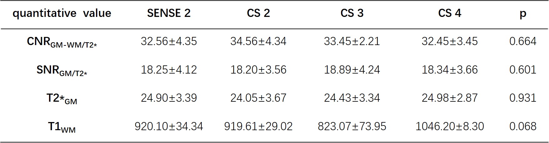
Table 3 Comparisons of quantitative
values between different sequences
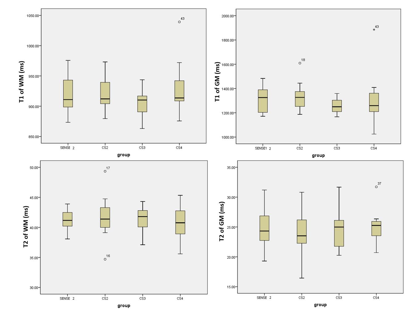
Figure 1 T1 and T2 value
comparison of STAGE sequences with different AF
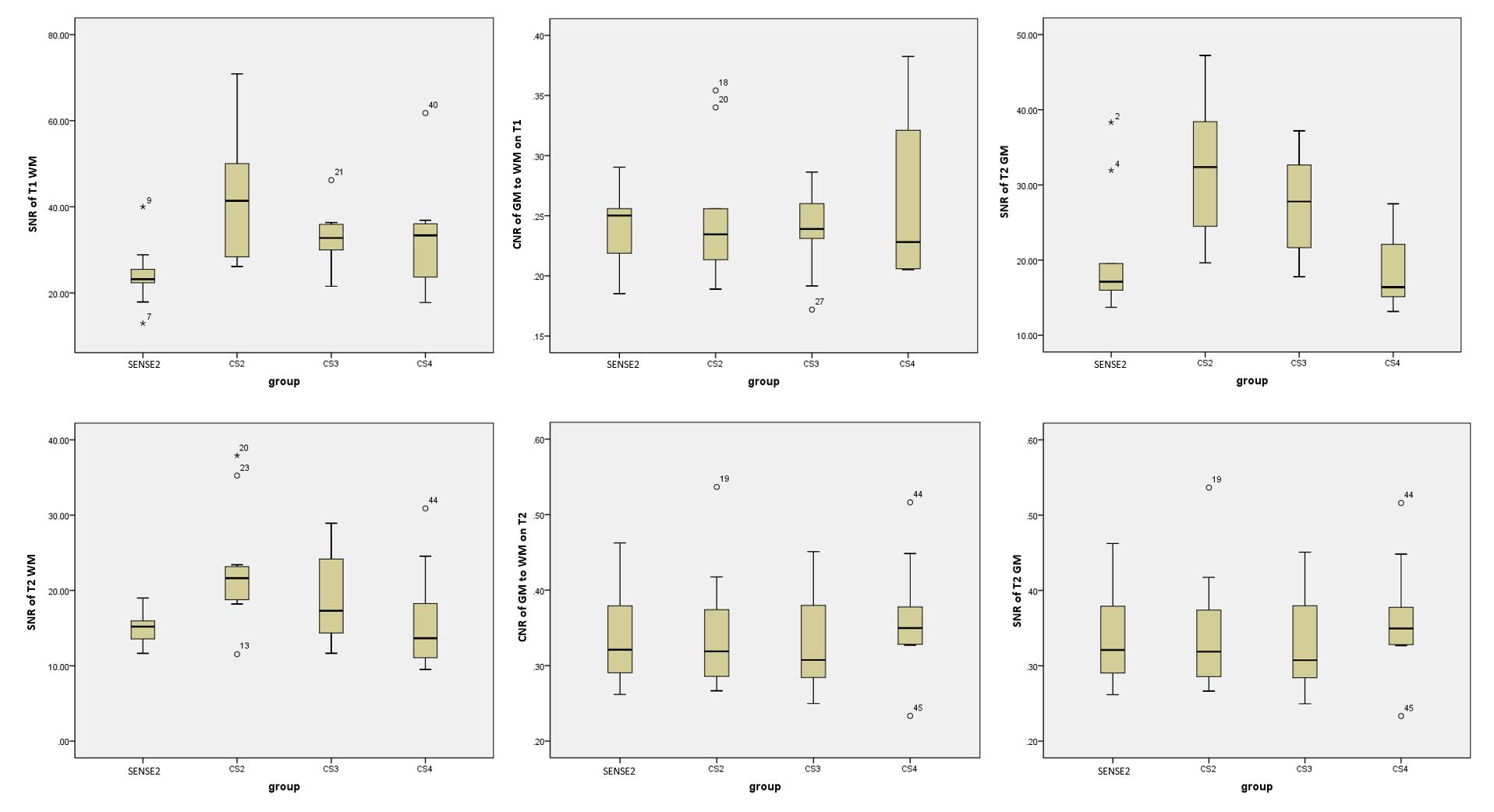
Figure 2 SNR and CNR comparison
of STAGE sequences with different AF
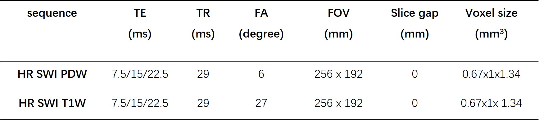
Table
1 MR parameters of STAGE imaging
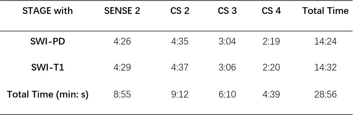
Table 2 the acquisition time of
scan protocol