0892
On the impact of B0 map resolution and undersampling on reconstructions using expanded encoding models1Medical Biophysics, Western University, London, ON, Canada, 2Center for Functional and Metabolic Mapping, Robarts Research Institute, London, ON, Canada, 3Techna Institute, University Health Network, Toronto, ON, Canada, 4Medical Biophysics, University of Toronto, Toronto, ON, Canada
Synopsis
Spiral diffusion images are known to suffer from image distortions due to the enhanced sensitivity to magnetic field inhomogeneities. Application of parallel imaging to accelerate readout times, and inclusion of robust B0 field maps are two techniques that generally contribute to spiral image improvements. In this work, the impact of field map resolution and acceleration factors are investigated to identify acquisition parameters that optimize image quality. A moderate acceleration factor (3 or 4) coupled with a field map on the order of the imaging resolution (1.5 mm in-plane) provided the best trade-off between geometric accuracy, blurring reduction, and noise amplification.
Introduction
Single-shot spiral MRI is a rapid acquisition technique that offers drastically shorter achievable echo times (TE) compared to EPI, which minimizes signal loss from T2 decay leading to higher signal-to-noise ratios (SNR)1. This can be particularly advantageous for diffusion MRI (dMRI) which generally experiences lower SNR due to signal attenuation of moving water molecules, and long diffusion encoding periods that prolong TE2. A limitation which prevents the technique’s implementation in clinical practice is its sensitivity to magnetic field inhomogeneities3, but recent work using field monitoring with external probes and an expanded encoding model have demonstrated the feasibility of high-quality spiral dMRI4. Static field (B0) maps are essential in single-shot spiral brain imaging, particularly at high field strengths, where images may suffer from susceptibility-induced distortions caused by the paranasal sinuses5. It is possible that field maps may be acquired at relatively low resolution given the typically slow spatial variation of static field offsets, but the trade-offs between B0 mapping resolution and single-shot spiral reconstruction performance are not well understood. At the same time, the deleterious effect of B0 inhomogeneity can be mitigated using undersampling. In this work, we investigate the effects of varying B0 map resolutions and acceleration factors on single-shot spiral dMRI image quality.Methods
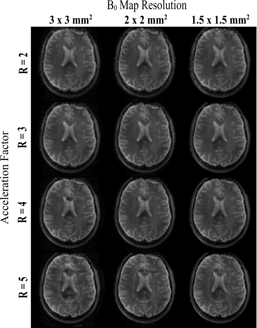
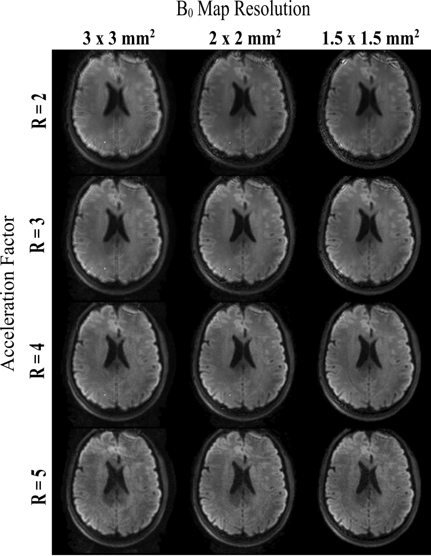
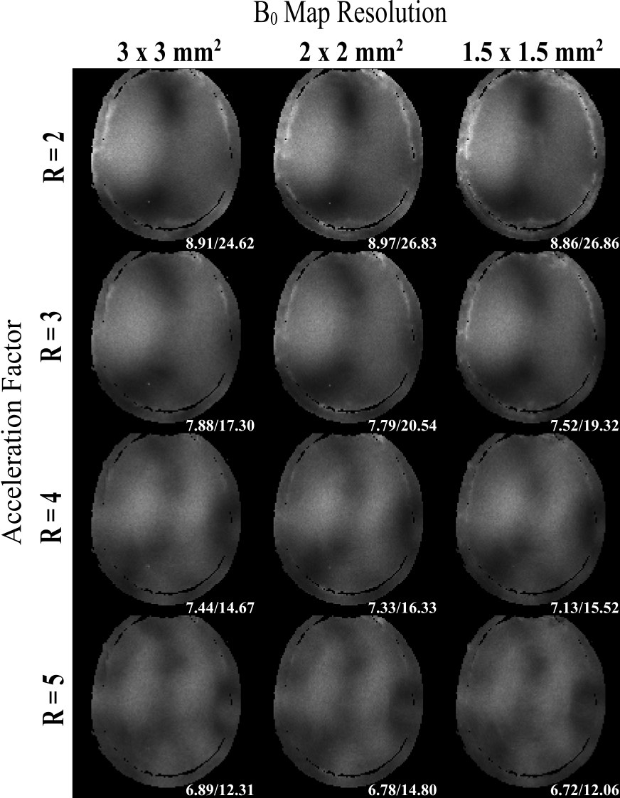
A healthy patient was scanned on a 7T head-only MRI (Siemens). Single-shot spiral acquisitions were performed at parallel imaging acceleration factors (R) of 2,3,4 and 5. A single b=0 s/mm2 scan and 6 diffusion acquisitions using b = 1000 s/mm2 were acquired with each acceleration factor. Field maps were acquired at 3 mm, 2 mm and 1.5 mm isotropic resolution. Remaining imaging parameters were as follows: in-plane resolution = 1.5 x 1.5 mm2, 3 mm slice thickness, TE = 33 ms, TR = 2500 ms, FOV = 192 x 192 mm2. To correct for eddy current effects appearing during image acquisition, the spatially varying field dynamics up to third order in space were measured after image acquisitions using a field monitoring system (Skope) consisting of 16 transmit/receive 19F field probes, and were integrated into an iterative expanded encoding model-base reconstruction with 30 iterations. The pseudo multiple replica method was implemented to evaluate the noise amplification for each R and B0 map resolution6. Synthetic uncorrelated Gaussian noise was generated and added to the respective k-space raw data prior to image reconstruction and repeated 100 times to produce a stack of image replicas. Normalized noise standard deviation (SD) maps were produced by calculating the SD of each pixel throughout the stack of images and dividing the maps by the square root of the acceleration factor, which accounts for the SNR loss due to fewer acquired signals6.Results
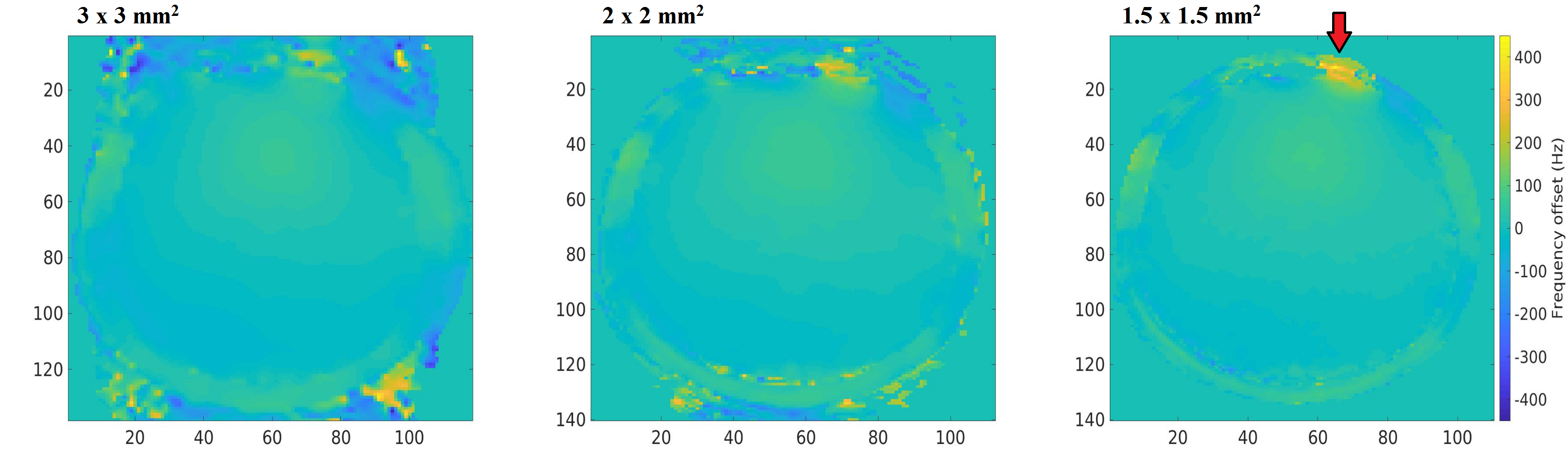
Increasing the B0 map resolution is seen to make the largest impact near the cortical surface (Figure 1). In Figures 2 and 3, increasing the B0 map resolution reduces blurring and artefacts for all acceleration rates. Increasing R generally decreases blurring and decreases B0 artefacts near the edge of the brain, particularly near the edge of the left frontal lobe (top right); however, at extremely high acceleration, residual aliasing artefacts are introduced near the center of the brain. Mean noise SD values show slightly reduced noise amplification with larger acceleration factors, and a generally low dependence on field map resolution (see Fig. 4).Discussion
The higher resolution field maps lead to a reduction in blurring that is pronounced for the acquisitions with longer readout times. For low resolution B0 maps, increasing acceleration can partially ameliorate artefacts and blurring, but the best image quality is observed for the B0 resolution matching the imaging resolution (1.5 mm) and moderate acceleration (R = 3 to 4). Notably, the B0 map resolution has no impact on blurring from T2* decay during the readout, which is evidenced by the decreased blurring for the highest B0 map resolution as the R is increased. At the highest accelerations, residual undersampling artefacts become noticeable; however, the degree of artefacts is much lower than what would be expected from EPI with similar R, stemming from the two-dimensional nature of the aliasing point spread function for spirals7. Although visibly reduced when using higher undersampling and high-resolution field maps, the dark stripes present in the left frontal lobe remain an issue for all cases. The field maps (Fig. 1) show excessive field offsets in the same region, which suggests that the encoding matrix is ill conditioned in that region. The unexpected trend for the noise SD to slightly decrease with increasing R is due to the use of the same number of iterations in the reconstruction for all R. This approach was used because there is no clear objective approach that we know of to choose the number of iterations. Nevertheless, the number of iterations balances the trade-off between residual artefacts and noise amplification, and higher numbers of iterations are required as the encoding matrix becomes less well conditioned with higher R. Alternatively, Tikhonov regularization with a high number of iterations could be used to balance this trade-off, but this requires lengthy reconstruction times. All things considered, the results suggest a weak dependence of noise amplification on R, in agreement with other recent findings7.Acknowledgements
This work has been supported by the Natural Sciences and Engineering Research Council of Canada (NSERC) and the Ontario Graduate Scholarship (OGS) Program.References
1. Botnar RM, Kim WY, Bö P, et al. 3D Coronary Vessel Wall Imaging Utilizing a Local Inversion Technique With Spiral Image Acquisition.; 2001.
2. Polders DL, Leemans A, Hendrikse J, Donahue MJ, Luijten PR, Hoogduin JM. Signal to noise ratio and uncertainty in diffusion tensor imaging at 1.5, 3.0, and 7.0 Tesla. Journal of Magnetic Resonance Imaging 2011;33:1456–1463.
3. Block KT, Frahm J. Spiral imaging: A critical appraisal. Journal of Magnetic Resonance Imaging 2005;21:657–668.
4. Wilm BJ, Barmet C, Gross S, et al. Single-shot spiral imaging enabled by an expanded encoding model: Demonstration in diffusion MRI. Magnetic Resonance in Medicine 2017;77:83–91 doi: 10.1002/mrm.26493.
5. Farahani K, Sinha U, Sinha S, Chiu LC, Lufkin RB. Effect of field strength on susceptibility artifacts in magnetic resonance imaging. Comput. Med. Imaging Graph. 1990;14:409–413.
6. Robson PM, Grant AK, Madhuranthakam AJ, Lattanzi R, Sodickson DK, McKenzie CA. Comprehensive quantification of signal-to-noise ratio and g-factor for image-based and k-space-based parallel imaging reconstructions. Magnetic Resonance in Medicine 2008;60:895–907.
7. Lee Y, Wilm BJ, Brunner DO, et al. On the signal-to-noise ratio benefit of spiral acquisition in diffusion MRI. Magnetic Resonance in Medicine 2020 doi: 10.1002/mrm.28554.
Figures