0879
Fast 3D whole Heart Imaging using low-discrepancy Sampling Strategies1Internal Medicine II, Ulm University Hospital, Ulm, Germany
Synopsis
Three-dimensional MRI is a valuable tool for the diagnosis of heart diseases, which furthermore offers advantages such as intrinsically higher signal-to-noise ratios, less scan planning efforts and the possibility to reconstruct arbitrary anatomical planes from one dataset. Despite these advantages, inherently long acquisition durations often limit clinical applications. This abstract highlights the possibility of acquiring qualitative 3D images at an isotropic resolution of 1.09 mm within 250 heartbeats (ECG-gating), using specifically generated k-space interleaves with low-coherent and low-discrepancy sampling properties, based on a generalised form of the previously introduced Seiffert-Spirals. Cardiac image segmentations are furthermore presented to emphasise image qualities.
Introduction
Cardiac and vascular anatomies of patients are often complex and require imaging in many non-standard anatomical planes1. Therefore, three-dimensional navigator based and ECG triggered sequences are often used to acquire images of the entire heart with the possibility of retrospectively reconstructing arbitrary anatomical planes. Unfortunately, these sequences suffer from intrinsically long acquisition times to achieve sufficient and diagnosable in-plane resolutions.This work applies the imaging concept of generalised Seiffert Spirals to cardiovascular imaging in order to achieve a fast and low-discrepancy coverage of k-space with the possibility of further accelerating the sequence in combination with non-linear optimisation techniques such as Compressed Sensing (CS), facilitating clinically acceptable scan duration.
Methods
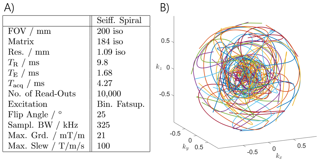
The 3D $$$\zeta$$$-based Spirals are iteratively constructed according to [2,3] and adapted to a gradient system with a maximum amplitude of 21 mT/m and a read-out duration of 4.27 ms per interleave to reduce off-resonance effects. Additionally, the radial increment2 was set to 1.3, based on the energy distribution of previously acquired Cartesian whole heart images. In total, the sequence consists of 10,000 interleaves to sample an isotropic volume of 2003 mm3 with a resolution of 1.09 mm. Further scan parameters are given in figure 1, which additionally shows the k-space coverage of 25 interleaves with the increased sampling density around the centre of k-space due to the radial increment.In order to minimise motion and off-resonance artefacts, the sequence was combined with ECG gating and a navigator based compensation of respiratory motion as well as with spectral selective fat suppression pulses. Enhanced $$$T_2$$$ contrast was generated by the application of $$$T_2$$$-preparation pulses once per heart cycle. Ten interleaves of the trajectory were acquired during each myocardial resting phase, leading to a total requirement of 1,000 heart beats.
Images were acquired on a 1.5T whole body MRI system (Achieva 1.5T, Philips Healthcare, Best, The Netherlands) using a 5-channel SENSE cardiac coil. Further acceleration was achieved by 4-fold (polar) undersampling in combination with a CS reconstruction, leading to only 250 required heart beats. All images were reconstructed using Matlab (R2020a) with a dedicated Voronoi tessellation for the calculation of the density compensation functions2. Image segmentation was achieved using Slicer 3D (V4.11).
Results
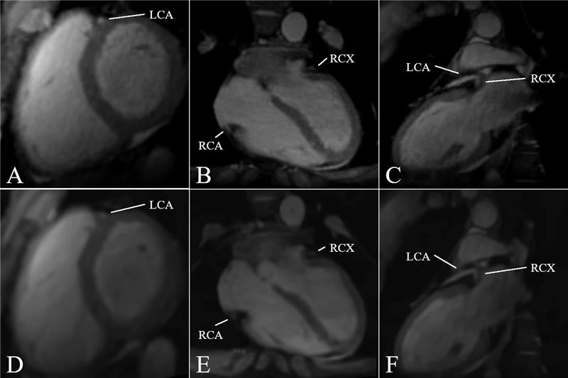
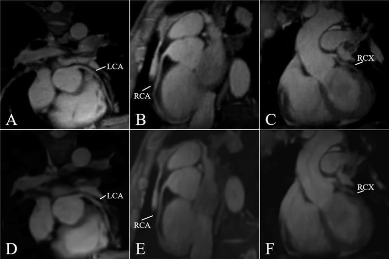
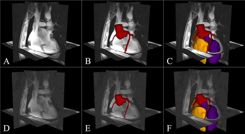
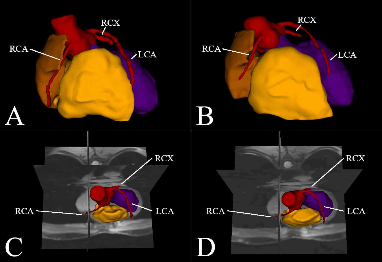
Figure 2 shows images in short axis, 2-chamber and 4-chamber orientation, reformatted from the isotropic whole-heart scan, reconstructed from 1,000 heart beats (fig.2 A,B,C)) and once with 4-fold undersampling in combination with CS (fig.2 D,E,F)). Figure 3 shows reformatted planes that follow the three coronary arteries LCA, RCA and RCX, which are clearly visible and differentiated, also in case of the undersampled dataset. Figure 4 and figure 5 show segmented and partially segmented images that are superimposed on the MRI data, to highlight an equal segmentation, arising from the fully and undersampled dataset. Furthermore, figure 5 shows two segmentations, based on the fully sampled and undersampled dateset, including RCA, RCX and LCA as well as the left and right ventricle and the right atrium.Discussion and Conclusion
The acquired whole-heart images, fully sampled and with 4-fold undersampling show clear delineation of the myocardium. Isotropic resolutions further enable arbitrary reconstructions of anatomical planes. Segmented images show that the course of the coronary arteries (LCA, RCA and LCX) can be reconstructed from only 250 heart beats, resulting in a fast diagnostic tool for the detection of e.g. congenital heart defects. Presented image qualities furthermore enable the generation of patient-specific 3D heart models for surgical planning e.g. in the presence of complex heart defects. A further decrease in scan duration can be achieved by restricting the sequence to anisotropic field of views, by adapting the sampling bandwidth of each interleave with respect to the position of the corresponding Fibonacci point2.Acknowledgements
This project has received funding from the European Union's Horizon 2020 research and innovation programme under FETOPEN grant agreement no. 858149. Furthermore, the author's acknowledge the support and sponsorship provided by Philips Healthcare and thank the Ulm University Centre for Translational Imaging MoMAN for its support.References
1. Greil, G., Tandon, A. A., Silva Vieira, M. and Hussain, T. 3D whole heart imaging for congenital heart disease. Frontiers in pediatrics. 5, 36, 2017.
2. Speidel T., Metze P. and Rasche V. Efficient 3D Low-Discrepancy k-Space Sampling Using Highly Adaptable Seiffert Spirals. IEEE Trans Med Imaging. 38, 1833-1840, 2019.
3. Speidel T., Metze P., Stumpf K., Huefken T. and Rasche V. On the possibility of reconstructing arbitrary FOVs using gradient waveforms with low-coherent aliasing properties. Abstract submitted to the ISMRM, #2368, 2020.
Figures