0873
Prediction of Wallerian Degeneration in the Corticospinal Tract after Cerebral Ischemic Stroke: A Pilot APT and DWI Study
junxin wang1, yanwei Miao1, and Jiazheng Wang2
1Department of Radiology, the First Affiliated Hospital of Dalian Medical University, Dalian, China, 2Phillips healthcare, dalian, China
1Department of Radiology, the First Affiliated Hospital of Dalian Medical University, Dalian, China, 2Phillips healthcare, dalian, China
Synopsis
This study aimed to exploit the early Wallerian degeneration (WD) along the corticospinal tract following cerebral ischemic stroke using amide proton transfer weighted (APTw) and diffusion weighted imaging (DWI). This study included 34 patients with acute cerebral infarction and 21 healthy adult controls. Our data suggested that APTw and apparent diffusion coefficient (ADC) values was significantly increased on ipsilateral compared to contralateral and the control group in the posterior limb of internal capsule. It proves the feasibility of APTw and ADC in early detection of WD following cerebral ischemic stroke.
Introduction
Amide proton transfer (APT) imaging has recently emerged as an important contrast mechanism for MRI in molecular imaging, and diffusion weighed imaging (DWI) has long been investigated for MR cellular imaging [1]. Wallerian degeneration (WD) is defined as progressive anterograde disintegration of axons and accompanying demyelination after an injury to the proximal axon or cell body [2-3]. The aim of this study was to evaluate the feasibility of APT imaging and DWI to detect the changes in early stage of Wallerian degeneration of the pyramidal tract after ischemic stroke.Methods
34 WD patients (23 men and 11 women; age range, 45-87 years) with middle cerebral artery stroke were examined 1-21 days after stroke, and 21 age-matched normal controls (12 men and 9 women; age range, 47-83 years) underwent APT, DWI, and traditional MRI examinations on a 3.0T MR scanner (Ingenia CX, Philips Healthcare, Best, the Netherlands). 3D amide proton transfer-weighted (APTw) images were collected with 3D-DIXON sequence (7 slices, voxel size = 2 x 2 x 7 mm3, B1 = 2 µT, t-sat = 2 s, TA = 6:50 min), while DWI were acquired with b values of 0 and 1000 s/mm2. Regions of interest (ROI) in the posterior limb of internal capsule (PLIC) and cerebral peduncle (CP) were manually placed on T2 FLAIR images based on anatomic knowledge about the location of the pyramidal tract (Figure 1). APT and apparent diffusion coefficient (ADC, calculated for DWI images) measurements were performed in the posterior limb of internal capsule and cerebral peduncle. The Wilcoxon test for paired samples was used to evaluate the difference between the ipsilateral and contralateral regions within each patient. The Mann–Whitney U test for independent samples was used to test the difference between patient and heathy groups. All values are presented as mean ± SD. All patients were evaluated by at least a neurologist with the National Institutes of Health Stroke Scale (NIHSS) score to assess the severity of the motor dysfunction.Results
At the PLIC, the increased APTw and ADC values were present on ipsilateral side of the ischemic lesion compared to contralateral side (P < 0.05), and also to the controls (P < 0.05). At the CP, only bilateral ADC values in patients were significantly increased than those in the controls (P < 0.05). Furthermore, the bilateral APTw values at the PLIC were positively correlated with interval time (ipsilateral r = 0.592, P < 0.001; contralateral r = 0.459, P = 0.006). Positive correlations were found between ADC values on ipsilateral side and the NIHSS score on discharge (r = 0.368, P = 0.032) (Table1, 2 and 3).Discussion and Conclusion
Our results shown that APTw and ADC values were promising imaging biomarkers for early detection of Wallerian degeneration after intracerebral ischemic stroke (within 3 weeks). The increase of mobile protein content in the process of myelination may result in the increase of APTw signal intensity. Protein-based APT imaging can provide additional information in assessing brain demyelination in the WD population. APT and DWI may further allow improved prognostic evaluation of patients recovered from ischemic stroke within acute to subacute phases.Acknowledgements
No acknowledgement found.References
[1] Zhou J, Heo HY, Knutsson L, van Zijl P, Jiang S. APT-weighted MRI: Techniques, current neuro applications, and challenging issues. J Magn Reson Imaging. 2019. 50(2): 347-364. [2] Johnson, A.C., McNabb, A.R., Rossiter, R.J., 1950. Chemistry of Wallerian degeneration. A review of recent studies. Arch. Neurol. Psychiatry 64, 105–121. [3] Lampert, P.W., Cressman, M.R., 1966. Fine-structural changes of myelin sheaths after axonal degeneration in the spinal cord of rats. Am. J. Pathol. 49, 1139–1155.Figures
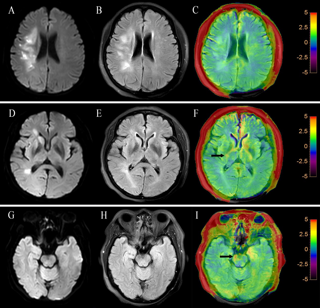
Fig.1 (A,D,G)
DWI, (B,E,H) FLAIR, and (C,F,I) FLAIR+APTw images of an ischemic stroke patient
(female; 56 years old; left limb inactivity for 15 days). DWI images
demonstrated that the patient developed an acute infarction of right frontal
parietal lobe cerebral infarction. The
APTw intensities in regions of the ipsilateral of the ischemic lesion (black
arrow) were higher than the contralateral side in both levels of posterior limb
of internal capsule and cerebral peduncle. DWI
and FLAIR images show no difference between two sides of the two levels.
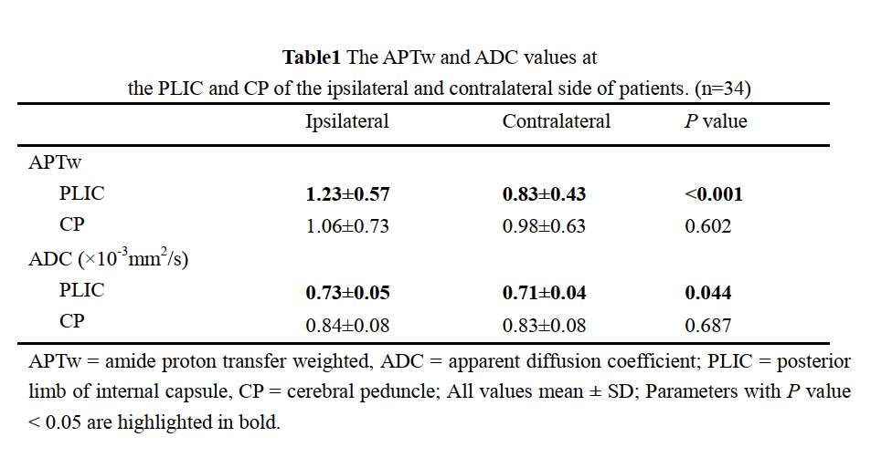
Table1
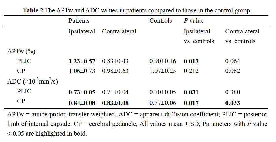
Table2
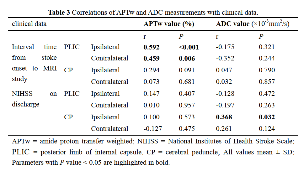
Table3