0856
In Vivo Susceptibility-based Positive Contrast Imaging of MR Compatible Metallic Devices Based on Modified Slab-Selective 3D SPACE Sequence1Shenzhen Institutes of Advanced Technology, Chinese Academy of Sciences, Shenzhen, China, 2Shenzhen College of Advanced Technology, University of Chinese Academy of Sciences, Shenzhen, China, 3Research Centre for Medical AI, Shenzhen Institutes of Advanced Technology, Chinese Academy of Science, Shenzhen, China
Synopsis
Susceptibility-based positive contrast MR imaging exhibits excellent efficacy for visualizing the MR compatible metallic devices, by taking advantage of their high magnetic susceptibility. In this work, a novel method is developed to realize the 3D susceptibility-based positive contrast MR imaging on in vivo experiments of one human patient with one tumour of the scapula. The method is based on a modified 3D SPACE sequence and a PDF background field removal to achieve positive contrast imaging.
Introduction
Susceptibility-based positive contrast MR imaging exhibits excellent efficacy for visualizing the MR compatible metallic devices, by taking advantage of their high magnetic susceptibility 1,2. However, data acquisition of the original technology is based on the Fast spin echo (FSE) sequence 2, resulting in a low sampling efficiency when using it realize the 3D imaging. Furthermore, it can not realize the good visualization of small metallic devices, such as the brachytherapy seeds, because of the existing slice gaps of the current 2D data sampling. To address this issue, a modified slab-selective SPACE (variable-flip-angle 3D FSE) sequence was proposed to realize the 3D positive contrast MR imaging. Here, the field map has been obtained in three dimension which was used to calculate the susceptibility map much accurately. The visualization of the positive contrast based on the modified SPACE sequence and PDF (projection onto dipole fields) background field removal algorithm were tested and preliminary validated on phantom and in vivo experiments of one human patient, compared with the previous 2D FSE-based method and the other two typical MR positive contrast techniques, i.e., susceptibility gradient mapping using the original resolution (SUMO) and the gradient echo acquisition for superparamagnetic particles (GRASP) methods.Methods
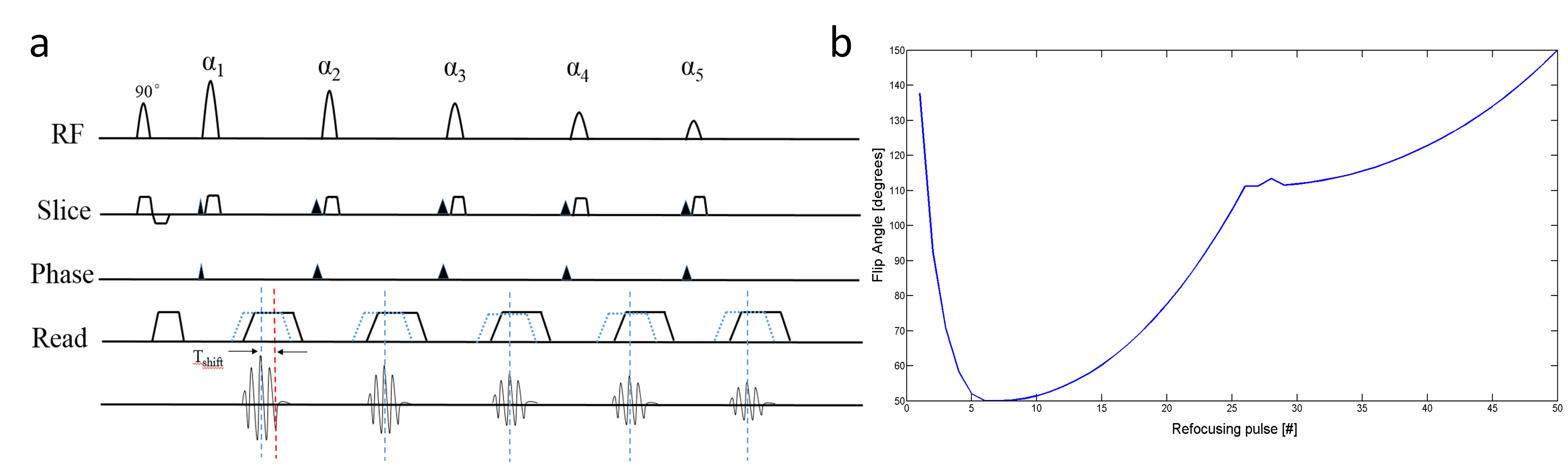
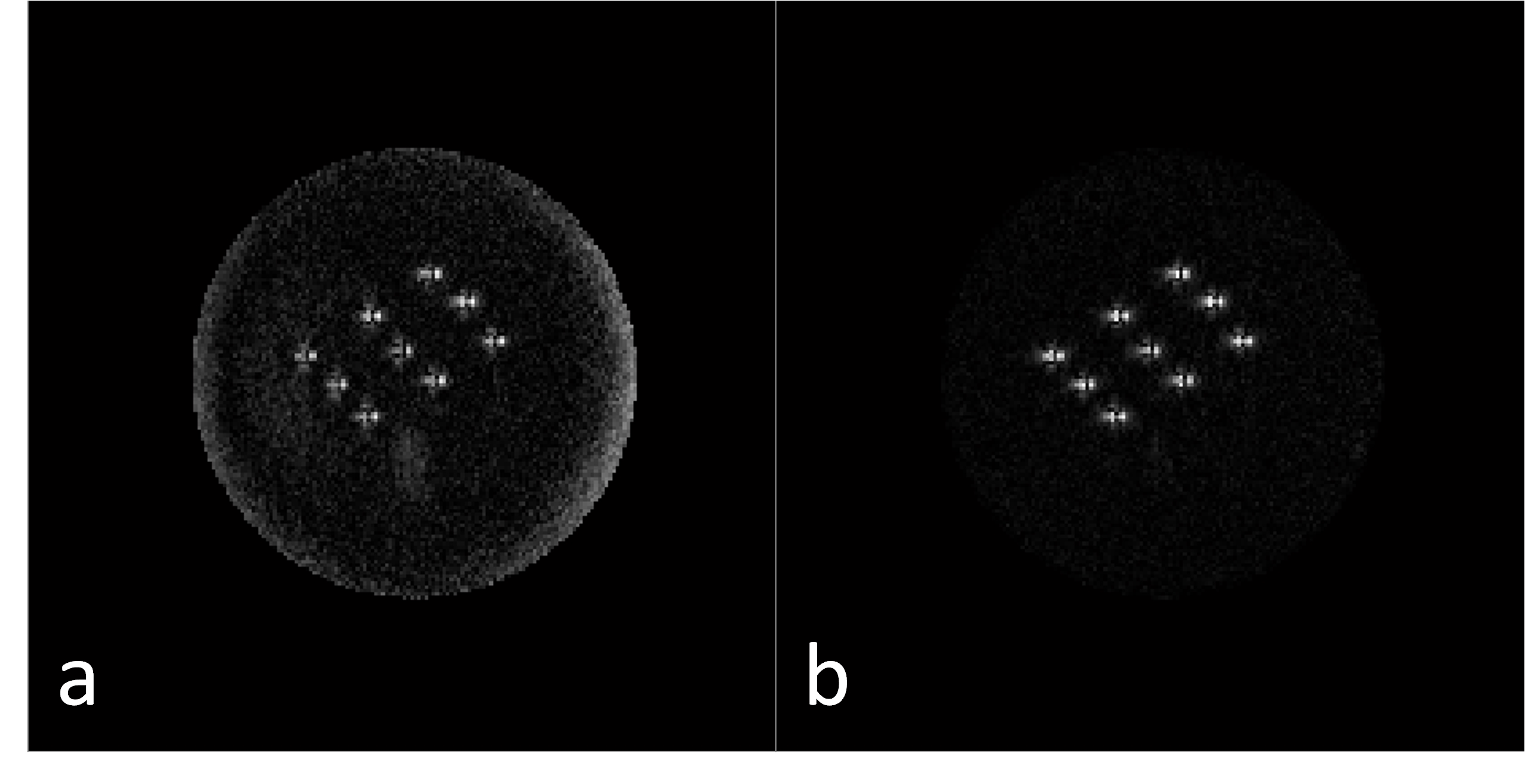
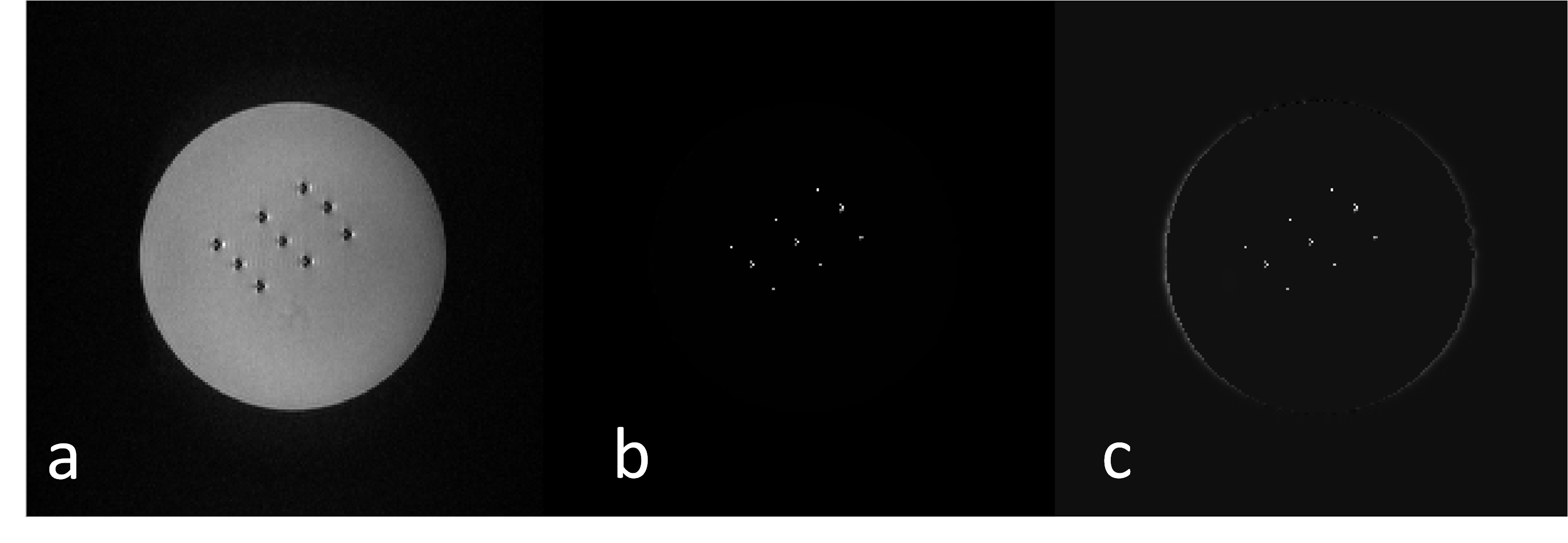
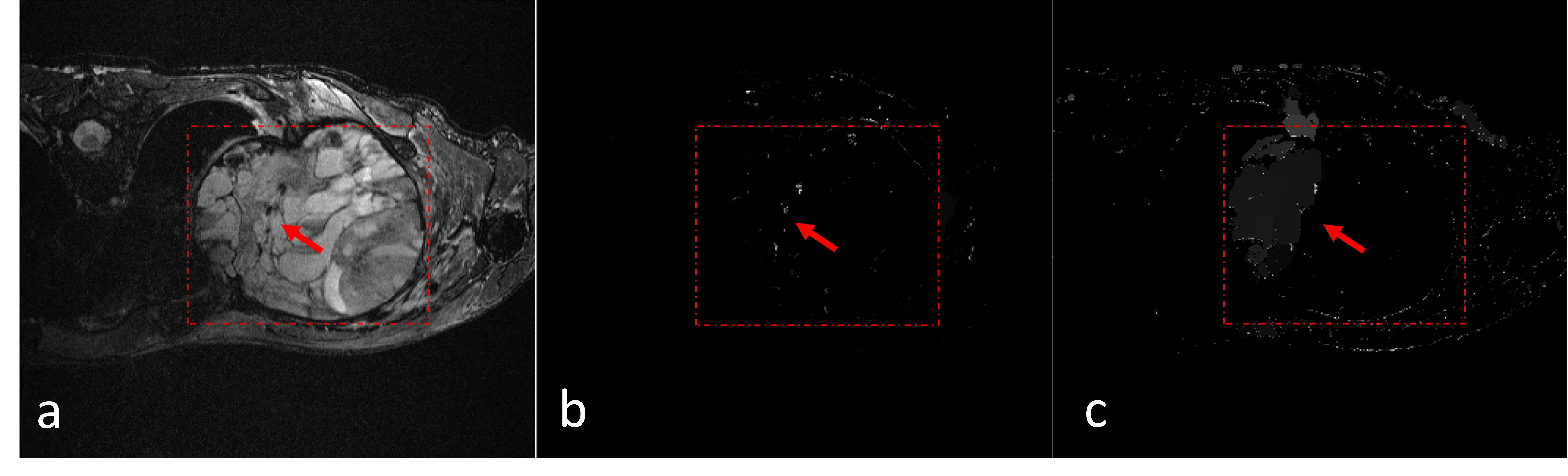
Sequence and Image Reconstruction: The 3D-SPACE sequence is a single-slab turbo-spin-echo sequence with a variable-flip-angle excitation pulse that has been described in detail elsewhere 3. In this study, the variable-flip-angle of the 3D SPACE with refocusing pulse is shown in Figure 1b. We shift each readout gradient of the 3D-SPACE by a small Tshift ,(0.2~0.7ms) (Figure 1). Two datasets are acquired (with or without echo shift) for measuring the total field map. The local field map induced only by the device is obtained after background field removal. The PDF method can be used to estimate the background susceptibility distribution 4.Then this local field map are used to calculate the susceptibility map by using a kernel deconvolution algorithm with a regularized ℓ1 minimization, which is similar to the references 1,2. To achieve faster reconstruction and better imaging quality of positive contrast MRI, a primal-dual (PD) formulation 5,6 is used to solve the minimization problem.Experiments: The 3D positive contrast MR imaging data were acquired on a 3T whole-body MRI scanner (u790, United imaging, China). The in vivo experiments were approved by our institutional review board, and written informed consent was obtained before this experiment. The data was acquired using the modified SPACE sequence with and without Tshift. In the phantom experiment, nine dummy brachytherapy seeds were placed into a gelatin phantom. Orientations of these seeds were placed perpendicular to the B0 field. The imaging parameters: TE/TR=15/1200 ms, ETL=60, FOV=144×144×30 mm3, spatial resolution = 0.7×0.7×1.5mm3. A patient with permanent implanted brachytherapy seeds in tumor on the scapula was imaged using a 24-channels flexible receiver coil and SPIR preparation pulse was used for fat suppression. The scan parameters: TE/ TR=128/1200 ms, ETL= 50, FOV=252×233×39 mm3, spatial resolution = 0.72×0.72×1.5mm3. In addition, the in vivo human patient experiment was also carried out to compare to the other MR positive contrast methods, i.e., 2D FSE based susceptibility-based method, SUMO and GRASP 7,8. These experimental data were acquired on a 3T whole-body MRI scanner (Tim Trio, Siemens Medical Solutions, Erlangen, Germany). In the 2D susceptibility-based method, TE/ TR=17/2660 ms, slice thickness =2.5 mm with no gap, spatial resolution = 0.7×0.7mm2. For the SUMO, TE/ TR=2.5/12 ms, slice thickness = 2.5 mm. For the GRASP, TE/ TR=5/3186 ms, slice thickness = 3 mm, and the gradient rephasing was previously optimized as 50% of that used in the ordinary GRE sequence. In-plane spatial resolution of 1×1 mm2 for both of the SUMO and GRASP.
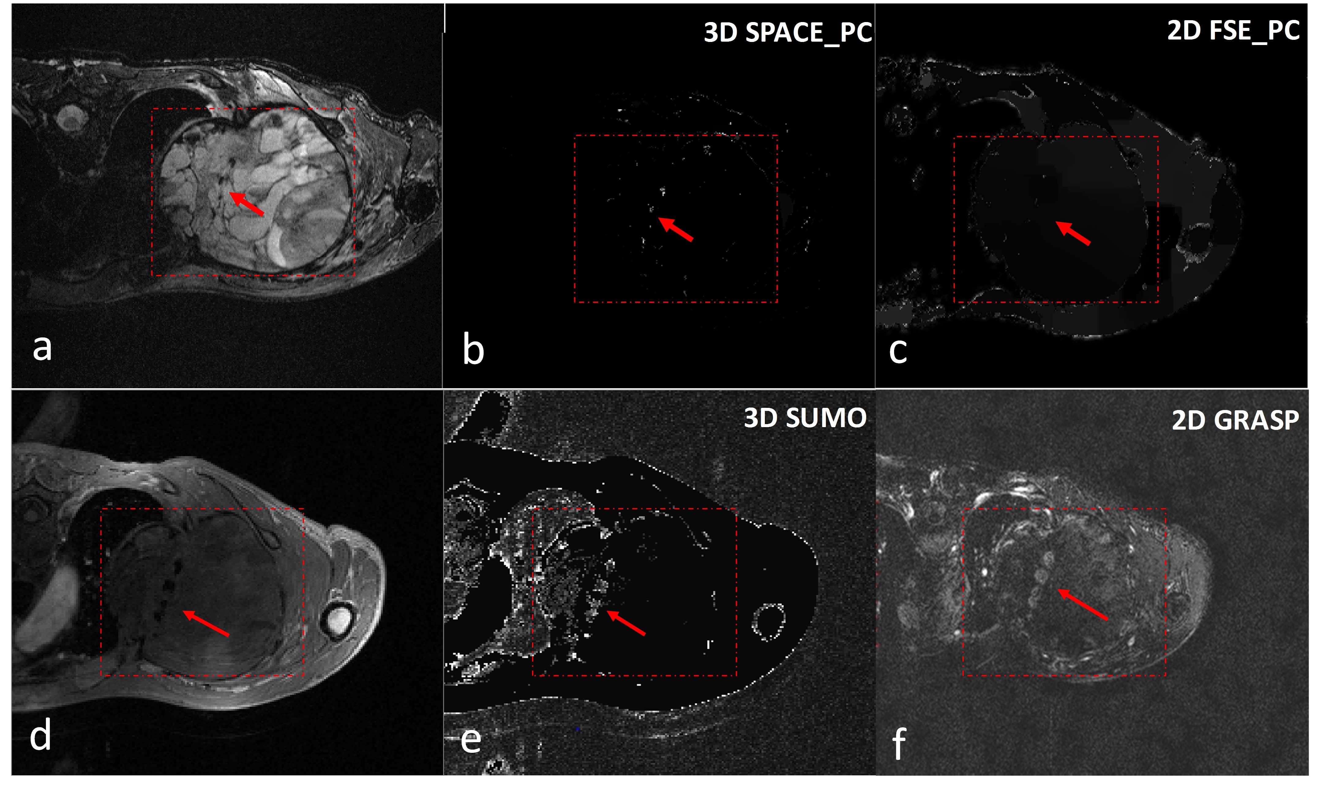
Results
In Figure 2b showed the PDF method successfully removed the background field, leaving the local fields from the brachytherapy seeds. Because the seeds were perpendicular to the B0 field, resulting in a spindle field distribution around the seeds 9 . The seeds were visually lower in the positive-contrast MR images without background field removal compare to that with background field removal (Figure 3 b&c). The results of 3D susceptibility-based positive contrast imaging on in vivo experiments were shown in Figure 4. As human tissue is less homogeneous than the gelatin phantom, the field map is much more inhomogeneous and noisier even the background field was removed using PDF technique. Therefore, this increased noise represents a more challenging scenario to visualize and localize the seeds. Anyway, the 3D SPACE based positive-contrast MR images can achieve better visualization and accuracy of the seeds in positive contrast imaging than the 2D FSE-based technique (Figure. 5b&c ). While, in comparison, SUMO and GRASP imaged the surrounding area of the seeds than its precise location ( Figure 5e&f). In contrast, the susceptibility-based method can localize the real location of the seeds.Conclusion
3D susceptibility-based positive contrast imaging technique was developed and preliminary evaluated with phantom and clinic in vivo patient studies. Compared to the 2D FSE-based technique, the proposed method provides better positive contrast imaging results with PDF background field removal. Therefore, the proposed 3D positive contrast imaging method have the ability to better imaging the smaller metals like brachytherapy seeds because of no slice gap.Acknowledgements
This work was supported in part by the National Science Foundation of China (NSFC, nos. 81901736, 81571669, 81729003, and 61871373), National Key R&D Program of China (2017YFC0108802) and Guangdong Provincial Key Laboratory of Medical Image Processing (nos. 2017A050501026 and 2018A0303130132). Any opinions, findings and conclusions or recommendations expressed in this material are those of the authors and do not necessarily reflect those of the NSFC.References
1. Dong Y, Chang Z, Xie G, Whitehead G, Ji JX. Susceptibility-Based Positive Contrast MRI of Brachytherapy Seeds. 2014;00:1–11.
2. Shi C, Xie G, Zhang Y, et al. Accelerated susceptibility-based positive contrast imaging of MR compatible metallic devices based on modified fast spin echo sequences. Physics in Medicine and Biology. 2017;62:2505–2520.
3. Kato Y, Higano S, Tamura H, et al. Usefulness of contrast-enhanced T1-weighted sampling perfection with application-optimized contrasts by using different flip angle evolutions in detection of small brain metastasis at 3T MR imaging: Comparison with magnetization-prepared rapid acquisition. American Journal of Neuroradiology. 2009;30:923–929.
4. Liu T, Khalidov I, de Rochefort L, et al. A novel background field removal method for MRI using projection onto dipole fields (PDF). NMR in Biomedicine. 2011;24:1129–1136.
5. Chambolle A, Pock T. A first-order primal-dual algorithm for convex problems with applications to imaging. Journal of Mathematical Imaging and Vision. 2011;40:120–145.
6. Wang H, Cai F, Shi C, et al. Positive Contrast Susceptibility MR Imaging Using GPU-based Primal-Dual Algorithm. 42nd Annual International Conference of the IEEE Engineering in Medicine & Biology Society (EMBC). 2020:1485–1488.
7. Varma G, Clough RE, Acher P, et al. Positive visualization of implanted devices with susceptibility gradient mapping using the original resolution. Magnetic Resonance in Medicine. 2011;65:1483–1490.
8. Mani V, Briley-Saebo KC, Itskovich V V., Samber DD, Fayad ZA. GRadient echo Acquisition for Superparamagnetic particles with Positive contrast (GRASP): Sequence characterization in membrane and glass superparamagnetic iron oxide phantoms at 1.5T and 3T. Magnetic Resonance in Medicine. 2006;55:126–135.
9. Whitehead G, Ji J. Positive contrast MRI of prostate brachytherapy seeds based on resonant frequency offset mapping. 32nd Annual International Conference of the IEEE EMBS. 2010:6641–6644.
Figures