0845
Simultaneous anatomical, pathological and T2 quantitative knee imaging with 3D submillimeter isotropic resolution using MIXTURE1Radiology, Eastern Chiba Medical Center, Tonage, Japan, 2Division of Health Sciences, Graduate School of Medical Sciences, Kanazawa University, Kanazawa, Japan, 3Philips Japan, Tokyo, Japan, 4General Medical Services, Chiba University Graduate School of Medicine, Chiba, Japan, 5Orthopaedic Surgery, Eastern Chiba Medical Center, Chiba, Japan, 6Philips Healthcare, Hamburg, Germany, 7Faculty of Health Sciences, Institute of Medical, Pharmaceutical and Health Sciences, Kanazawa University, Kanazawa, Japan
Synopsis
We propose a new sequence called MIXTURE (Multi-Interleaved X-prepared TSE with inTUitive RElaxometry). MIXTURE is a 3D TSE that can set arbitrary echo times using the T2 preparation pulses, and enables several image contrasts (such as PDW, T2W) and T2 mapping by acquiring at least two echo time images. In this study, we evaluated the clinical feasibility of morphological and pathological MRI with two contrasts images and quantitative MRI with T2 mapping using MIXTURE.
PURPOSE
The progression of knee osteoarthritis has a close relation to cartilage degeneration. Cartilage degeneration can be evaluated non-invasively by qualitative T2 mapping MRI1. Recently, an isotropic 3D T2 mapping technique of knee cartilage with a clinically feasible acquisition time has been developed. It’s allows for reformats in any plane and minimization of partial volume effects, which results in a more accurate T2 relaxation time quantification2,3. Therefore, it is possible to make a morphological diagnosis for 3D MRI and a quantitative diagnosis for T2 mapping simultaneously. However, clinical knee joint MRI requires multiple contrasts to diagnose various diseases. Thus, we propose a new sequence called MIXTURE (Multi-Interleaved X-prepared TSE with inTUitive RElaxometry). MIXTURE is a 3D TSE that can set arbitrary echo times using the T2 preparation pulses, and enables several image contrasts (such as PDW, T2W) and T2 mapping by acquiring at least two echo time images. The purpose of this study was to evaluate the clinical feasibility of morphological and pathological MRI with two contrasts images and quantitative MRI with T2 mapping using MIXTURE.METHODS
All subjects were examined on a 3.0T whole-body clinical system (Ingenia CX, Philips Healthcare) with a 16ch knee coil. The study was approved by the local IRB, and written informed consent was obtained from all subjects. The pulse sequence diagram of the MIXTURE method for 3D simultaneous morphological, pathological and quantitative T2 imaging is shown in Figure 1. T2-mapping was performed using T2-prepared 3D segmented turbo spin- echo (TSE) with variable refocusing pulse trains. Two images with different TE (TE = 0 and 50ms) were acquired with interleaved acquisition. In this study, referring to the contrast used for clinical diagnosis by the conventional methods4, 1st echo time was set for PDWI (without any pre-pulses) and 2nd echo time was set for fat suppressed T2WI (by applying spectral fat suppression (SPAIR) and T2prep) [Fig.2].We compared T2 values between conventional Multi-echo TSE and MIXTURE in a sucrose phantom with various T2 values. Furthermore, the following evaluations were performed in thirteen patients (14-84y, 4 females, 9 males) with knee joint pain and various pathologies such as osteoarthritis, osteochondrosis, meniscus tear and ganglion using MIXTURE. We compared T2 values between inside and outside in knee cartilage, and evaluated ability of diagnostic imaging for multi contrast MRI.Imaging parameters for Multi-echo TSE were; Coronal, voxel size=0.7×0.7×2.0mm3, FOV=150×150mm2, 9 slices. band width=289.7Hz/px, TR=2500ms, TE=15, 30, 45, 60, 75, 90ms, and total acquisition time=5m34s. Imaging parameters for MIXTURE were; Coronal, voxel size=0.7×0.7×0.7mm3, FOV=150×150mm2, 260 slices. band width=867.8Hz/px, fat suppression=SPAIR, TR=1200ms, TE=25ms, T2prep time=0, 50ms, CS-SENSE factor=3.5 and total acquisition time=6m47s.
RESULTS AND DISCUSSION
The obtained T2 values from Multi-echo TSE and MIXTURE in the sucrose phantom showed a high correlation [Fig. 3A]. T2 values in patients with knee joint pain was prolonged on the impaired side such as narrowing of the joint gap and deformation or defect of cartilage [Fig. 3B]. Clinical images taken by MIXTURE are shown [Fig. 4, 5]. 3D PDWI contributed to the anatomical evaluation of meniscus, ligaments and cartilage, and 3D FS T2WI contributed to diagnosis of various specific pathologies such as inflammation, edema, bone contusion and tumor. For knee joints where fine tissue is the object of observation, thin slice 3D images are useful. One of the major advantages of the MIXTURE method is its isotropic spatial resolution, from which multi-planer reconstruction (MPR) of all obtained contrasts and images including PDWI, FS T2WI and T2 maps can be derived.CONCLUSION
MIXTURE successful offers multi-contrast and quantitative images within one single scan, which could provide information for potential anatomical and pathological assessment simultaneously for knee imaging. The achieved volumetric isotropic high-resolution is promising for evaluation of early cartilage degeneration. Further clinical studies are warranted.Acknowledgements
No acknowledgement found.References
1. Nieminen MT, et al. T2 relaxation reveals spatial collagen architecture in articular cartilage: a comparative quantitative MRI and polarized light microscopic study. Magn Reson Med. 2001 Sep; 46(3): 487-93.
2. Roberto Colotti, et al. Isotropic three-dimensional T 2 mapping of knee cartilage: Development and validation. J Magn Reson Imaging. 2018 Feb;47(2):362-371.
3. Roberto Colotti, et al. Simultaneous fat-free isotropic 3D anatomical imaging and T 2 mapping of knee cartilage with lipid-insensitive binomial off-resonant RF excitation (LIBRE) pulses. J Magn Reson Imaging. 2019 May;49(5):1275-1284.
4. C G Peterfy, et al. The osteoarthritis initiative: report on the design rationale for the magnetic resonance imaging protocol for the knee. Osteoarthritis Cartilage. 2008 Dec;16(12):1433-41.
Figures
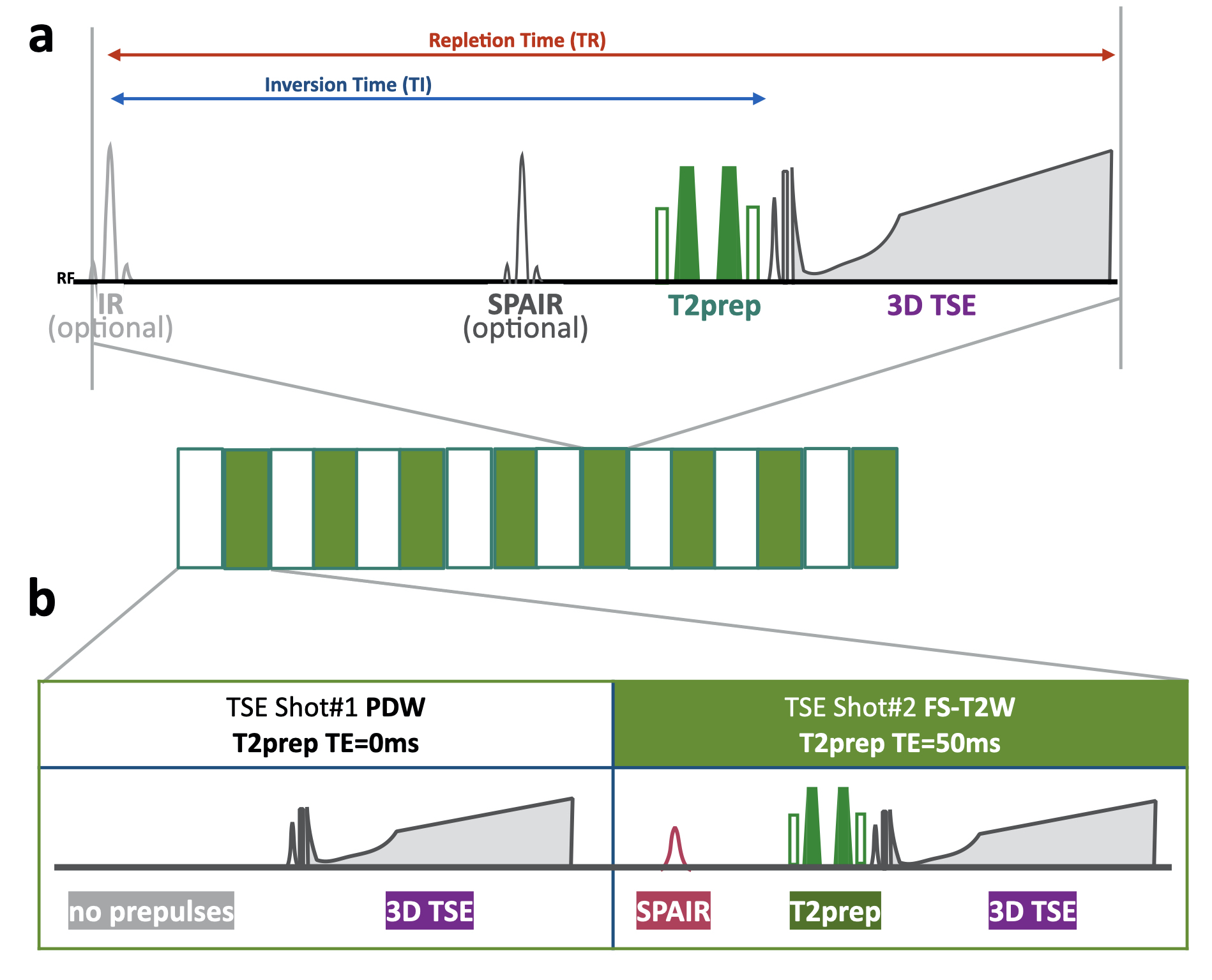
Fig.1 Scheme of the MIXTURE (Multi-Interleaved X-prepared tse with inTUitive RElaxometry).
(a) T2-mapping was performed using T2-prepared 3D segmented turbo spin- echo (TSE) with variable refocusing pulse trains.(b) Two images with different TE (TE = 0 and 50ms) were acquired with interleaved acquisition. To obtain the compatible contrasts with routine TSE images, TSE shot#1 did not apply any pre-pulses (as “PDW) and shot#2 applied both SPAIR and T2pre (as “fat-suppressed T2W”).
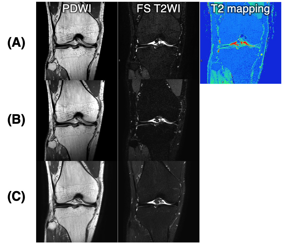
Fig.2 Comparison of MIXTURE and conventional methods (2D TSE and 3D TSE).
(A) MIXTURE, Multiple Interleaved X-prepared TSE with Universal Relaxation-mapping Extensions,(B) Conventional 3D TSE, (C) Conventional 2D TSE
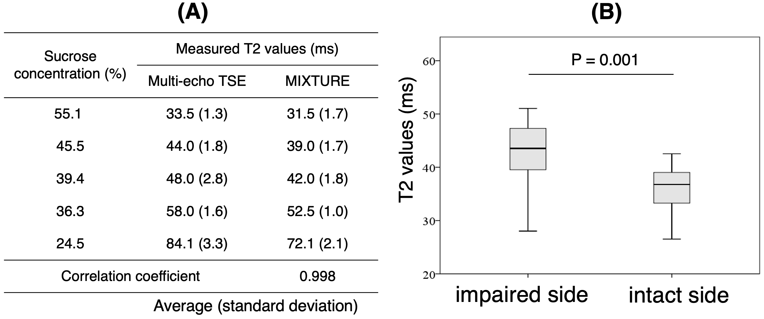
Fig.3 T2 values measured in the sucrose phantom (A) and knee cartilages of thirteen patients using MIXTURE (B).
MIXTURE, (Multi-Interleaved X-prepared tse with inTUitive RElaxometry).
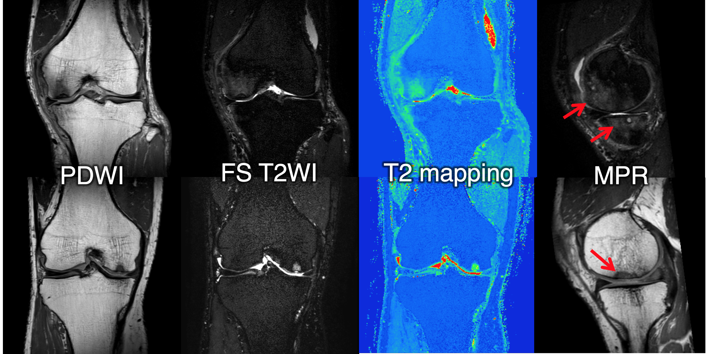
Fig.4 Simultaneous multi-contrast and quantitative images of MIXTURE from a patient with knee joint pain.
Upper: 57y male, knee osteoarthritis, osteonecrosis, bone bruise, intraosseous cyst.Lower: 65y male, idiopathic osteonecrosis.
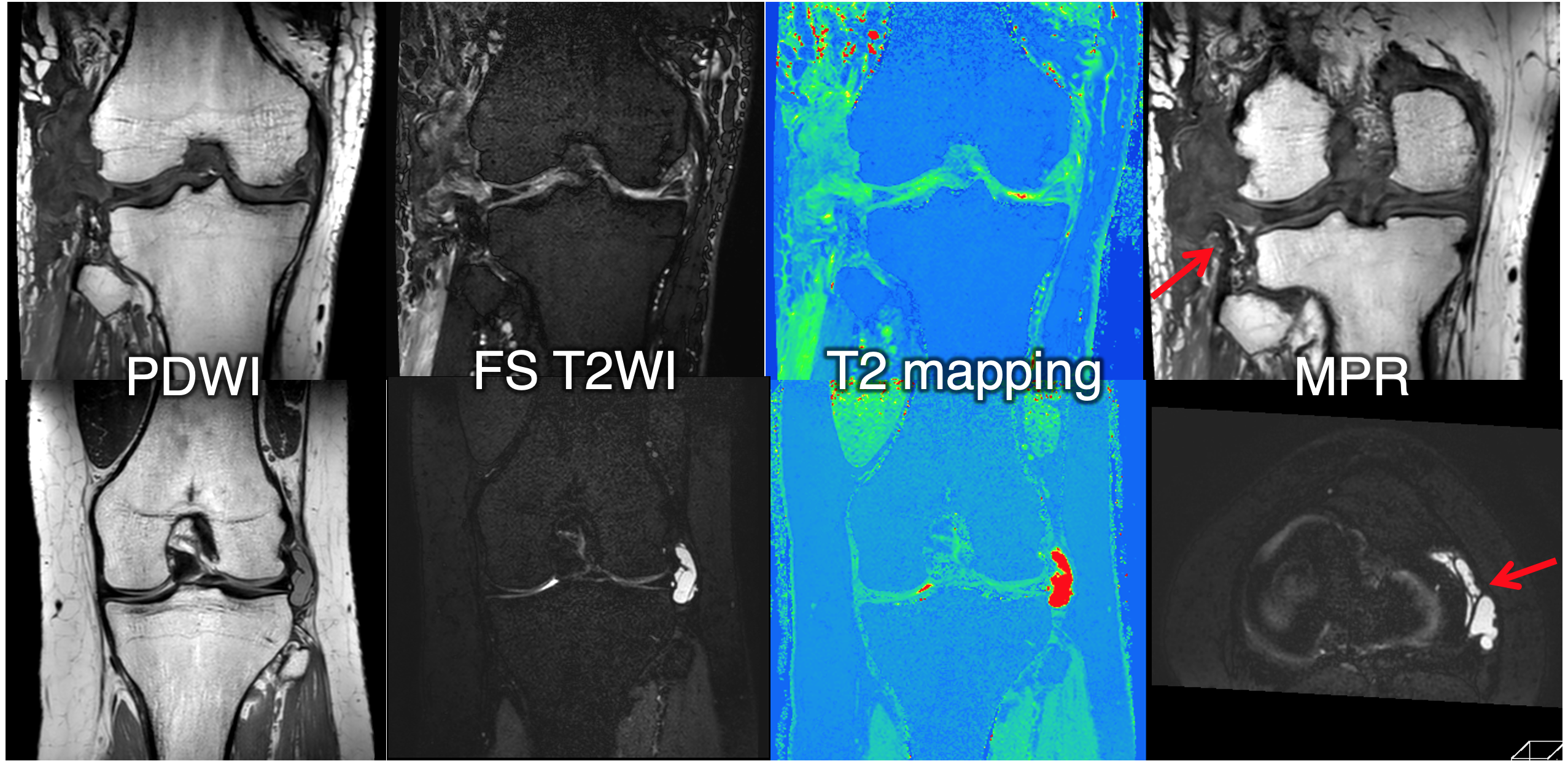
Fig.5 Simultaneous multi-contrast and quantitative images of MIXTURE from a patient with knee joint pain.
Upper: 63y male, lateral collateral ligament and medial meniscus injury.Lower: 18y female, ganglion.