0839
Spiral Cardiac bSSFP with Phase-Modulation to Enable Long Repetition Times1Radiology, Johns Hopkins University School of Medicine, Baltimore, MD, United States
Synopsis
Recently, it was shown that phase-modulated balanced steady-state free precession (bSSFP) offers banding-free radial cine bSSFP images. Because this novel approach for bSSFP overcomes the need for short repetition times (TR), this work proposes to use more efficient spiral readouts with long TR of 15 ms. Banding free bSSFP cine images are demonstrated for either high temporal or high spatial resolution. The long TR furthermore facilitates fat suppression with spatial-spectral water-only excitation pulses which could improve depiction of the coronary arteries. Out of slice signal pileup at dark band frequencies is shown to be reduced with pre-saturation of those frequencies.
INTRODUCTION
Balanced steady-state free precession (bSSFP) is the method of choice for cardiac cine imaging at 1.5T, thanks to its robustness and high contrast-to-noise ratio between blood and myocardial tissue.1 However, at 3T, bSSFP requires short repetition times (TR) and careful shimming / frequency determination to keep dark banding and flow artifacts from obstructing the heart.1,2 In a phase-modulated bSSFP acquisition, also called dynamically phase cycled3 or frequency-modulated4,5, the phase of the excitation pulse is modulated so that the location of the banding artifact is dynamically shifted across the entire field-of-view (FOV) and banding free images can be obtained when the data are combined. Recently, Slawig et al. used an elliptical signal model6 in a model-based reconstruction to achieve banding-free radial cine bSSFP images.7 Because this novel approach for bSSFP overcomes the need for short TR, this work proposes to use more efficient spiral readouts with comparatively long TR of up to 15ms to achieve robust high-resolution cine imaging.METHODS
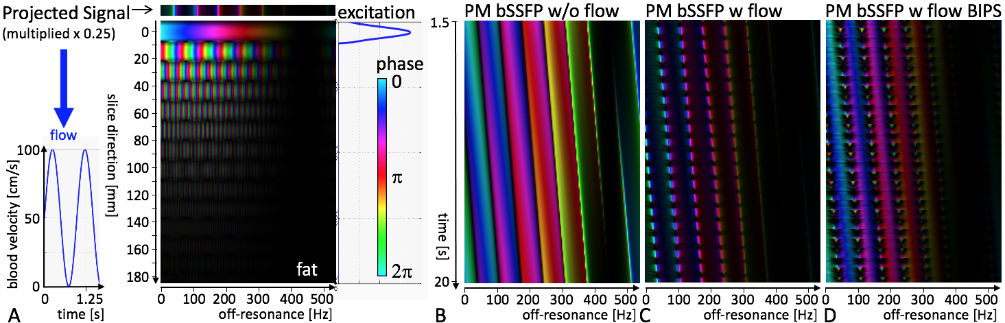
Simulation: Numerical Bloch equation calculations incorporating phase-modulation and through-plane flow were performed. As a potential solution to out of slice signal pileup2,8 the steady-state is interrupted every 40 TRs and tissue at dark band frequencies including in- and outflowing blood are saturated with a non-localized binomial pre-saturation (BIPS) pulse followed by a Kaiser-Bessel shaped train of three start-up TRs to prime the steady-state.9–11Acquisition: Spiral bSSFP data were acquired in a 3T scanner (Achieva R5.1.7, Philips) from one subject who provided written consent for this IRB approved study. 1-2-1 spectral-spatial water-only excitation pulses had 180.324º phase added every TR, with TR = 15ms. 1200 variable density spiral readouts were acquired and consecutive spiral arms were rotated by the golden angle.12 Acquisitions were repeated with and without a BIPS module inserted every 40 TRs. Other acquisition parameters were: 100º excitation angle, FOV = 350mm, acquired voxel size 2.2 or 0.89mm, slice thickness = 8mm, 20-s breathhold. Conventional Cartesian bSSFP cine with 1.6x1.3mm2 voxels and 40 cardiac phases (68% phase percentage) were acquired for comparison.
Reconstruction: Images were reconstructed offline using GPI.13 Data were retrospectively binned into a 2-dimensional matrix of frequency bins and 60 or 40 cardiac phases. The number of frequency bins was the number of heartbeats during the acquisition, and each bin contained data from three neighboring heartbeats using sliding window. Images were reconstructed for each frequency bin separately using k-t sparse SENSE14,15, minimizing parallel imaging data consistency and temporal total variation sparsity constraints. Images from different frequency bins were then combined with either complex summation or root-mean-square (RMS).
RESULTS and DISCUSSION
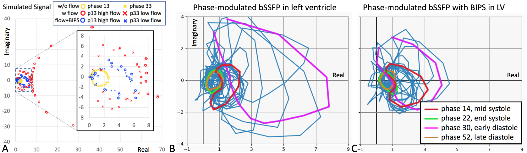
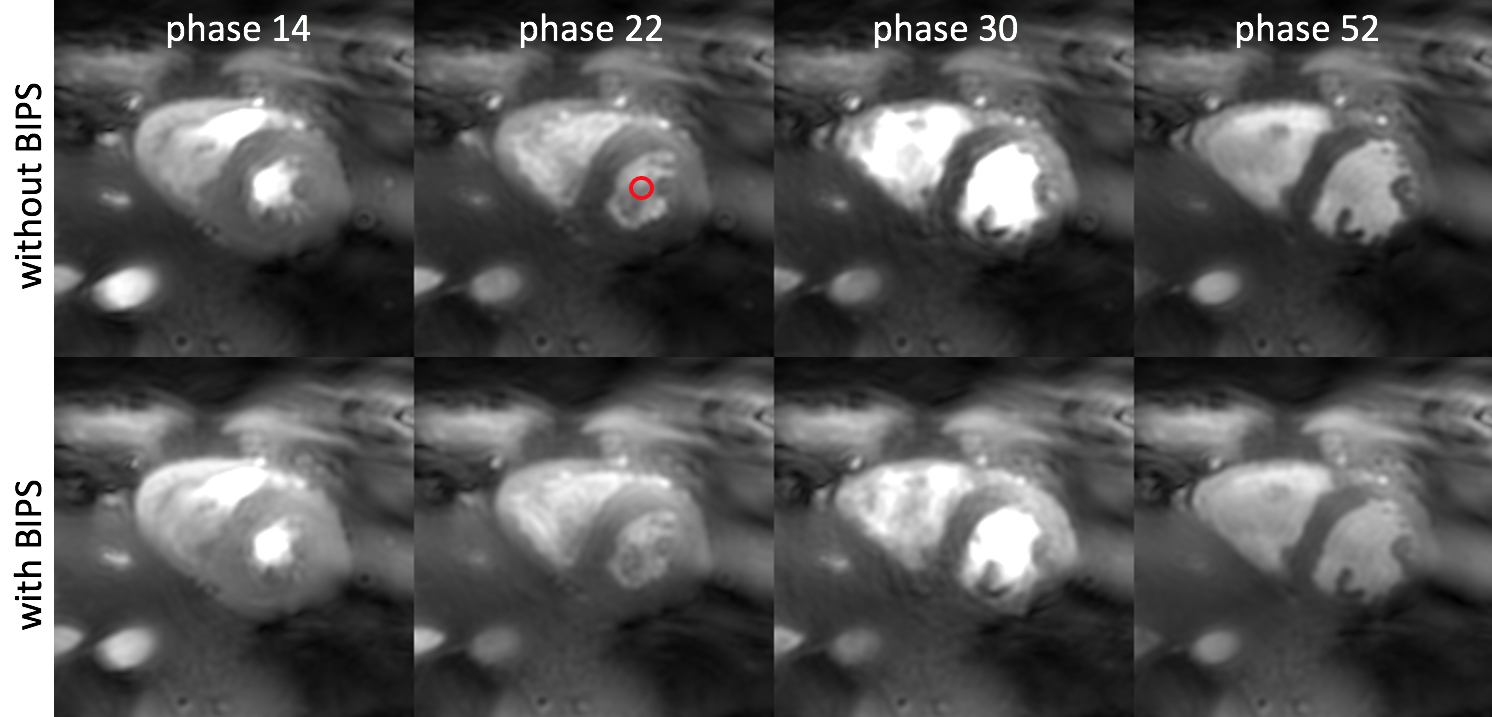
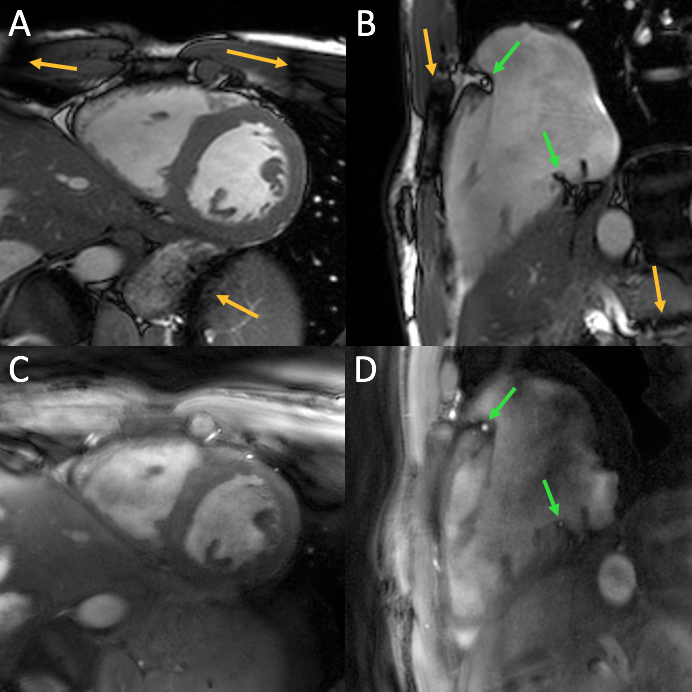
Simulations: Figure 1 A) explains the setup of the simulation, and B)-D) show the simulated magnetization for the 20-s breathhold for a range of off-resonance frequencies. Without flow (B) the dark bands shift one full period and fat frequencies remain suppressed. With blood flowing in and out of the excitation slice (C), signal pileup at locations of prior dark bands dominates. This signal pileup can be suppressed with BIPS modules (D). After binning the on-resonant signal shown in Figure 1 into 40 cardiac phases, the signal of each cardiac phase without flow forms an ellipse (yellow, Figure 2A) as expected,6 but with flow, the ellipse is enlarged with more than 20-fold higher signal amplitude (red circles for high flow). Using BIPS, the simulation predicts that the signal ellipse is only about 3-times larger (blue circles and crosses for both low and high flow).Human data: Figure 3 shows the movie of a mid-ventricular short-axis cine with 60 frames acquired with the proposed phase-modulated spiral bSSFP technique with BIPS. Figure 4 shows four frames acquired with and without BIPS. Figure 2B and C show the signal from the center of the left ventricle (red circle in Figure 4) for all 60 frames, and the four colored “ellipses” correspond to the frames shown in Figure 4: normal ellipses correspond to normal contrast for the frames at end-systole and at end-diastole, but enlarged ellipses and very bright blood signal for frames with high flow at mid systole and early diastole that are improved with BIPS. Figure 5 show diastolic frames of cine acquisitions acquired with only 40 frames but high spatial resolution comparing conventional Cartesian acquisitions (A, B) with the proposed technique in (C, D). The Cartesian images have excellent quality but suffer from dark band artifacts (yellow arrows), which might affect image quality if shimming or frequency determination are not optimal. The spiral images are at this point still blurrier and noisier in comparison but do not suffer from dark band artifacts and have the potential to be a more robust alternative at high field. Furthermore, because of the fat suppression, spiral images do not suffer from Indian ink artifacts and may be preferred to image the coronary arteries (green arrows).
CONCLUSION
This proof-of-concept study shows the potential of phase-modulated bSSFP cine imaging with long TR enabling more efficient readouts such as spirals and longer excitation pulses such as spectral-spatial pulses. Signal pileup from blood flow out of the imaging slice at dark band frequencies can be reduced with a pre-saturation module, or could foreseeably be taken into account by the reconstruction model.Acknowledgements
No acknowledgement found.References
1. Kramer CM, Barkhausen J, Bucciarelli-Ducci C, Flamm SD, Kim RJ, Nagel E. Standardized cardiovascular magnetic resonance imaging (CMR) protocols: 2020 update. J Cardiovasc Magn Reson. 2020;22:17.
2. Schär M, Kozerke S, Fischer SE, Boesiger P. Cardiac SSFP imaging at 3 Tesla. Magnetic Resonance in Medicine. 2004;51:799–806.
3. Benkert T, Ehses P, Blaimer M, Jakob PM, Breuer FA. Dynamically phase-cycled radial balanced SSFP imaging for efficient banding removal: Dynamically Phase-Cycled Radial bSSFP. Magnetic Resonance in Medicine. 2015;73:182–194.
4. Foxall DL. Frequency-modulated steady-state free precession imaging. Magn Reson Med. 2002;48:502–508.
5. Slawig A, Wech T, Ratz V, Tran-Gia J, Neubauer H, Bley T, Köstler H. Multifrequency reconstruction for frequency-modulated bSSFP: Multifrequency Reconstruction for Frequency-Modulated bSSFP. Magnetic Resonance in Medicine. 2017;78:2226–2235.
6. Xiang Q-S, Hoff MN. Banding artifact removal for bSSFP imaging with an elliptical signal model: Banding Artifact Removal for bSSFP Imaging. Magn Reson Med. 2014;71:927–933.
7. Slawig A, Koehler T. The total ellipse of the heart: Cardiac CINE imaging using frequency-modulated bSSFP and the elliptical signal model. In: Proc of the 2020 ISMRM Virtual Conference. 2020. p. 600.
8. Markl M, Alley MT, Elkins CJ, Pelc NJ. Flow effects in balanced steady state free precession imaging. Magnetic Resonance in Medicine. 2003;50:892–903.
9. Foxall DL. Starter sequence for steady-state free precession imaging. Magn Reson Med. 2005;53:919–929.
10. Le Roux P. Simplified model and stabilization of SSFP sequences. Journal of Magnetic Resonance. 2003;163:23–37.
11. Schär M, Nezafat R, Stuber M, Boesiger P, Kozerke S. Tailoring Transient Balanced Gradient Echo Coronary MRA at 3.0T. In: Proc. Intl. Soc. Mag. Reson. Med. 14. 2006. p. 370.
12. Winkelmann S, Schaeffter T, Koehler T, Eggers H, Doessel O. An optimal radial profile order based on the Golden Ratio for time-resolved MRI. IEEE Trans Med Imaging. 2007;26:68–76.
13. Zwart NR, Pipe JG. Graphical programming interface: A development environment for MRI methods. Magn Reson Med. 2015;74:1449–1460.
14. Otazo R, Kim D, Axel L, Sodickson DK. Combination of compressed sensing and parallel imaging for highly accelerated first-pass cardiac perfusion MRI. Magn Reson Med. 2010;64:767–776.
15. Schär M, Bonanno G, Hays AG, Wang D, Li Z, Pipe JG, Weiss RG. Golden-Angle Spiral Sparse Parallel-Imaging for Coronary Lumen Area Measurements in Short Breath-Holds. Washington, D.C.: 2017. p. P321.
Figures