0782
Lipid Artifact Removal by Dynamic Shimming (LARDS) with multi-coil B0 shim arrays1State key Laboratory of Modern Optical Science and Engineering, Zhejiang University, Hangzhou, China, 2A. A. Martinos Center for Biomedical Imaging, Massachusetts General Hospital, Charlestown, MA, United States, 3Harvard Medical School, Boston, MA, United States
Synopsis
We show that a switched B0 offset field can be used to improve lipid suppression pulse performance in 2D imaging by pushing water and lipids apart in the frequency domain. The method is realized using multi-coil B0 shim arrays with rapidly switchable output currents that can be turned on during the lipid suppression pulse. Convex optimization is used to jointly solve for the shim currents and the lipid suppression pulse center frequency to optimize lipid suppression while minimize water signal loss. Applications to brain and body imaging are considered.
Synopsis
We show that a switched B0 offset field can be used to improve lipid suppression pulse performance in 2D imaging by pushing water and lipids apart in the frequency domain. The method is realized using multi-coil B0 shim arrays with rapidly switchable output currents that can be turned on during the lipid suppression pulse. Convex optimization is used to jointly solve for the shim currents and the lipid suppression pulse center frequency to optimize lipid suppression while minimize water signal loss. Applications to brain and body imaging are considered.Introduction
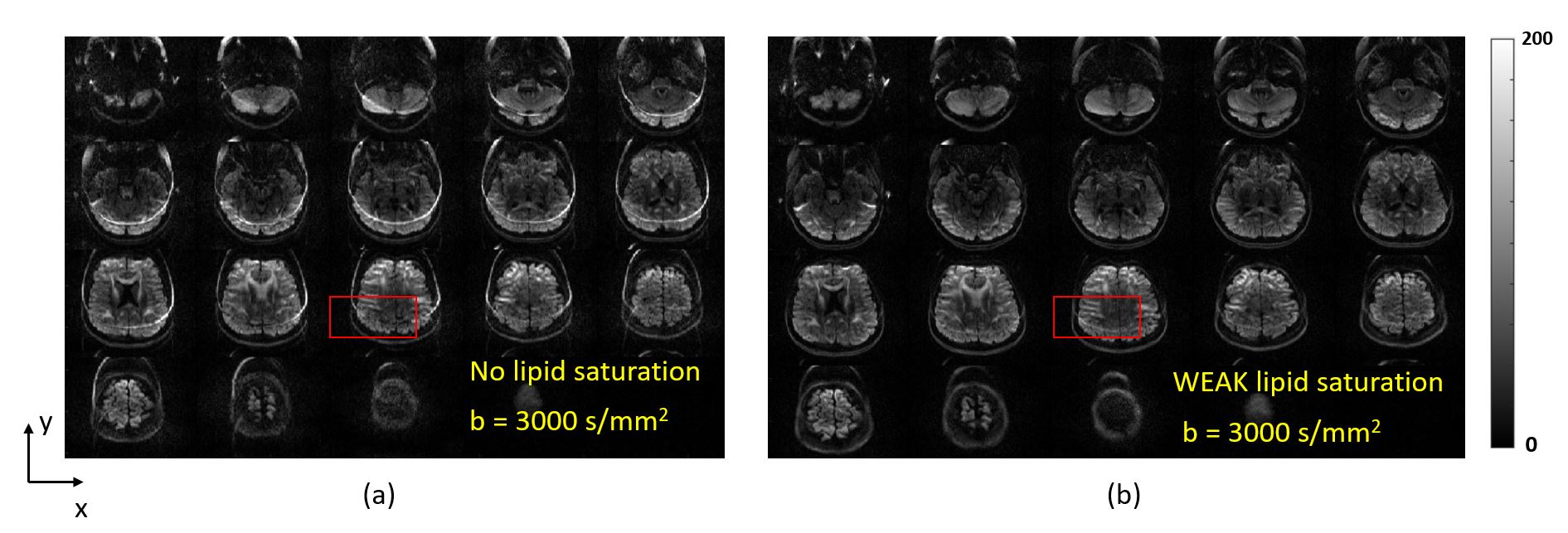
Incomplete lipid suppression results in chemical shift artifacts in a variety of brain and body pulse sequences. For example, residual lipids routinely create artifacts in body imaging, where severe off-resonance and inter-subject anatomic variability make it difficult to achieve good lipid saturation across the whole imaging volume1-2. Lipid suppression also remains a challenge in brain imaging3 .One such recent example is the g-SLIDER method4. Due to the nature of the RF pulses used, g-SLIDER is restricted to weak fat suppression, which commonly results in “fat ring” artifacts in the diffusion-weighted images, as shown in Fig.1, presenting a confound in applications such as tractography.Our group has previously shown that multi-coil (MC) B0 shim arrays5 that can be used to improve lipid suppression in MR Spectroscopic Imaging by rapidly switching on a tailored field offset during the lipid suppression pulse6. The MC fields and baseline B0 field in the body are fed into a convex optimization routine, which jointly solves for the currents in each shim channel to push lipid and water voxels further apart in the frequency domain. In the present work, we extend this approach to 2D imaging by refining the optimization problem to account only for the lipid voxels which would fall within the bandwidth of the water excitation pulse for the slice being acquired. In this way, the number of lipid voxels fed into the optimizer for each slice is dramatically reduced, enabling more complete spectral separation of water and lipids. The new method – “Lipid Artifact Removal by Dynamic Shimming (LARDS)” – is compared to conventional global B0 shimming in simulations. For brain imaging, we simulate using an existing “AC/DC” 32-ch brain MC shim array, and for body imaging we simulate a proposed 64-ch body MC shim array (single-turn loops in both cases).
Methods
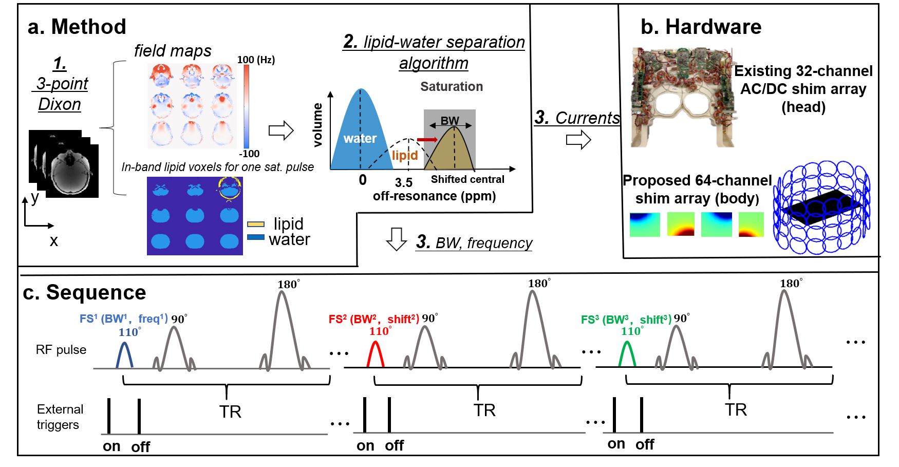
Fig. 2 shows the proposed LARDS methodology. Step 1: Acquire water and lipid masks as well as baseline B0 map using 3-point Dixon method (TE = 2.46ms, 3.69ms, 4.92ms). Step 2: Convex optimization is used to improve water-lipid spectral separation. Optimizer inputs: (i) water mask over the whole brain, (ii) lipid mask of voxels excited by the RF excitation pulse, and (iii) the B0 field map basis set for the MC shim array (subject to limit of 3.5A/channel). Optimizer outputs: (i) current amplitude for each MC shim channel, (ii) center frequency and bandwidth of lipid saturation pulse. For consistency, we simulate a gaussian lipid pulse (1.92 time-bandwidth-product and 110° flip angle) to match the pulse used in vendor-provided EPI sequences on our scanner platform (3 Tesla Siemens Prisma).LARDS simulations use brain and body DIXON7 images acquired on 2 volunteers. In simulations, LARDS is compared against baseline B0 shimming using the vendor-provided shim routine (“Case 1”). We also simulate using the 32-ch brain and 64-ch body shim arrays for global shimming (“Case 2”) using a conventional least-squares objective function on ΔB0. For case 1 and case 2, the same lipid mask is used. For case 3, the lipid mask is different for each simulated slice acquisition. Performance is summarized using metrics of unsaturated lipid fraction and saturated water fraction.
Results
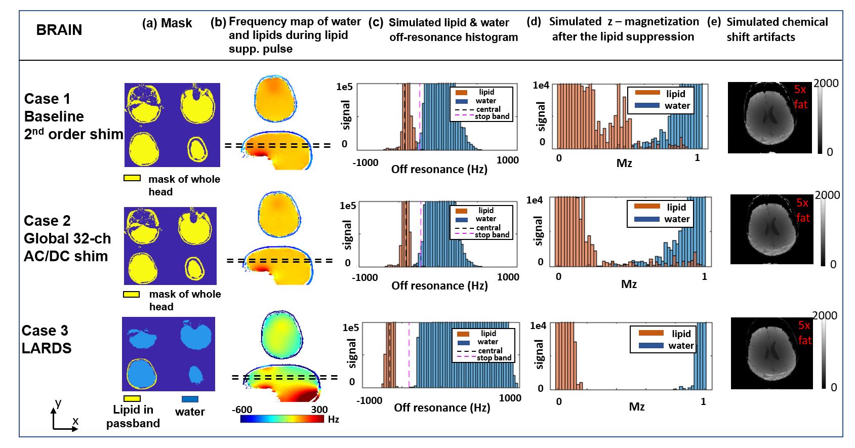
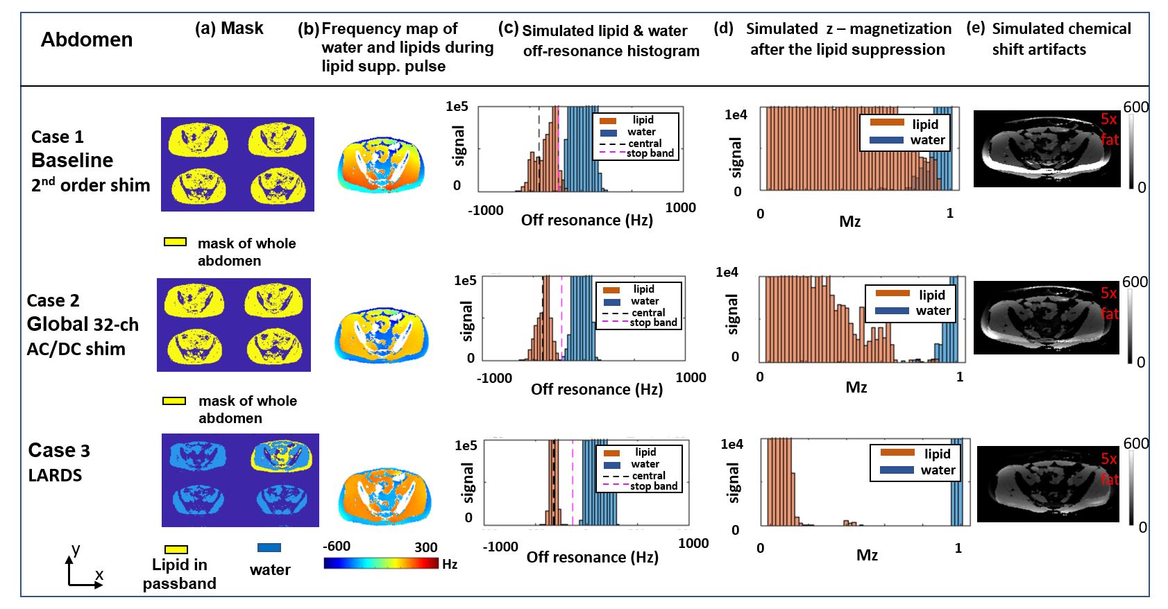
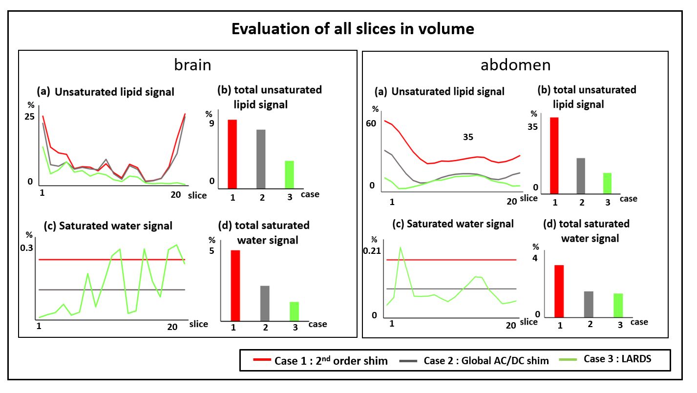
Fig.3 and Fig.4 show simulation results for multi-slice 2D imaging of the brain and body, respectively. The water and lipid frequency histograms and z-magnetization plots show that Case 2 (MC global shimming) does not significantly outperform Case 1 (baseline 2nd-order global shimming) for brain imaging, and provides only modest improvements for body imaging. By contrast, Case 3 (LARDS) substantially improves water-lipid spectral separation, resulting in less residual lipid signal while also minimizing unwanted water saturation. The histograms in Figs. 3(c) and 4(c) show that the lipid pulse center frequency and stop band are shifted in order to optimally suppress lipids while minimizing water saturation. Fig.5 summarizes performance on a slice-wise basis as well as over the whole imaging volume. For brain and body imaging, LARDS reduces residual lipid signal by over 50% while also modestly reducing unwanted water signal loss.Discussion and Conclusion
Simulations suggest that the benefits of LARDS are greater for 2D abdominal imaging relative to brain imaging, since the baseline Case 1 histograms show more water-lipid overlap for abdominal slices compared the brain slices, which arises from the far greater difficulty of global homogeneity shimming in the abdomen and its anatomical variability and complexity.Experimental validation of LARDS for brain imaging using the 32-ch AC/DC array has been delayed due to COVID-19, but will proceed in the near future. we will also consider the benefits of other lipid saturation pulses, for example maximum-phase Shinnar-LeRoux pulses8, to achieve a sharper transition band than the gaussian pulse.
Acknowledgements
Funding support from NIH NIBIB R01EB028797, U24EB028984, the National Natural Science Foundation of China (No: U1809204, 61525106, 61427807, 61701436), the National Key Technology Research and Development Program of China (No: 2017YFE0104000, 2016YFC1300302), and by Shenzhen Innovation Funding (No: JCYJ20170818164343304, JCYJ20170816172431715)References
[1] Del Grande, Filippo, et al. "Fat-suppression techniques for 3-T MR imaging of the musculoskeletal system." Radiographics 34.1 (2014): 217-233.
[2] Huang, Susie Y., et al. "Body MR imaging: artifacts, k-Space, and solutions." Radiographics 35.5 (2015): 1439-1460.
[3] Hernando, D., et al. "Removal of olefinic fat chemical shift artifact in diffusion MRI." Magnetic Resonance in Medicine 65.3 (2011): 692-701.
[4] Liao, Congyu, et al. "High‐fidelity, high‐isotropic‐resolution diffusion imaging through gSlider acquisition with and T1 corrections and integrated ΔB0/Rx shim array." Magnetic Resonance in Medicine 83.1 (2020): 56-67.
[5] Stockmann, Jason P., et al. "A 32‐channel combined RF and B0 shim array for 3T brain imaging." Magnetic resonance in medicine 75.1 (2016): 441-451.
[6] Arango, Nicolas. Sequence-phase optimal (SPO)Δ B₀ field control for lipid suppression and homogeneity for brain magnetic resonance spectroscopic imaging. Diss. Massachusetts Institute of Technology, 2020.
[7] Glover, Gary H., and Erika Schneider. "Three‐point Dixon technique for true water/fat decomposition with B0 inhomogeneity correction." Magnetic resonance in medicine 18.2 (1991): 371-383.
[8] Pauly, John, et al. "Parameter relations for the Shinnar-Le Roux selective excitation pulse design algorithm (NMR imaging)." IEEE transactions on medical imaging 10.1 (1991): 53-65.
Figures