0779
Off-resonance Robustness in Reduced FOV Imaging using Sheared 2DRF Excitation
Bahadir Alp Barlas1,2, Cagla Deniz Bahadir1,2,3, Sevgi Gokce Kafali1,2,4, Ugur Yilmaz2, and Emine Ulku Saritas1,2,5
1Department of Electrical and Electronics Engineering, Bilkent University, Ankara, Turkey, 2National Magnetic Resonance Research Center (UMRAM), Bilkent University, Ankara, Turkey, 3Department of Biomedical Engineering, Cornell University, New York, NY, United States, 4Department of Bioengineering, University of California Los Angeles, Los Angeles, CA, United States, 5Neuroscience Graduate Program, Bilkent University, Ankara, Turkey
1Department of Electrical and Electronics Engineering, Bilkent University, Ankara, Turkey, 2National Magnetic Resonance Research Center (UMRAM), Bilkent University, Ankara, Turkey, 3Department of Biomedical Engineering, Cornell University, New York, NY, United States, 4Department of Bioengineering, University of California Los Angeles, Los Angeles, CA, United States, 5Neuroscience Graduate Program, Bilkent University, Ankara, Turkey
Synopsis
The use of 2D echo-planar radiofrequency (2DRF) excitation has been widely applied in reduced field-of-view imaging of targeted regions, especially for diffusion weighted imaging (DWI). This work proposes effective and efficient coverage of the excitation k-space by shearing the 2DRF trajectory. This approach enables significant improvement in off-resonance robustness of 2DRF pulses by minimizing the pulse duration. It also promises improved clinical utility by increasing SNR in problem regions, eliminating slice coverage limitations and providing inherent fat suppression capability.
Introduction
Reduced field-of-view (FOV) imaging techniques excite only the region of interest to enable aliasing-free imaging with fewer phase encoding (PE) lines and increased resolution1-8. The reduction in PE lines improves the in-plane off-resonance robustness during echo-planar imaging (EPI). However, the conventional 2DRF pulses used for reduced-FOV imaging are considerably longer than their 1D counterparts, which makes them sensitive to through-plane off-resonance issues. Furthermore, the replicas along the blipped axis of the excitation k-space trajectory can limit the slice coverage1. Previously, tilted 2DRF excitation was proposed to overcome the slice coverage limitations while causing further increase in pulse duration2,3. In this work, we propose a sheared 2DRF pulse excitation to achieve significantly reduced pulse durations with improved off-resonance robustness and enhanced SNR. Additionally, the sheared 2DRF profile eliminates slice coverage limitations while providing fat suppression capability.Methods
2DRF Pulse DesignFirst, a standard 2DRF pulse was produced with slab and slice thicknesses FOVSlab=40mm and Δz=4mm, respectively, 16.0ms pulse duration, and time-bandwidth products (TBWs) in SS and PE directions TBWSlice=3 and TBWSlab=8, respectively. These parameters allow a maximum of 16 slices to be imaged without partial saturation caused by replicas along SS direction. Next, a sheared trajectory was designed to cover the same excitation k-space extent. Based on the shearing angle, the number of trajectory lines (NLines) were automatically adjusted to minimize the 2DRF pulse duration, while ensuring that the replicas are pushed outside the potential slice locations. Accordingly, the shearing angle θ that completely eliminates the slice coverage limitation can be found as:
$$ θ≥tan^{−1}\bigg( \frac{(FOV_{Slab})(TBW_{Slice})}{(N_{Lines}-1)(Δz)}\bigg) (1) $$
The 2DRF pulses were designed in MATLAB assuming GMAX=2.8G/cm maximum gradient amplitude and SMAX=15G/cm/s maximum slew rate, and were shortened with respect to these hardware limits using variable-rate selective excitation (VERSE)9.
Analysis on Effects of TBWSlab , FOVSlab and Δz
The shearing operation shortens the pulse durations, with its performance depending on the 2DRF parameters. To analyze this dependency, the following “reference” parameters were chosen: FOVSlab=40mm, Δz=4mm, TBWSlab=8. Each parameter was then varied individually, and the corresponding standard and sheared 2DRF pulses with θ ranging between 30°-80° were generated. TBWSlab was varied as [6, 8, 10]. FOVSlab was varied as [30, 40, 50, 60] mm. Δz was varied as [3, 4, 5] mm.
In Vivo Imaging Experiments
In vivo reduced-FOV T2-weighted imaging was conducted in the cervical spinal cord of a healthy subject, on a 3T MRI scanner (Siemens Magnetom Trio) using an 8-channel spine array coil. The standard 2DRF pulse explained above was compared with a sheared 2DRF pulse with θ=73° and a pulse duration of 9.5ms, the minimum duration pulse with inherent fat suppression (i.e., clear separation of water and fat excitation profiles for the 2DRF pulse). Single-shot, T2-weighted EPI images of the spinal cord were acquired with 200X50mm2 FOV, 128X32 acquisition matrix, TE=22ms (28ms for standard 2DRF due to its longer duration). A standard 3ms 1DRF excitation with additional fat suppression was used for comparison, with 200X162.5mm2 full-FOV, 128X104 acquisition matrix, TE=52ms. Common parameters were Δz=4mm, 1.6X1.6mm2 in-plane resolution, 6/8 partial Fourier acquisition, TR=6500ms.
Results and Discussion
The trajectories and waveforms of the standard and sheared 2DRF pulses are shown in Fig. 1, and the corresponding 2D excitation profiles are given in Fig. 2. For both 2DRF pulses, a subsequent 180° refocusing RF pulse suppresses the signal from fat, providing inherent fat suppression capability. The 1D cross-sectional profiles in Fig. 2c demonstrate that the sheared 2DRF pulse preserves the sharpness in both slab and slice profiles, while effectively eliminating the replicas in SS direction.The effects of pulse parameters on the sheared 2DRF pulse duration are shown in Fig. 3. Accordingly, an increase in FOVSlab causes a decrease in the standard 2DRF pulse duration, while the sheared 2DRF pulse duration increases with FOVSlab, as a wider FOVSlab requires NLines to be increased for fixed θ (see Eqn. 1). Increasing TBWSlab causes an increase in the duration of both the standard and sheared 2DRF pulses at a similar rate. An increment in Δz has almost no effect on the duration of the standard 2DRF pulse, while decreasing the duration of the sheared 2DRF pulse. Importantly, the sheared 2DRF pulse provides significant reduction in pulse duration, especially for smaller FOVSlab cases where reduced-FOV imaging is needed.
Figure 4a shows the standard and sheared 2DRF pulse durations used for in vivo imaging, where θ=73° was chosen. As shown in Fig. 4, full-FOV imaging suffers from extensive in-plane off-resonance artifacts, whereas both versions of the 2DRF pulse successfully alleviate this problem to achieve higher resolution images with reduced in-plane distortion. The sheared 2DRF pulse further improves the SNR, especially in the central part of the cervical spine that typically experiences high off-resonance effects. In short, the shorter duration of the sheared 2DRF pulse provides robustness against through-plane off-resonance effects that otherwise cause reduced signal intensity.
Conclusion
The proposed sheared 2DRF pulse design technique achieves considerable reduction in pulse duration, improving the off-resonance robustness of 2DRF-pulse-based reduced-FOV imaging. The in vivo imaging results in the cervical spine demonstrate that the sheared 2DRF pulse improves SNR, eliminates the slice coverage limitations, and maintains inherent fat suppression capability.Acknowledgements
This work was supported by the Scientific and Technological Research Council of Turkey (TUBITAK 117E116).References
- Saritas EU, Shankaranarayanan A, Zaharchuk G, Nishimura DG. Reduced-FOV single-shot diffusion-weighted EPI: extended slice coverage with tailored RF pulse design. In Proceedings of the 19th Annual Meeting of ISMRM, Montreal, Quebec, Canada, 2011, p. 1953.
- Banerjee S, Nishimura DG, Shankaranarayanan A, Saritas EU. Reduced field-of-view DWI with robust fat suppression and unrestricted slice coverage using tilted 2D RF excitation. Magn Reson Med. 2016;76(6):1668–1676. DOI: 10.1002/mrm.26405.
- Saritas EU, Cunningham CH, Lee JH, Han ET, Nishimura DG. DWI of the spinal cord with reduced FOV single-shot EPI. Magn Reson Med 2008;60(2):468–473.
- Finsterbusch J. Improving the performance of diffusion-weighted inner field-of-view echo-planar imaging based on 2D-selective radiofrequency excitations by tilting the excitation plane. J Magn Reson Imaging 2012;35(4):984–992.
- Wheeler-Kingshott CA, Parker GJ, Symms MR, Hickman SJ, Tofts PS, Miller DH, Barker GJ. ADC mapping of the human optic nerve: increased resolution, coverage, and reliability with CSF-suppressed ZOOM-EPI. Magn Reson Med 2002;47(1):24–31.
- Wilm BJ, Svensson J, Henning A, Pruessmann KP, Boesiger P, Kollias SS. Reduced field-of-view MRI using outer volume suppression for spinal cord diffusion imaging. Magn Reson Med 2007;57(3):625–630.
- Heidemann RM, Anwander A, Feiweier T, Knösche TR, Turner R. Turner. k-space and q-space: combining ultra-high spatial and angular resolution in diffusion imaging using ZOOPPA at 7 T. NeuroImage 2012;60(2):967–978.
- Finsterbusch J. High-resolution diffusion tensor imaging with inner field-of-view EPI. J Magn Reson Imaging 2009;29(4):987–993.
- Lee D, Lustig M, Grissom WA, Pauly JM. Time‐optimal design for multidimensional and parallel transmit variable‐rate selective excitation. Magn Reson Med. 2009;61(6):1471-1479
Figures
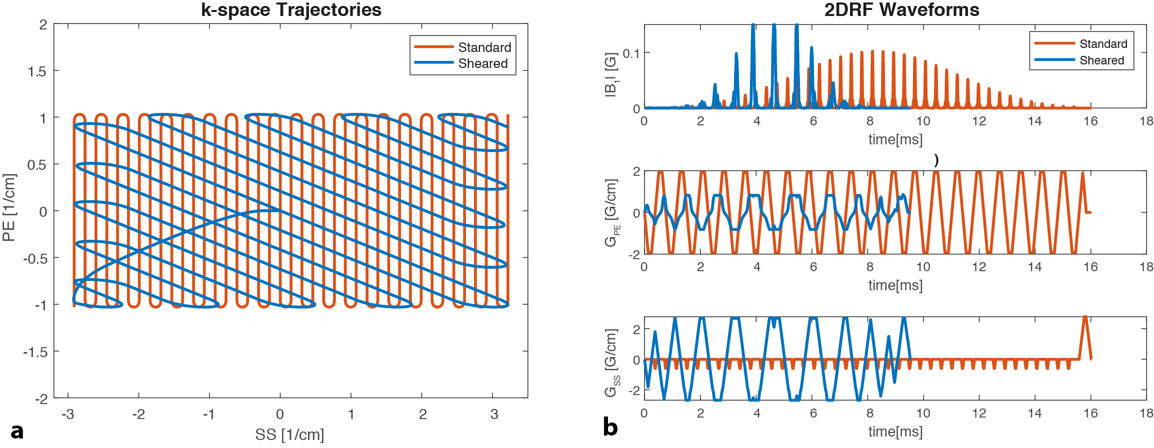
FIGURE 1. (a) Excitation k-space trajectories for the standard and sheared 2DRF designs. (b) The corresponding RF pulses and gradient waveforms for the standard and sheared 2DRF designs. The sheared 2DRF pulse with a shearing angle of 73° has significantly shorter duration (9.5 ms) when compared to the standard 2DRF pulse (16.0 ms), while providing identical excitation k-space coverage.
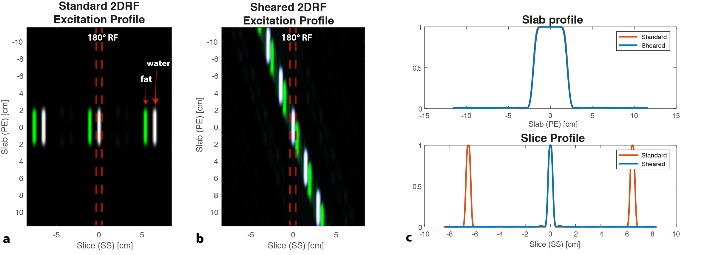
FIGURE 2. Simulated 2DRF excitation profiles for (a) the standard and (b) the sheared 2DRF designs. For the standard 2DRF pulse, the profile is periodic in the SS direction, limiting the slice coverage due to partial saturation effects. In contrast, the sheared 2DRF pulse places the replicas along the sheared axis, effectively pushing them outside the potential slice locations. (c) 1D cross-sectional excitation profiles along SS and PE directions demonstrate identical sharpness, with a slab thickness of 40 mm and a slice thickness of 4 mm.
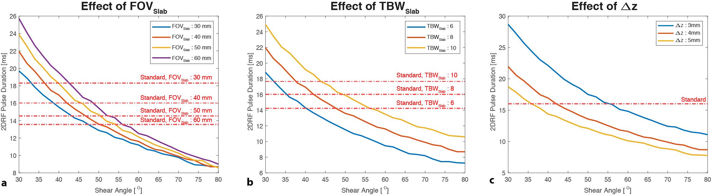
FIGURE 3. Effects of design parameters on the duration of the sheared 2DRF pulse. The effect of (a) slab thickness (FOVSlab), (b) slab sharpness (TBWSlab), and (c) slice thickness (Δz) is shown for shearing angles ranging between 30°- 80°. The corresponding standard 2DRF pulse durations are also marked. Increasing TBWSlab and FOVSlab causes the sheared 2DRF pulse duration to increase, while Δz has the reverse effect. Note that the sheared 2DRF pulse provides significant reduction in pulse duration, especially for smaller FOVSlab cases where reduced-FOV imaging is needed.
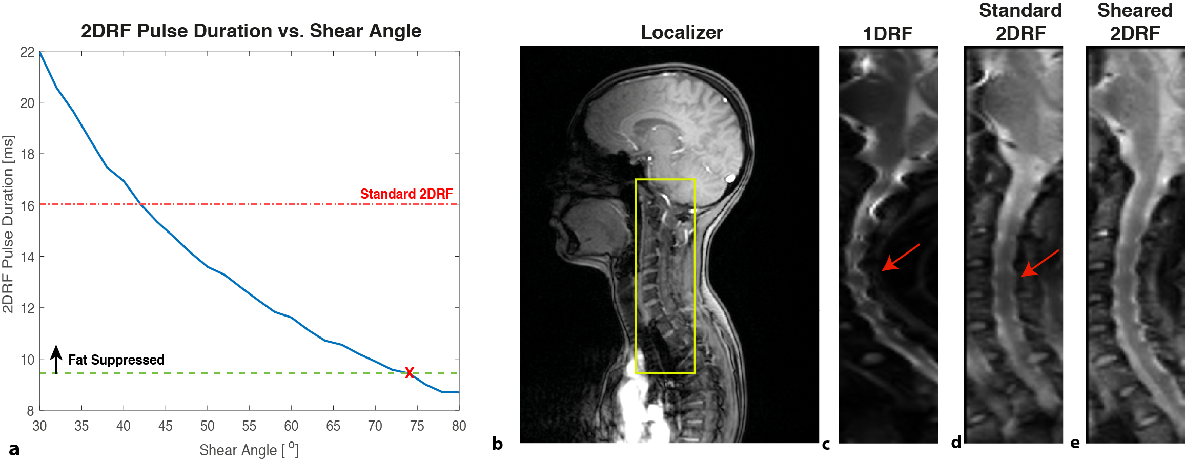
FIGURE 4. In vivo imaging results in the cervical spinal cord. (a) The standard and sheared 2DRF pulses had 16.0 ms and 9.5 ms durations, respectively. (b) The localizer showing the reduced-FOV region corresponding to 200 mm X 50 mm FOV. T2-weighted EPI images acquired using (c) 1DRF pulse with additional fat suppression (cropped full FOV of 200 mm X 162.5 mm), and (d) standard and (e) sheared 2DRF pulses. Reduced-FOV imaging achieves higher resolution with reduced in-plane distortion. The sheared 2DRF pulse further improves SNR in regions with high off-resonance effects (red arrows).