0775
3D APTw Brain Tumor Imaging with Compressed SENSE: Comparison of Different Acceleration Factors and with Conventional Parallel Imaging
Nan Zhang1, Qingwei Song2, Ailian Liu2, Haonan Zhang2, Renwang Pu2, Jiazheng Wang3, and Zhiwei Shen3
1The First Affilliated Hospital of Dalian Medical University, Dalian, China, 2The First Affiliated Hospital of Dalian Medical University, Dalian, China, 3Philips Healthcare, Beijing, China, Beijing, China
1The First Affilliated Hospital of Dalian Medical University, Dalian, China, 2The First Affiliated Hospital of Dalian Medical University, Dalian, China, 3Philips Healthcare, Beijing, China, Beijing, China
Synopsis
Amide proton transfer weighted (APTw) imaging is a novel and promising MRI method for brain tumor imaging, but it can be time-consuming. Common parallel imaging methods, like SENSE, can lead to reduced image quality and increased artifact at high acceleration factors. Here, the compressed SENSE (CS) technique with combined strength from both compressed sensing and SENSE was evaluated for the acceleration of APTw imaging in brain. Results showed that it is feasible to apply an CS accelerator factor of 5 to APTw imaging of brain tissue and tumor, which could reduce the scan time to less than 1 min.
Introduction:
Amide proton transfer weighted (APTw) imaging is a type of MR molecular imaging technique based on chemical exchange between free bulk water protons and the amide protons (-NH) of endogenous mobile proteins and peptides in tissue. [1, 2] Clinical applications of APTw imaging on brain tumors were very promising, but the long scan could be a limitation especially for 3D APTw imaging. Some studies have been carried out to evaluate the application of compressed sensing for accelerating the acquisition of APTw images [3, 4], but the effects of different accelerators on APTw images needs to be further explored. Moreover, the compressed SENSE (CS) technique with combined strength from both the compress sensing and conventional SENSE method has been evaluated for accelerated scan of different sequences in different tissues. The aim of the current study was to evaluate the performance of compressed SENSE for 3D brain APTw imaging with different acceleration factors, and the results were compared with those by SENSE.Methods:
MR scans were performed on a 3.0 T scanner (Ingenia CX, Philips Healthcare, Best, the Netherlands) using a body coil for RF transmit and a 32‐channel head coil for signal receiving in our hospital in 34 brain tumor patients (44.00±19.65, range:11-64 years ,18 women). The tumors included 12 meningiomas, 11 metastatic tumors, and 11 gliomas. APTw imaging were accelerated by compressed SENSE with acceleration factors (AF) of 2, 3, 4 and 5 (CS2-CS5) and by conventional SENSE with an AF of 1.6 (SENSE1.6, as a reference scan). The other scan parameters were shown in Table 1. The APTW images were transfer to the Philips IntelliSpace Portal for the APTmean, APTmax, APTmax-min and APTmin measurements. Five regions of interest (ROIs) were draw on the APTw image by SENSE1.6 with co-registration and fusion on the Gd-T1 weighted image and copied to the other APTw images by CS-SENSE; the size of each ROI was fixed at 15 pixels (Fig 2-4). The ROIs were draw with exclusion of necrosis, cystic cavities, large vessels, calcification, and hemorrhagic components. For each patient, the APTw values in five ROIs (APTw1, APTw2, … APTw5) were recorded. Then, the maximum value (APTmax), the minimum value (APTmin), the maximum–minimum value (APTmax-min), and (APTw1 + APTw2 + … + APTw5 )/5(APTmean) were calculated. The multivariate analysis of variance was used to identify APT values under different AF in brain tumor patients.Results:
The APTmean, APTmax, and APTmin values obtained from the CS scans with AFs of 2、3、4、5 for different types of brain tumors were well maintained as compared with the reference scan (SENSE1.6). No significant difference of APTmean and APTmax was observed between the reference and CS-accelerated scans. The APTmin measured by CS-accelerated scans were significantly different from those by the reference scan. There was no significant difference in APTmean, APTmax, and APTmin among different scans for each of three brain tumors. (Table2、3、4) The scan time of CS-accelerated APTw imaging was dramatically reduced to 106 S (CS 5)as compared to SENSE1.6 (3:39 mins).Discussion and Conclusion:
In the current study, brain APTw imaging accelerated by compressed SENSE with increased acceleration factors (AF 2-5) could be comparable to results by SENSE with AF of 1.6. According to our result, the acceleration factor 5 for compressed SENSE technique was recommended in future study, saving at least 70 % scan time compared to the SENSE acceleration. In summary, compressed SENSE could be successfully extended to accelerated 3D APTw imaging without compromising image quality, and it would be beneficial for a wide range of clinical applications.Acknowledgements
No acknowledgement found.References
[1] Zhou J, Heo HY, Knutsson L, van Zijl P, Jiang S. APT‐weighted MRI: techniques, current neuro applications, and challenging issues. Magn Reson Imaging. 2019,50(2):347-364. [2] By S, Barry RL, Smith AK, et al. Amide proton transfer CEST of the cervical spinal cord in multiple sclerosis patients at 3T. Magn Reson Med. 2018;79:806–814. [3]Bratke G, et al. Accelerated MRI of the Lumbar Spine Using Compressed Sensing: Quality and Efficiency. J Magn Reson Imaging 2018; 49: e164-e175; [4]Vu KN, Gilbert G, Chalut M, et al. MRI-determined liver proton density fat fraction, with MRS validation: Comparison of regions of interest sampling methods in patients with type 2 diabetes. J Magn Reson Imaging, 2016, 43(5): 1090-1099.Figures
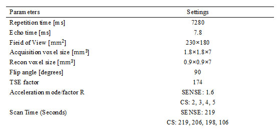
Table1 MR Parameters of APT
Sequences of brain imaging
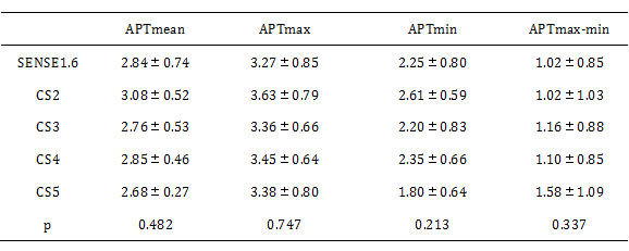
Table3 Comparisons of quantitative
APT values for meningiomas among CS and SENSE scans
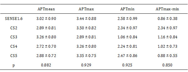
Table4 Comparisons of quantitative
APT values for metastatic tumors among CS-SENSE and SENSE scans
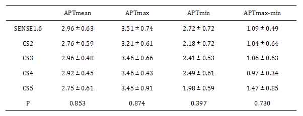
Table5 Comparisons of Qualitative APT Values for gliomas Between CS and SENSE Imaging
Figure
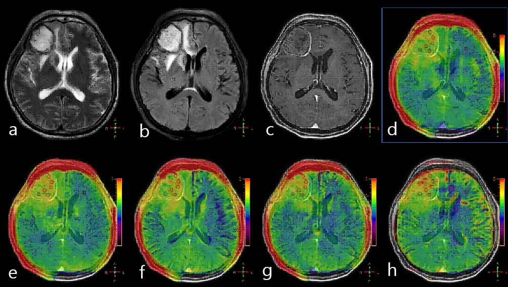
1a 54-year-old
female patient with meningiomas in brain: a. T2WI; b.
T2WI FLAIR; c. T1-Gd; d. APTw image by SENSE-1.6 fused with T2WI FLAIR image; e-h.
APTw images by CS-SENSE (with factors of 2, 3, 4, and 5) fused with T1-Gd image.
The ROIs were
obtained manually on the SENSE APTw image and copied to the others as shown.
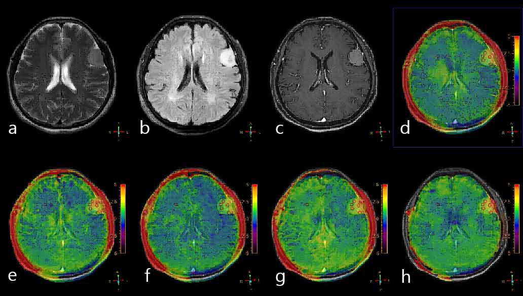
Figure 2 a 64-year-old
female patient with metastatic tumors in brain: a.
T2WI; b. T2WI FLAIR; c. T1-Gd; d. APTw image by SENSE-1.6 fused with T2WI
FLAIR image; e-h. APTw images by CS-SENSE (with factors of 2, 3, 4, and 5) fused
with T1-Gd image. The
ROIs were obtained manually on the SENSE APTw image and copied to the others as
shown
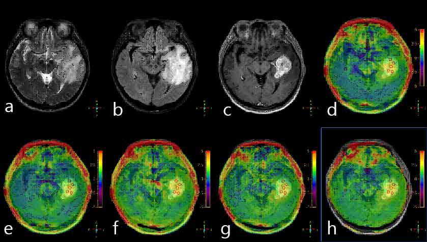
Figure 3 a
45-year-old female patient with gliomas tumors in brain: a. T2WI; b. T2WI FLAIR; c. T1-Gd; d.
APTw image by SENSE-1.6 fused with T2WI FLAIR image; e-h. APTw images by CS-SENSE
(with factors of 2, 3, 4, and 5) fused with T1-Gd image. The ROIs were obtained
manually on the SENSE APTw image and copied to the others as shown