0751
Removal of Subcutaneous Lipid Signals from Spin-Echo 1H-MRSI Brain Data Using an FID Reference and Machine Learning
Yunpeng Zhang1, Yibo Zhao2,3, Yudu Li2,3, Rong Guo2,3, Yao Li1, and Zhi-Pei Liang2,3
1School of Biomedical Engineering, Shanghai Jiao Tong University, Shanghai, China, 2Beckman Institute for Advanced Science and Technology, University of Illinois at Urbana-Champaign, Urbana, IL, United States, 3Department of Electrical and Computer Engineering, University of Illinois at Urbana-Champaign, Urbana, IL, United States
1School of Biomedical Engineering, Shanghai Jiao Tong University, Shanghai, China, 2Beckman Institute for Advanced Science and Technology, University of Illinois at Urbana-Champaign, Urbana, IL, United States, 3Department of Electrical and Computer Engineering, University of Illinois at Urbana-Champaign, Urbana, IL, United States
Synopsis
Spin-Echo (SE) MRSI can encode J-coupling information and is desirable for brain imaging applications. But it uses long TR, leading to long scan time and thus low resolution. Consequently, removal of subcutaneous lipid signals from low-resolution SE data is challenging. This paper presents a novel method to solve this problem. The proposed method uses a high-resolution FID reference and a neural network to transform it into SE signals, which are then used to construct a generalized-series model for lipid signal removal from the SE 1H-MRSI data. The proposed method has been tested using in vivo data, producing very encouraging results.
Introduction
Spin-Echo (SE) acquisitions have several unique advantages over FID acquisitions, including the capability for encoding J-coupling and diffusion weighting1. SE-MRSI is, therefore, often used to obtain J-resolved spectral information desired for accurate separation of brain metabolites and neurotransmitters. However, SE acquisitions use long TR, leading to long scan time and thus low spatial resolution. Consequently, removal of subcutaneous lipid signals from low-resolution SE 1H-MRSI data is challenging2. Conventional MRSI methods use lipid suppression to alleviate the problem but suppression pulses are often susceptible to system imperfections such as B0 inhomogeneity3. Various post-processing methods have also been proposed, which use prior information such as spatial support of lipid signals4, spectral support of metabolite signal5, sparsity6, and spatiospectral structures of lipid signals in the form of a union-of-subspaces model7. These post-processing methods work well with MRSI data acquired with lipid suppression; for MRSI data acquired with no lipid suppression and only a small number of spatial encodings as often is the case in J-resolved MRSI experiments, the lipid removal problem remains very challenging. This paper presents a novel method to solve this problem. The proposed method uses a high-resolution FID reference that can be acquired quickly and a neural network to transform it into SE signals. The generated SE lipid signals are then used to construct a generalized series (GS) model for reconstruction and removal of the subcutaneous lipid signals from the SE 1H-MRSI data.Methods
We assume that we have two data sets: one set of SE MRSI data, $$$d_{\rm SE}\left( {\bf k} ,t\right)$$$, with a small number of spatial encodings (i.e., limited k-space coverage), and a high-resolution reference containing only the subcutaneous lipid signals, $$$d_{\rm ref}\left({\bf k},t\right)$$$. The reference data set can be acquired quickly using an FID sequence, or can be obtained using a machine learning-based generative model. In this work, we assume that $$$d_{\rm ref}({\bf k},t)$$$, is obtained using an FID sequence. The proposed method, as illustrated in Fig. 1, consists of two key components: a) a neural network trained to map $$$\rho_{\rm ref}\left( {\bf x} ,t\right)$$$, the spatiotemporal function corresponding to $$$d_{\rm ref}\left({\bf k},t\right)$$$, to SE data, $$$\widetilde{\rho}_{\rm SE}\left( {\bf x} ,t\right)$$$, and b) a GS model constructed based on $$$\widetilde{\rho}_{\rm SE}\left( {\bf x} ,t\right)$$$.To map $$$\rho_{\rm ref}( {\bf x} ,t)$$$ to $$$\widetilde{\rho}_{\rm SE}( {\bf x} ,t)$$$, a 1D ResNet-based convolution neural network (CNN) was used. The architecture of the network is shown in Fig. 2. Specifically, the network consisted of 10 residual blocks followed by a convolutional output layer. Each residual block included a convolution layer, a deconvolution layer, and a batch normalization layer. The network was trained using pairs of high-resolution FID and SE lipid data acquired from healthy volunteers. After proper training, the network was capable of producing the desired SE lipid reference $$$\widetilde{\rho}_{\rm SE}( {\bf x} ,t)$$$ from the corresponding FID lipid spectra voxel-by-voxel.
With $$$\widetilde{\rho}_{\rm SE}( {\bf x} ,t)$$$ obtained, a GS model was constructed to represent the lipid signals8. More specifically, we expressed the spatiotemporal distribution of the lipid signals that match a specific SE MRSI data set as:
$$\rho_{\rm lipid}=\Sigma_{m=-M/2}^{M/2}h_m(t)\widetilde{\rho}_{\rm SE}( {\bf x} ,t)e^{-i2{\pi}m{\bf \triangle k \cdot x}}$$
where $$$M$$$ is the order of GS model and $$$h_m$$$ are the GS coefficients. The GS coefficients were estimated by solving the following least-squares problem:
$$\hat h_{m}(t)={\arg}\mathop\min_{h_m(t)}\| d_{\rm SE}\left({\bf k},t\right)-\Omega {\rm F}\left(\Sigma_{m=-M/2}^{M/2}h_m(t)\widetilde{\rho}_{\rm SE}({\bf x},t)e^{-i2{\pi}m{\bf \triangle k\cdot x}}\right)\|_2^2$$
where $$$\Omega$$$ and $$${\rm F}$$$ are the (k,t)-space sampling and Fourier encoding operators, respectively. The GS model can effectively absorb the high-resolution SE lipid reference into the reconstruction and compensate the discrepancies between the CNN-generated SE reference and real SE data.
Results
We have evaluated our proposed method using in vivo data collected from healthy human subjects. Figure 3 shows a set of representative prediction results by the neural network. As can be seen from both error map and selected spectra from individual voxels, the discrepancies between the predicted and acquired SE lipid data were at a very low level. Figure 4 compares the effectiveness in lipid removal of the proposed method with conventional methods including Fourier-based and Papoulis-Gerchberg (PG) reconstructions. As can be seen from the lipid integral map, the proposed method achieved the best performance in lipid signal removal, especially for the most challenging areas close to the subcutaneous region. To demonstrate the benefits of effective lipid removal for metabolite reconstruction, we also compared the associated metabolite maps as shown in Fig. 5. As expected, the proposed method outperformed conventional PG reconstruction with better preservation of neurometabolic signals within the brain.Conclusions
A new method has been proposed for effective removal of lipid signals from spin-echo 1H-MRSI brain data acquired with no lipid suppression and a limited number of spatial encodings. The proposed method has been validated using in vivo data, producing very encouraging results. With further development, the proposed method may provide an effective solution to the lipid removal problem associated with 1H-MRSI, especially J-resolved 1H-MRSI, of the brain.Acknowledgements
Y. L. is funded by National Science Foundation of China (No.61671292 and 81871083) and Shanghai Jiao Tong University Scientific and Technological Innovation Funds (2019QYA12).References
- Pohmann R, Von Kienlin M, Haase A. Theoretical evaluation and comparison of fast chemical shift imaging methods. J Magn Reson. 1997;129(2):145-160.
- Ho R J, Lam F. High-Resolution 3D Spin-Echo MRSI Using Interleaved Water Navigators, Sparse Sampling and Subspace-Based Processing. Proc IEEE Eng Med Biol Soc. 2020;1465-1468.
- Duyn J H, Gillen J, Sobering G, et al. Multisection proton MR spectroscopic imaging of the brain. Radiology. 1993;188(1):277-282.
- Haupt C I, Schuff N, Weiner M W, et al. Removal of lipid artifacts in 1H spectroscopic imaging by data extrapolation. Magn Reson Med. 1998;35(5):678-687.
- Hernando D, Haldar J, Sutton B, et al. Removal of lipid signal in MRSI using spatial-spectral constraints. Proc IEEE Int Symp Biomed Imaging. 2007;1360-1363.
- Bilgic B, Gagoski B, Kok T, et al. Lipid suppression in CSI with spatial priors and highly undersampled peripheral k-space. Magn Reson Med. 2013;69(6):1501-1511.
- Ma C, Lam F, Johnson C L, et al. Removal of nuisance signals from limited and sparse 1H MRSI data using a union-of-subspaces model. Magn Reson Med. 2016;75(2):488-497.
- Liang ZP, Lauterbur PC. A generalized series approach to MR spectroscopic imaging. IEEE Trans Med Imaging. 1991;10(2):132–137.
Figures
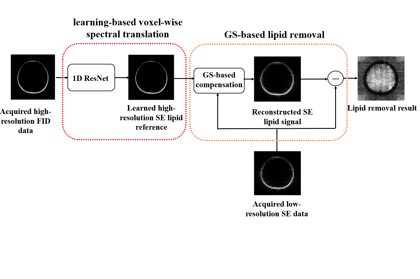
Figure 1. Illustration of the
proposed method. A neural network was trained to transform high-resolution FID
reference into SE signals. A GS model was then constructed to incorporate the
generated SE signals into the reconstruction and removal of the subcutaneous lipid
signals from the SE 1H-MRSI data
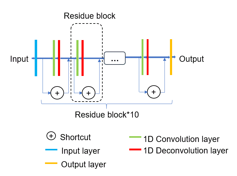
Figure 2. The neural network architecture to learn
the relationship between the FID and SE lipid
spectra
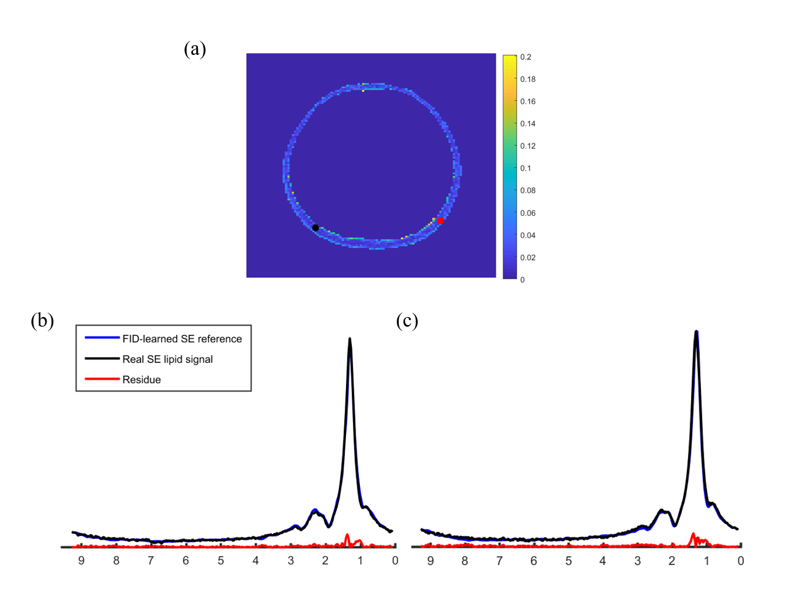
Figure 3. Representative neural network prediction results. (a)
Spatial distribution of the difference between the generated SE lipid signals
and acquired SE lipid signals. (b-c) Network prediction and real SE spectra
from the locations marked by a black and red dot, respectively. Corresponding
RMSEs were 0.90%, 1.07%, respectively.
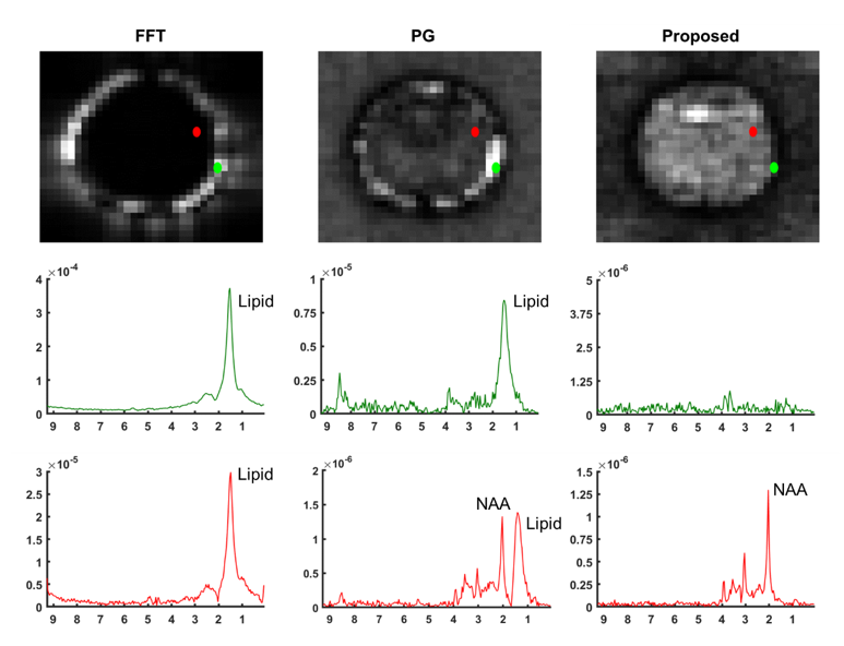
Figure 4. Comparison of lipid
removal performance using Fourier reconstruction (FFT), Papoulis-Gerchberg (PG)
reconstruction, and Machine-learning-based generalized series reconstruction
(proposed). Top panel: lipid integral map; Middle panel: lipid residue spectra
from the location marked by a green dot; Bottom panel: metabolite spectra from
the location marked by a red dot.
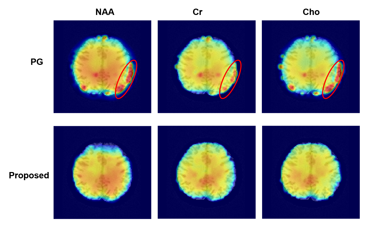
Figure 5. Comparison of NAA, Cr
and Cho maps from the lipid-removed data using Papoulis-Gerchberg (PG)
reconstruction (top) and proposed Machine-learning-based generalized series
reconstruction (bottom). Note the significant artifacts caused by residual
lipid signals in PG reconstruction as circled, which were effectively removed
by the proposed method.