0750
Comparing Zero Time of Echo (ZTE) MRI sequence and Computed Tomography for assessing bony lesions of skull base and calvarium : A Pilot study1Imaging Sciences and Interventional Radiology, Sree Chitra Tirunal Institute for Medical Sciences and Technology, Thiruvananthapuram, India, 2GE Healthcare, GE Healthcare, Bangalore, India
Synopsis
ZTE is a novel MRI sequence which can be used for the imaging of the bone. In this pilot study we present our initial experience with this sequence. The pseudoCT images generated after post-processing of raw ZTE data were similar to the real CT images in bone windows and were able to give the relevant clinical information about the lesion size, margins, erosions and bony expansion. As this technique is free of ionising radiation, it has a huge potential for bone imaging. Thus the MRI becomes one stop shop for the imaging of patients with bone lesions.
PURPOSE
Computed Tomography (CT) is considered the gold standard for evaluation of bone lesions. This, however comes at a cost of ionisation radiation exposure. MRI is radiation free but known to be poor in visualisation of bone and its lesions. ZTE is an emerging MRI technique which can be used to image the bone(1). ZTE has produced ‘pseudoCT’ images which are well suited to demonstrate bony details. In the present study we compared image quality and diagnostic accuracy of pseudoCT generated from ZTE MRI sequence in characterisation of bony lesions involving skull base and calvarium.METHODS
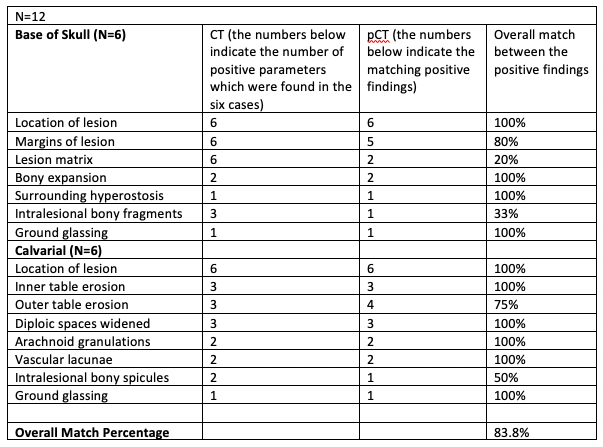
MRI had lacked the capacity to image bones with the details needed for optimal clinical decisions, mainly due to low proton density (20% of water) and short signal life (2). With ZTE imaging technique it is now possible to image the structures with very low proton content and short T2 times such as bones with greater clarity as it achieves a near-zero time interval between radiofrequency excitation and data acquisition(3). After post processing with inverse logarithmic scaling and histogram analysis, virtual CT like images are generated.Experiment: We imaged consecutive patients with lytic lesion involving the skull base or calvarium with ZTE as an additional sequence to routine MRI sequence. Most of the patients already had a CT scan done previously, which served as a control image.The ZTE sequence only took less than 2 minutes to complete on a 3T MR scanner (GE Healthcare, Chicago, IL, USA) using the following protocol: 3D imaging mode with axial acquisitions, TE = 0 msec, flip angle = 1 degree, receiver bandwidth = ±62.5 kHz, FOV = 30 cm, resolution = 1.5x1.5x1.5 mm3 number of excitations = 2, slices = 200 and Spokes per segment = 512. Total time for scanning was 1 minute 52 seconds and 32 channel head coil was used. Raw ZTE images were converted into CT like HU map or ‘pseudoCT’ images using a deep learning (DL) based algorithm. The DL algorithm consists of a modified UNet architecture designed to maintain overall structural accuracy with an increased focus on accurate bone depiction. The DL model was trained on numerous co-registered ZTE & CT image pairs. The post processed pseudoCT images were studied and compared with the original CT images. Two radiologists (VC and KC with more than 8 and 23 years of experience in radiology each), compared the pseudoCT images with CT images, by consensus, for lesion visualization, spatial resolution, contrast resolution, artifacts and overall image quality and assessed the percentage match. For the skull base lesions parameters used were location of lesion, margins, matrix, bony expansion, surrounding hyperostosis, intralesional bony fragments and ground glassing. For calvarial lesions parameters used were location of lesion, involvement of inner or outer table, matrix, widening of diploe, intralesional bony spicules and ground glassing. The informed consent for this additional sequence was taken from the patient/ relative undergoing the scan. We present our experience with initial12 cases done till now in this ongoing project.RESULTS
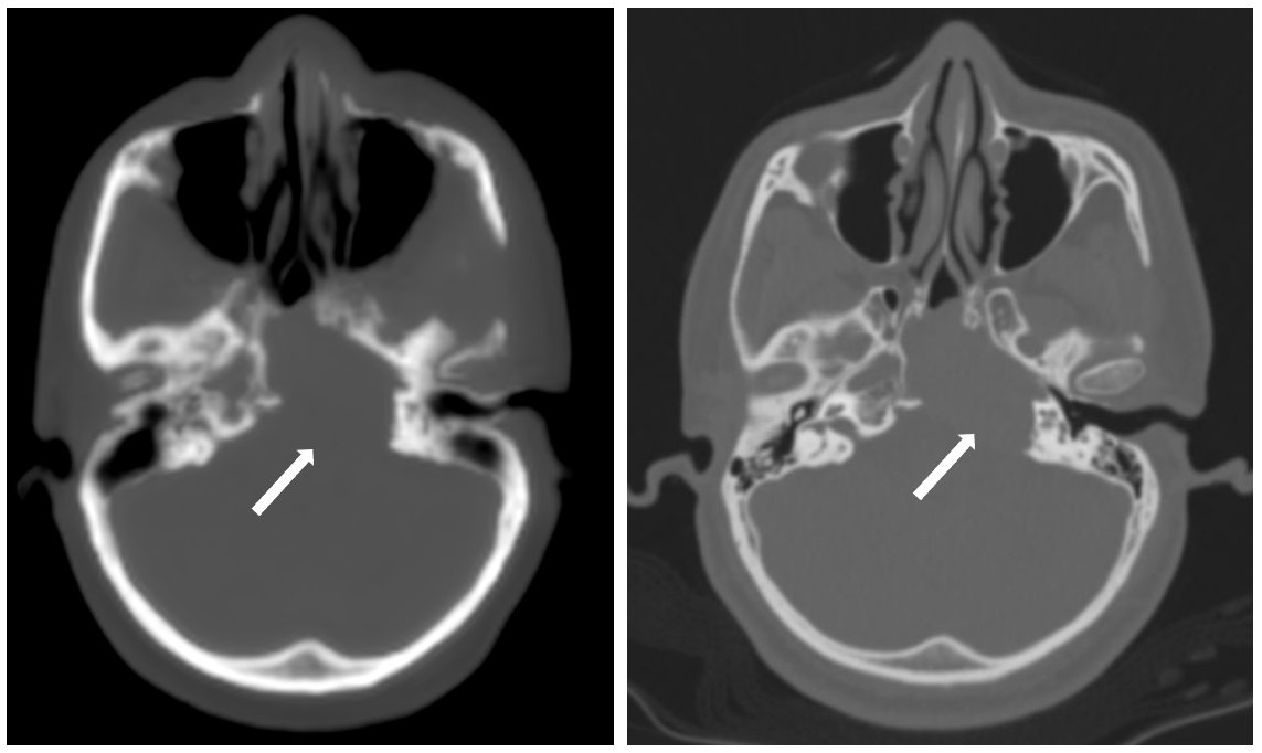
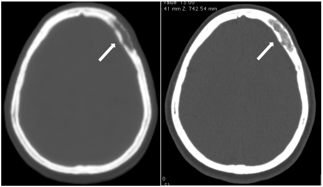
The results of the image analysis is given in Table 1. As reported in the data in table, ZTE was excellent in picking up the lesion, details of the margins and surrounding bone density changes, it was still not as clear in depicting the matrix composition of the lesions and was not able to show the exact details of the intralesional bony fragments. In one case pseudoCT overestimated the erosion of outer table, giving a false positive result. Image quality of the pseudoCT images of ZTE are shown with comparative CT images in Figure 1 & 2.DISCUSSION
ZTE pseudoCT images showed the lesion accurately and the margins were clearly defined. The contrast resolution was as good as CT and the overall image quality was satisfactory. This clinical study did not show significant difference in visualisation of the bony lesions of the skull base and calvarium. However the pseudoCT images lacked the spatial resolution of the CT images. There were also blurring of the bony margins in upper neck region due to patient motion as the inherent longer duration of image acquisition time as compared to CT. However for the non-moving parts like calvarium excellent delineation of the margins was achieved. Artefacts were also seen due to dental amalgam, which lead to blooming in the region of teeth. PseudoCT images gave the required information which was needed in the further management of the patients. This pilot study demonstrates that Zero TE MR imaging is a promising novel technique that offers an alternative to CT with rapid, high-resolution, silent imaging(4).This sequence avoids ionising radiation exposure which is inherent with CT scans.Acknowledgements
The authors acknowledge GE Healthcare for research support.References
1. Wiesinger F, Sacolick LI, Menini A, Kaushik SS, Ahn S, Veit-Haibach P, et al. Zero TEMR bone imaging in the head: Zero TE Bone Imaging. Magn Reson Med. 2016 Jan;75(1):107–14.
2. Du J, Carl M, Bydder M, Takahashi A, Chung CB, Bydder GM. Qualitative and quantitative ultrashort echo time (UTE) imaging of cortical bone. J Magn Reson San Diego Calif 1997. 2010 Dec;207(2):304–11.
3. Wiesinger F, Bylund M, Yang J, Kaushik S, Shanbhag D, Ahn S, et al. Zero TE-based pseudo-CT image conversion in the head and its application in PET/MR attenuation correction and MR-guided radiation therapy planning. Magn Reson Med. 2018 Oct;80(4):1440–51.
4. Lu A, Gorny KR, Ho M-L. Zero TE MRI for Craniofacial Bone Imaging. AJNR Am J Neuroradiol. 2019;40(9):1562–6.
Figures