0733
JET - A Matlab toolkit for automated J-difference-edited MR spectra processing of in vivo mouse MEGA-PRESS study at 9.4T1Department of Electrical Engineering and the Taub Institute, Columbia University, New York, NY, United States, 2Columbia University, New York, NY, United States, 3Department of Biological Sciences and the Taub Institute, Columbia University, New York, NY, United States, 4Department of Biomedical Engineering, Columbia University, New York, NY, United States, 5University of California, Los Angeles, Los Angeles, CA, United States, 6Grenoble Institut Neurosciences (GIN), Grenoble, France, 7Herbert Irving Comprehensive Cancer Centre, Columbia University, New York, NY, United States, 8Department of Neurosurgery, University of California, Los Angeles, Los Angeles, CA, United States, 9Grenoble MRI Facility IRMaGe, France, France, 10Department of Psychiatry, Columbia University, New York, NY, United States, 11Department of Neurology, Columbia University, New York, NY, United States, 12Taub Institute for Research on Alzheimer's Disease and the Aging Brain, Columbia University, New York, NY, United States, 13Radiology and Biomedical Imaging and of Biomedical Engineering, Yale University, New Haven, CT, United States, 14Mortimer B. Zuckerman Mind Brain Behavior Institute, Columbia University, New York, NY, United States
Synopsis
Spectral editing studies in mice brains have been limited due to difficulty in spectrum processing and lack of software package analysis. However, in preclinical studies, mouse models play an important role in understanding effects of drugs and its impact on the nervous system. JET is a fully automated software that performs raw data conversion, spectrum registration, spectral quality assessment and metabolite quantification of MEGA-PRESS mouse data at 9.4T. In this work, we first introduce the automated spectra processing pipeline of JET and further demonstrate its utilities in mouse studies.
Introduction
Spectral editing is an important technique in clinical studies to perform metabolite quantification which is a difficult stage in MRS. The technique operates on individual frequency ranges to simplify the overlapping spectra of different metabolites (e.g. GABA, glutamate) for easier quantification1.MRS spectral editing studies of mice have been limited due to difficulties in spectrum processing and a lack of standardized software package for analysis. Factors such as using the same editing sequence, MRS scans from different subjects, or different sessions of the same subject longitudinally, can result in differences in receiver gain, water suppression, phase and frequency shift. Therefore, a direct comparison between metabolite quantities can be biased by the inter-scan variances leading to loss in detecting small metabolic changes in vivo, which can affect small animals as the targeting metabolites have intrinsically low concentrations in the brain2.J-difference editing toolkit (JET) is a software package designed for the batch analysis of raw MRS data that can be acquired from all major clinical and preclinical MRI scanners3. From the use of the current version of JET, various studies and findings have been facilitated resulting in multiple publications4,5,6.Material and Methods
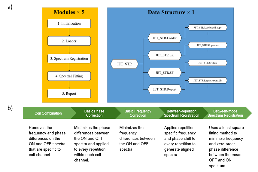
2.1 JET RoutineJET quantified MRS data in a sequence of five modules. The Initialization module set up the folder hierarchy and defined methods to access data. The Loader module either bundled the data from external raw data files or converted them from data structures processed by other softwares. The Spectrum Registration module generated an ON and OFF spectra after editing and correcting frequency and phase differences. Later, the Spectral Fitting module estimated a best fitting line over the DIFF and OFF spectrum to compute metabolite concentrations. Finally, the Report module generates summarized reports of each individual scan. All these modules contain data structures where MATLAB function files are saved. An overview of the JET procedure is demonstrated in Fig.1a.
2.2 Spectrum Registration Demonstration
A close up analysis was conducted on the Spectrum Registration module. From given data, an ON and OFF spectra was constructed where the ON spectra was generated using frequency-editing pulses. Spectrum registration began by removing the frequency and phase differences that were specific to each coil-channel. Phase correction followed by frequency correction was then performed and applied to every repetition within each coil channel to generate an aligned spectra. Between-repetition spectrum registration followed by between-mode spectrum registration was then conducted to minimize differences within the OFF and ON spectra by correcting parameters. A summarized demonstration of the flow of spectrum registration is shown in Fig.1b and the changes in the spectra from each parameter adjustments can be seen in Fig.2a.
2.3 Spectra Fitting Demonstration
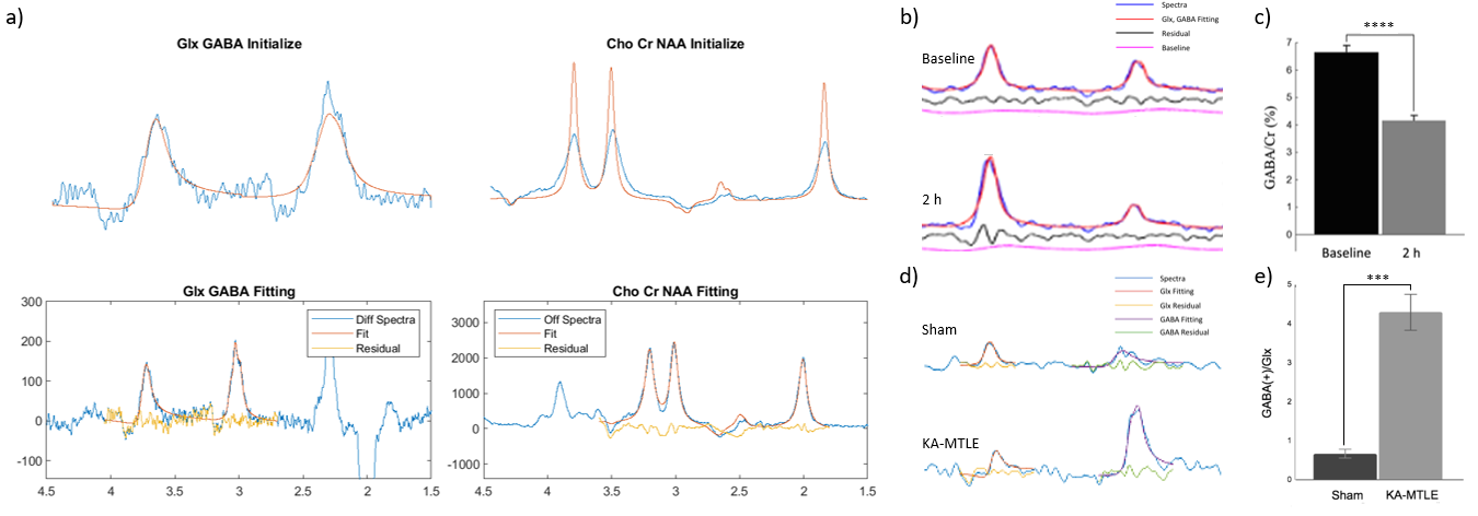
Spectra fitting of JET was conducted to perform metabolite quantification on mice. A sample result of GABA fitting is shown in Fig.3a. GABA quantification in an in vivo mouse brain under the influence of isoflurane over a timespan of 2 hours was analyzed using JET. Furthermore, spectra fitting of mice influenced by kainic acid was also performed using JET. The spectra fitting result of each experiment is depicted in Fig. 3b and Fig.3d, respectively.
Results
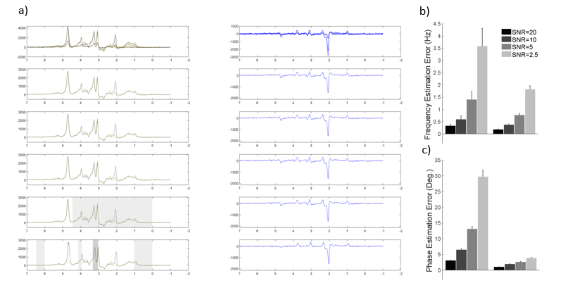
3.1 Spectrum registration of in vivo mouse studySpectrum registration correction is observed in Figure 2a. It can be observed that after each parameter correction, the more areas in the ON and OFF spectra overlap, producing an easier quantifiable DIFF spectra. Additionally, JET was also able to demonstrate a decrease in frequency and phase estimation errors with larger SNR. The results can be seen in Fig.2b and Fig.2c.
3.2 Spectra fitting of in vivo mouse study
JET was found to accurately demonstrate the effects of isoflurane towards GABA content using spectral fitting. A decrease in GABA content was observed from the use of isoflurane overtime. This result is depicted in Fig.3c. Conversely, JET was also able to demonstrate the increase in GABA content when using kainic acid. The results are depicted in Fig.3c and Fig.3e, respectively.
3.3 Software output reports
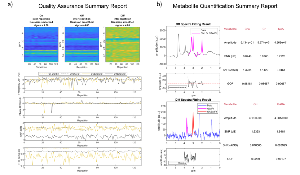
A sample report of an individual scan using JET generated a Quality Assurance Summary Report (Fig.4a) depicting information of the spectrum registration correction and a Metabolite Quantification Summary Report (Fig.4b) that provided metabolite information and the analysis parameters used in the research.
Discussion and Conclusion
The updated spectrum registration and spectral fitting routine generated an easily quantifiable ON, OFF and DIFF spectra for analysis. In addition, JET was able to accurately demonstrate effects of administration of drugs such as isoflurane and kainic acid to GABA and Glx concentration of the brain. The report module also demonstrated an ease to store essential spectral quantity and quality information for each individual scan that can be retrieved anytime for future investigation. We present a software toolkit JET, designed for automated analysis of MEGA-PRESS spectra of the mouse brain. With Bruker raw data supported, our toolkit is fully automated and rater-independent. Furthermore, it is specifically designed to deal with low SNR spectra and can perform group wise study. We anticipate that it will be a useful tool for the adoption of J-difference editing studies in transgenic mice.Acknowledgements
This study was performed at the Zuckerman Mind Brain Behavior Institute at Columbia University and Columbia MR Research Center site.References
1. Guo, J. et. al. In vivo detection and automatic analysis of GABA in the mousebrain with MEGA‐PRESS at 9.4 T. NMR In Biomedicine. 2017. DOI: 10.1002/nbm.38372.
2. Guo, J. et. al. MRSMouse - A Matlab Toolkit for Automated MR Spectrum Processing of In Vivo Mouse MEGA-PRESS Study at 9.4T.3.
3. Chen, R. et. al. TECHNICAL WHITE PAPER: J-difference Editing Toolkit (JET) and the Novel Spectrum Registration Methods.
4. Riggle, Brittany A., et al. "MRI demonstrates glutamine antagonist-mediated reversal of cerebral malaria pathology in mice." Proceedings of the National Academy of Sciences 115.51(2018): E12024-E12033.
5. Provenzano, Frank A., et al. "Hippocampal pathology in clinical high-risk patients and the onset of schizophrenia." Biological Psychiatry 87.3 (2020): 234-242.
6. Hamelin, S., et. al. In vivo γ‐aminobutyric acid increase as a biomarker of the epileptogenic zone: An unbiased metabolomics approach. Epilepsia (Copenhagen).2020. DOI: 10.1111/epi.16768
Figures