0716
Confounding of Macromolecular and Paramagnetic Tissue Content in Quantitative MTI Remedied by Explicit Estimation of Bound Pool Relaxation
Alexey Samsonov1 and Aaron S. Field1
1Radiology, University of Wisconsin-Madison, Madison, WI, United States
1Radiology, University of Wisconsin-Madison, Madison, WI, United States
Synopsis
We study the effect of macromolecular proton fraction (MPF) and R1 interdependency in quantitative MT experiments. We hypothesize that the two-pool model with properly calibrated relaxation constraints on the bound proton pool can separate interdependency of both metrics, potentially improving the specificity of both, specifically, MPF to macromolecular content and R1 to paramagnetic ions. The simulation and in vivo results support feasibility of such refinement.
Introduction
Two-pool modeling of magnetization transfer (MT) is a popular approach to assess tissue macromolecules. One parameter of the model, macromolecular proton fraction (MPF), is strongly associated with myelin in neural tissues. Another parameter is the longitudinal relaxation rate R1 of tissue water, which correlates with MPF1 and is often considered a myelin marker. However, unlike MPF, it provides only secondary correlations with clinical variables2, likely owing to other sources affecting the parameter3. For example, modified measurement and modeling techniques yielded R1 estimates with increased correlations to tissue iron accumulation and reduced contributions from macromolecules, potentially improving its specificity4. In this work, we study the effect of MPF and R1 interdependency within fast MPF/R1 mapping framework5,6; we hypothesize that the two-pool model with properly calibrated constraints can separate these metrics, potentially improving the specificity of both.Theory
Variations in tissue water R1 (as measured by a single-pool model) can be empirically modeled in the fast exchange limit as:$$R1=R_{1,0}+fr_b+cr_p,$$where $$$R_{1,0}$$$ is the value for pure saline, $$$r_b$$$ and $$$r_p$$$ are the relaxivities of macromolecular and paramagnetic (e.g. iron) content, respectively, $$$f$$$ and $$$c$$$ are their concentrations7. The two-pool MT model aims to separate the contribution of macromolecules from that of the water compartment. However, the longitudinal relaxation rate of macromolecular (bound) protons (R1b), a fixed parameter of the model, was shown to be significantly underestimated in standard approaches, which arbitrarily choose R1b=1 s-1. This may result in incomplete separation of macromolecular contributions from R1 of free water protons (R1f). Recent studies provided initial measurements of R1b and the current consensus is that R1b is underestimated by several times8,9, with different studies reporting values from 2 to 5 s-1.Methods
Methods: We formulated our R1b estimation around a histogram-based optimization previously described9. We assumed that modeling interactions of free and bound protons with correct estimates of their longitudinal relaxation should eliminate macromolecule-induced spatial variations from observed R1f values, reducing their information content, which can be measured by the histogram entropy function10. The gradient-based search was run by varying R1b to minimize R1f histogram entropy.Data Acquisition: Experiments were performed on a 3.0T GE Discovery MR750 (Waukesha, WI) using an 8-channel head array in two healthy and three multiple sclerosis (MS) subjects after informed consent. The protocol included two SPGR and one MT-weighted SPGR measurements with optimized Z-spectra sampling and additional model constraints for fast MPF mapping5,6. Healthy volunteers were scanned with 1x1x2mm resolution in 18 min; MS subjects were scanned with 2x2 GRAPPA-accelerated 1.3 mm-3 isotropic in 7.5 min11. Additionally, a B1 map was collected with AFI12 to correct excitation flip angles and MT saturation powers13.
Simulations: To estimate the effect of R1b assumptions, simulated data were generated using a full MPF mapping protocol14 for a range of model parameters in WM, GM, and MS lesions, and a two-pool model with updated R1b constraint. Parameters obtained fitting the model with R1b=1 s-1 were compared to ground-truth values.
Results
Our fit yielded R1b=6.5+/-0.08 s-1 across all subjects. Mean R1f was 0.3045 s-1, consistent with previous studies1,4. The simulations (Fig.1) demonstrate that underestimation of R1b, common in previous approaches, causes near-linear correlation of apparent tissue water R1f with macromolecular content (measured by MPF), which explains the previous use of standard R1 as a surrogate myelin marker. The effect of R1b underestimation on apparent MPF values is smaller but non-negligible (~10% for maximum nominal MPF). Figure 2 demonstrates in vivo MPF/R1f mapping in a healthy volunteer. Note the striking flattening of the R1f map estimated with our R1b constraint, consistent with anticipated removal of the macromolecular component from this measure. The whole brain histograms (Fig. 3) further show the reduced variability and mean value of R1f observed in Fig. 2. Figure 4 compares several representative quantitative maps in an MS subject. Patterns of elevated R1f values correspond to known iron distribution in the basal ganglia7 and to patterns of MS-induced deposition of non-heme iron (e.g. ferritin) as previously suggested15. At the same time, conventional R1f is primarily reflecting macromolecular content (as evidenced by its resemblance to MPF), with none of the variations expected from paramagnetic iron detectable in deep GM on this map.Discussion and Conclusions
Our results support the finding of others7 that standard R1 is dominated by macromolecular content, which may hide information about other substrates affecting R1, e.g. paramagnetic substances. The two-pool model with correctly calibrated R1b, as proposed here, may remove this uncertainty by separating macromolecular and paramagnetic contributions into MPF and water-only R1f. This may potentially increase specificity for myelin and paramagnetic depositions (e.g., iron), which are confounded even when using advanced methods for iron quantification such as QSM16, limiting our understanding of the role of iron in aging and neurodegenerative disorders. Given the availability of fast, high-resolution MPF mapping protocols, our approach may be a valuable clinical tool for studying macromolecules and paramagnetic tissue properties independently, in order to better understand the pathophysiology of neurodegenerative processes. Moreover, in studies involving exogenous paramagnetic substances such gadolinium-based contrast agents, our water-only R1 mapping may allow estimation of Gd concentration in areas of leaky vasculature unbiased by macromolecular effects, which are known to compromise the accuracy of contrast-enhanced imaging7.Acknowledgements
The work was supported by NIH (R01EB027087, R24NS104098) and GE Healthcare.References
- Yarnykh VL. Correction of B1 non-uniformity errors in fast macromolecular proton fraction and R1 mapping without B1 maps. In: Proc of ISMRM; 2020; Sydney, Australia. p 887.
- Yarnykh VL, Bowen JD, Samsonov A, Repovic P, Mayadev A, Qian P, Gangadharan B, Keogh BP, Maravilla KR, Jung Henson LK. Fast whole-brain three-dimensional macromolecular proton fraction mapping in multiple sclerosis. Radiology 2015;274(1):210-220.
- Stuber C, Morawski M, Schafer A, Labadie C, Wahnert M, Leuze C, Streicher M, Barapatre N, Reimann K, Geyer S, Spemann D, Turner R. Myelin and iron concentration in the human brain: a quantitative study of MRI contrast. Neuroimage 2014;93 Pt 1:95-106.
- Wang Y, van Gelderen P, de Zwart JA, Duyn JH. B0-field dependence of MRI T1 relaxation in human brain. Neuroimage 2020;213:116700.
- Yarnykh VL. Fast macromolecular proton fraction mapping from a single off-resonance magnetization transfer measurement. Magn Reson Med 2012;68(1):166-178.
- Samsonov AA, Mossahebi P, Anderson A, Velikina JV, Johnson KM, Johnson SC, Fleming JO, Field AS. High Resolution, Motion Corrected Mapping of Macromolecular Proton Fraction (MPF) In Clinically Acceptable Time Using 3D Undersampled Radials. In: Proc of ISMRM; 2014; Milan, Italy. p 3337.
- Rooney WD, Johnson G, Li X, Cohen ER, Kim SG, Ugurbil K, Springer CS, Jr. Magnetic field and tissue dependencies of human brain longitudinal 1H2O relaxation in vivo. Magn Reson Med 2007;57(2):308-318.
- Helms G, Hagberg GE. In vivo quantification of the bound pool T1 in human white matter using the binary spin-bath model of progressive magnetization transfer saturation. Phys Med Biol 2009;54(23):N529-540.
- van Gelderen P, Jiang X, Duyn JH. Effects of magnetization transfer on T1 contrast in human brain white matter. Neuroimage 2016;128:85-95.
- Likar B, Viergever MA, Pernus F. Retrospective correction of MR intensity inhomogeneity by information minimization. Ieee Transactions on Medical Imaging 2001;20(12):1398-1410.
- Griswold MA, Jakob PM, Heidemann RM, Nittka M, Jellus V, Wang J, Kiefer B, Haase A. Generalized autocalibrating partially parallel acquisitions (GRAPPA). Magn Reson Med 2002;47(6):1202-1210.
- Yarnykh VL. Actual flip-angle imaging in the pulsed steady state: a method for rapid three-dimensional mapping of the transmitted radiofrequency field. Magn Reson Med 2007;57(1):192-200.
- Mossahebi P, Yarnykh VL, Samsonov A. Analysis and correction of biases in cross-relaxation MRI due to biexponential longitudinal relaxation. Magn Reson Med 2014;71(2):830-838.
- Yarnykh VL, Yuan C. Cross-relaxation imaging reveals detailed anatomy of white matter fiber tracts in the human brain. Neuroimage 2004;23(1):409-424.
- Boyko OB, Burger PC, Shelburne JD, Ingram P. Non-heme mechanisms for T1 shortening: pathologic, CT, and MR elucidation. AJNR Am J Neuroradiol 1992;13(5):1439-1445.
- Hametner S, Endmayr V, Deistung A, Palmrich P, Prihoda M, Haimburger E, Menard C, Feng X, Haider T, Leisser M, Kock U, Kaider A, Hoftberger R, Robinson S, Reichenbach JR, Lassmann H, Traxler H, Trattnig S, Grabner G. The influence of brain iron and myelin on magnetic susceptibility and effective transverse relaxation - A biochemical and histological validation study. Neuroimage 2018;179:117-133.
Figures

Figure 1. Effect
of underestimated macromolecular relaxation rate (R1b = 1 s-1) in standard two-pool MT modeling methods on
the apparent R1 of the tissue water (R1f) (a) Estimated vs true MPF values for three levels of B1
inhomogeneity (no error, +/- 30%). The sign and the level of the error is
varying both with B1 field and MPF but does not exceed 0.02. (b) The presence of non-zero
macromolecular fraction causes significant deviation of the apparent R1f from its nominal value (0.3045 s-1)
reaching ~300% error.
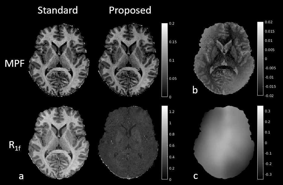
Figure 2. Quantitative
maps estimated using MT modeling with standard and proposed R1b in a healthy volunteer. (a) MPF and R1f. As predicted by simulations (Fig. 1b), standard R1f is dominated by
macromolecular content and therefore resembles MPF. The proposed R1f is much more uniform, likely due
to removing the effects of MPF. Note slightly elevated values in the basal
ganglia, likely due to iron accumulation. (b)
Error between the proposed and standard MPF. Note its high variability with macromolecular
content and B1 field (c), also consistent
with simulations (Fig. 1a).

Figure 3. Whole brain histograms of R1f reconstructed using the two approaches under investigation. Note
three modes in the standard map corresponding to WM, GM and CSF. WM and GM
merge into a single peak in the new map, as the differences in macromolecular
content between WM and GM are removed by the proposed method.

Figure 4. Quantitative
mapping in an MS subject. (a) Proposed
R1f is elevated in deep GM. The
increase is consistent with the known distribution of iron in these regions,
including areas with high (globus pallidus, #1), medium (putamen, #2), and low
(thalamus, #3) iron content. Note elevated R1f
in areas affected by diffuse lesional changes (#4) (as seen on T2-FLAIR and MPF).
(b) The R1f is increased on the rim of the heavily demyelinated lesion (as
revealed by MPF) (#1) and in WM areas affected by more diffuse disease (#2). (c) Example of a lesion without noticeable
increase in R1f.