0684
Preclinical Hyperpolarized 129Xe Ventilation Imaging Using 3D Spiral (FLORET) Encoding1Center for Pulmonary Imaging Research, Cincinnati Children's Hospital Medical Center, Cincinnati, OH, United States, 2Imaging Research Center, Cincinnati Children's Hospital Medical Center, Cincinnati, OH, United States, 3Department of Pediatrics, University of Cincinnati Medical Center, Cincinnati, OH, United States, 4Department of Biomedical Engineering, University of Cincinnati, Cincinnati, OH, United States
Synopsis
Center-out trajectories are often used in preclinical HP gas MRI to reduce the impact of physiological motion and magnetization decay on image quality. Recently, implementation of 3D spiral (FLORET) imaging for human 129Xe ventilation imaging demonstrated higher accuracy in detecting ventilation abnormalities than traditional sequences. Here we show FLORET sequences provide superior SNR, consume less xenon, and reduce scan time by more than five times when used to image ventilation in mice.
Introduction
In humans, Gradient Recalled Echo (GE) sequences are used commonly for hyperpolarized (HP) 129Xe ventilation imaging, because they can be implemented in a single breath and are ubiquitous across scanner platforms(1). In contrast, preclinical images are typically acquired over 10s to 100s of breaths, increasing the deleterious impact of respiratory and cardiac motion on image quality. Of necessity, only a small number of k-space lines are encoded per breath, and signal is intrinsically small. Thus, large flip-angle RF pulses (20-90º) are needed, which cause substantial in-breath signal decay. Further, the high field strengths used in preclinical imaging leads to more rapid T2* relaxation (~5 ms at 7T), further degrading image quality(2). Because of these challenges, radial sequences have displayed utility in preclinical HP gas imaging, because they oversample low-frequency imaging data, making them robust to motion, data undersampling, view-to-view magnetization variations, and T2* decay(3-5). Slice-selective 2D strategies have been employed with success, but they generate coarse resolution (~1 mm) in the slice dimension relative to the minute scale of mouse lungs. As such, true 3D sequences are preferable, because they provide an anatomically equivalent to images obtained in human subjects. To date, 3D radial 129Xe ventilation imaging had been implemented in both rats and mice to achieve high quality images with high isotropic resolution. These 3D sequences allow magnetization dynamics to be tracked throughout acquisition, allowing signal decay to be mapped and corrected, without addition data collection, to improve quantitative image accuracy(6). However, 3D Radial is inefficient at encoding k-space and requires a large gas volume or a high degree of undersampling. In contrast, 3D spiral sequences, such as Fermat Looped Orthogonally Encoded Trajectories (FLORET), can encode k-space with an efficiency that exceeds conventional Cartesian sequences while retaining the advantages of center-out encoding(7). The utility of FLORET was recently demonstrated in human HP 129Xe imaging, and here we extend it to mouse imaging and demonstrate benefits of reduced scan time, decreased 129Xe consumption, and increased SNR relative to 3D radial.Methods
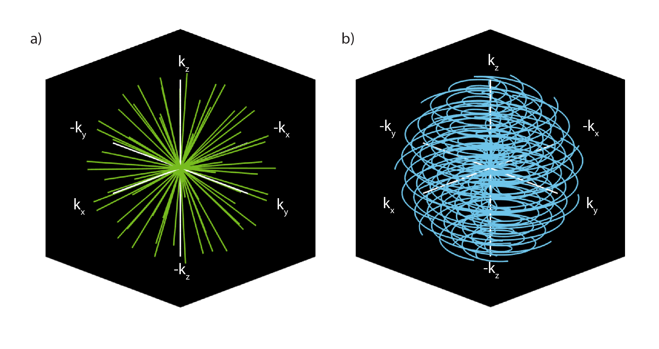
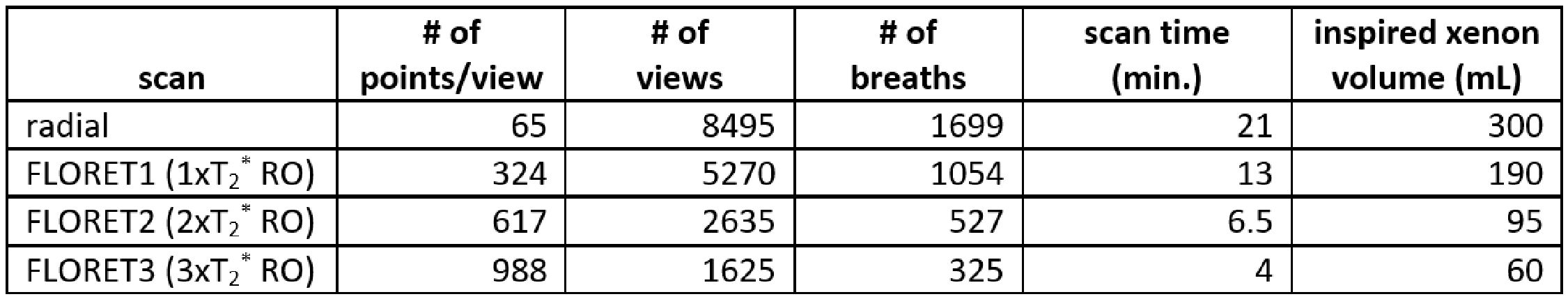
3-D radial and FLORET images (Figure 1) were acquired using a 7 T Bruker BioSpin (Billerica, MA). Acquisition parameters consistent in all scans were: matrix=1043, FOV=26 mm3, bandwidth=64 kHz, dummy pulses=20, α=30°, TE/TR=0.77/30ms. Slab-selection was used to prevent wraparound artifacts. Three separate fully encoded FLORET images, named for readout (RO) duration relative to expected T2* (i.e., FLORET1: 5ms; FLORET2: 10ms; FLORET3: 15ms), were used. The 3D radial readout duration was 1ms. Points, views, and number of breaths, etc. are summarized in Table 1. 129Xe was hyperpolarized (HP) to 35-45 % (Model 9820, Polarean PLC., Durham, NC). Shorter scan durations enabled all three FLORET scans to be acquired in a single 350mL batch of HP 129Xe, while a full batch was needed for the 3D radial scan. The order of the FLORET scans was randomized within each mouse to reduce T1 dependent systematic bias. Five C57BL/6J mice (2 male, 3 female, mass 27.5 ±4.5g, age 36 weeks ±1 week, Jackson Laboratory, Bar Habor, ME) were anesthetized using intraperitoneal injections of 90/9/3 mg/kg ketamine/xylazine/acepromazine. Mice were intubated with 22-ga all plastic catheters, placed on a custom-built, HP-gas compatible, small animal ventilator, and ventilated as described previously, 79/21% mixture of HP 129Xe gas/O2 at expiration or inspiration. Image reconstruction and statistical analysis were performed in MATLAB 2019b (MathWorks, Natick, MA). Signal thresholds (5× standard deviation of the background) followed by erosion-dilation with a spherical structuring element (radius 2 voxels) were used to delineate ventilated volume. Noise was calculated from the standard deviation of artifact-free background, and signal-to-noise ratios (SNR) were adjusted for Rician noise distribution. A slight correction for tidal volume and number of breaths was made to the SNR normalization technique previously reported. Mean polarization, SNR and normalized SNR were compared using ANOVA with post hoc Tukey test.Results and Discussion
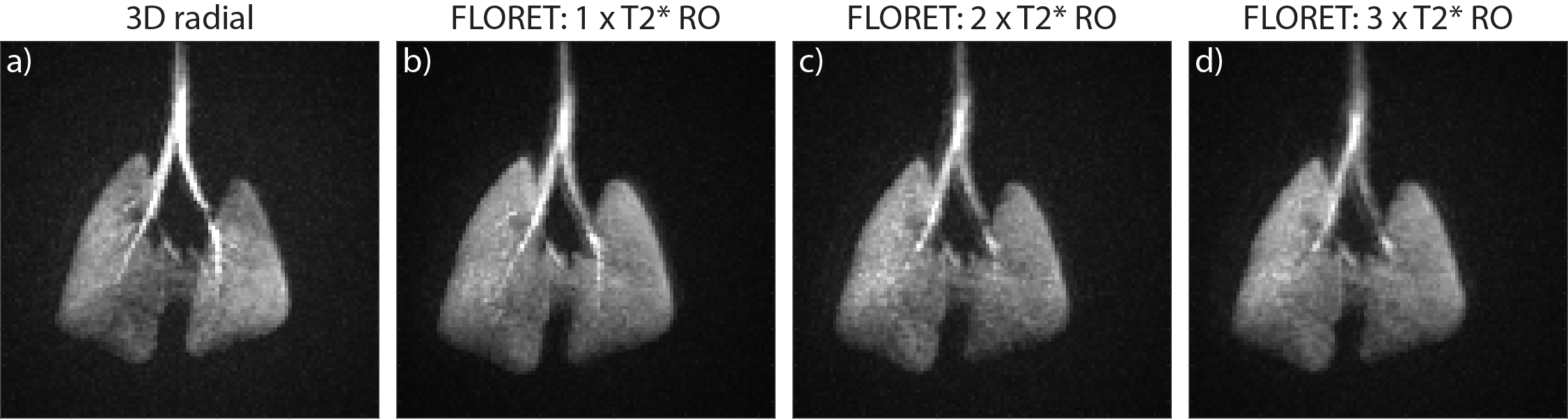
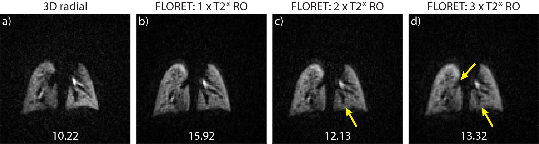
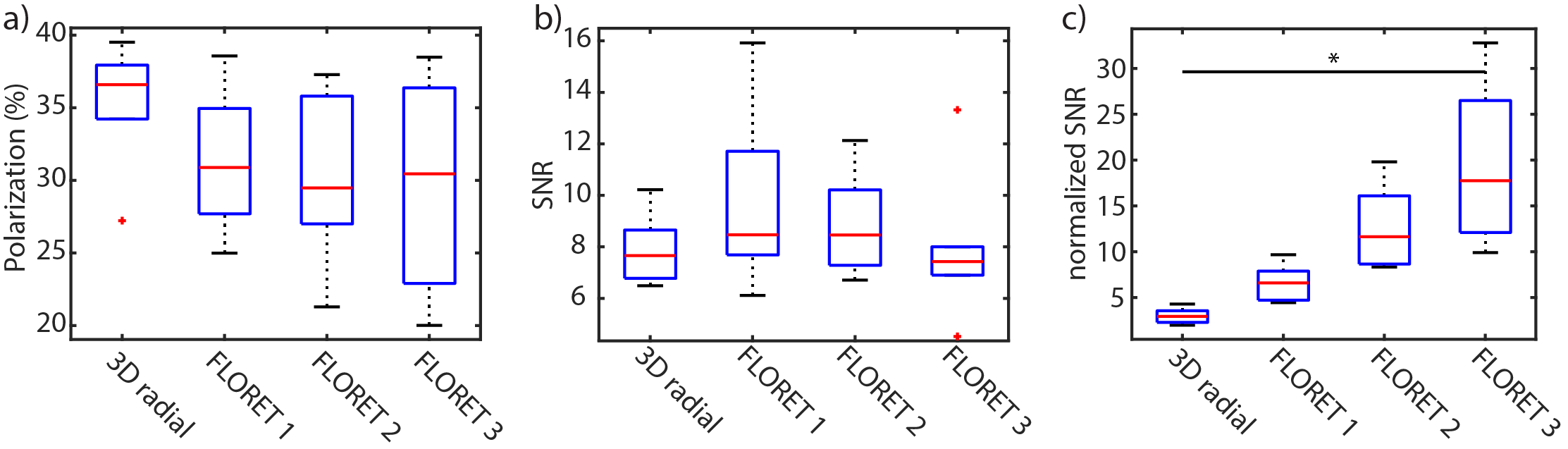
FLORET greatly reduces preclinical scan time while enabling full k-space encoding and high image quality. This can be seen qualitatively, in Figure 2, which displays maximum intensity projections in the coronal plane for 4-fold undersampled radial and all three FLORET scans. Of note, the trachea and several generations of the large airways are apparent in all images, and uniform signal is observed from the lung parenchyma. Representative coronal slices in Figure 3 reveal only slight blurring with longer RO durations in FLORET2 and 3. Figure 4a shows initial 129Xe polarization estimated from T1 and polarizer calibrations(8). The highest mean polarization, observed in the 3D radial scan, did not result in superior SNR. No significance difference in mean SNR was observed for these four scans. It should be noted that FLORET1 compared to quarter-sampled radial imaging reduces scant time by seven minutes and xenon consumption by 100mL with no apparent change in image quality. By normalizing the SNR, as noted in the methods, significantly higher SNR is observed for FLORET3 (p=0.0008). This highlights the >5x reduction in xenon consumption with long RO durations.Conclusion
By implementing 3D FLORET, high-resolution images with superior SNR to 3D radial scans, are acquired with comparable image quality while reducing both scan time and xenon consumption by greater than five times.Acknowledgements
The authors thank the following sources for research funding and support: NIH R01 HL143011 and NIH T32 HL007752.References
1. He M, Robertson SH, Kaushik SS, Freeman MS, Virgincar RS, Davies J, Stiles J, Foster WM, McAdams HP, Driehuys B. Dose and pulse sequence considerations for hyperpolarized 129Xe ventilation MRI. Magn Reson Imaging 2015;33(7):877-885.
2. Niedbalski PJ, Cochran AS, Akinyi TG, Thomen RP, Fugate EM, Lindquist DM, Pratt RG, Cleveland ZI. Preclinical hyperpolarized 129Xe MRI: ventilation and T2* mapping in mouse lungs at 7 T using multi-echo flyback UTE. NMR Biomed 2020;33(7):e4302.
3. Mistry NN, Thomas A, Kaushik SS, Johnson GA, Driehuys B. Quantitative analysis of hyperpolarized 3He ventilation changes in mice challenged with methacholine. Magn Reson Med 2010;63(3):658-666.
4. Cleveland ZI, Virgincar RS, Qi Y, Robertson SH, Degan S, Driehuys B. 3D MRI of impaired hyperpolarized 129Xe uptake in a rat model of pulmonary fibrosis. NMR Biomed 2014;27(12):1502-1514. 5. Santyr GE, Lam WW, Ouriadov A. Rapid and efficient mapping of regional ventilation in the rat lung using hyperpolarized 3He with Flip Angle Variation for Offset of RF and Relaxation (FAVOR). Magn Reson Med 2008;59(6):1304-1310.
6. Niedbalski PJ, Willmering MM, Robertson SH, Freeman MS, Loew W, Giaquinto RO, Ireland C, Pratt RG, Dumoulin CL, Woods JC, Cleveland ZI. Mapping and correcting hyperpolarized magnetization decay with radial keyhole imaging. Magn Reson Med 2019;82(1):367-376.
7. Willmering MM, Niedbalski PJ, Wang H, Walkup LL, Robison RK, Pipe JG, Cleveland ZI, Woods JC. Improved pulmonary 129Xe ventilation imaging via 3D-spiral UTE MRI. Magn Reson Med 2020;84(1):312-320.
8. Plummer JW, Emami K, Dummer A, Woods JC, Walkup LL, Cleveland ZI. A semi-empirical model to optimize continuous-flow hyperpolarized 129Xe production under practical cryogenic-accumulation conditions. J Magn Reson 2020;320:106845.
Figures