0675
Hyperpolarized 13C MRI reveals age-related changes in lactate metabolism in the human brain1Medical Biophysics, University of Toronto, Toronto, ON, Canada, 2Sunnybrook Health Sciences Centre, Toronto, ON, Canada, 3Radiation oncolocy, Sunnybrook Health Sciences Centre, Toronto, ON, Canada, 4GE Healthcare, Toronto, ON, Canada, 5Physical sciences, Sunnybrook Health Sciences Centre, Toronto, ON, Canada, 6Radiology, Sunnybrook Health Sciences Centre, Toronto, ON, Canada, 7Medical Biophysics, Sunnybrook research institute, Toronto, ON, Canada
Synopsis
In this study, hyperpolarized 13C MRI was used to investigate age-related changes to lactate and bicarbonate production in the brain in a healthy aging population. A whole-brain parcellation method was used to investigate regional changes. A global reduction in the production of both 13C-lactate and 13C-bicarbonate was observed vs. age, with certain regions showing increased rates of change in comparison to the rest of the brain. Our results suggest an age-related change in the underlying metabolic processes, and paves the way for future analysis of pathological aging as seen in disorders like Alzheimer's disease.
Introduction
Healthy normal aging of the human brain results in multi-faceted physiological changes that can result in reduced cognitive ability, and results in a higher risk of neurodegenerative disease onset.1 Cerebral blood flow and the metabolic rate of oxygen have been shown to change with age, resulting in a net decrease in venous deoxygenation.2 Fluorodeoxyglucose positron emission tomography has also shown reduced glucose uptake in the brain.3 These changes are further exacerbated with pathological aging, especially with diseases where age is a key risk factor like Alzheimer’s disease.4 Recent developments in hyperpolarized 13C MRI methodology (HP-13C) makes it a promising technique for characterization of metabolic changes in the aging brain. HP-13C following intravenous injection of 13C-pyruvate allows for the measurement of 13C-lactate production in the brain, which depends on several potentially age varying parameters such as the expression level of monocarboxylate transporters MCT1, MCT2 and MCT4, which affect pyruvate uptake, and the availability of NADH which is consumed during conversion of pyruvate to lactate by the enzyme lactate dehydrogenase in the cellular cytosol. Additionally, 13C-bicarbonate signal is detectable and provides a measure of acetyl-CoA formation from pyruvate through the pyruvate-dehydrogenase complex (PDC) in mitochondria. Previously, we have shown the high degree of concordance between regional [1-13C] lactate patterns across healthy subjects.6 In this work, we assessed global and regional age-related changes in 13C metabolite signals.Methods
Volunteers (N=19, ages 23-77) were enrolled in the study. All enrolled volunteers showed normal cognitive ability when screened through the Montreal Cognitive Assessment (MoCA). The study was conducted under a protocol approved by the Sunnybrook Research Ethics board, and by Health Canada as a Clinical Trial Application. Informed written consent was acquired from all subjects. Prior to the start of scanning, a 20 gauge intravenous catheter was inserted into the volunteers forearm, after which they were positioned into a GE MR750 3.0T MRI scanner. Localization scans and multi-echo reference data, to be used for reconstruction, were acquired using the scanner’s body coil. A custom 13C birdcage coil was then placed into the head-coil base of an 8-channel neurovascular receive array (Invivo Inc.) without moving the volunteer. A 0.1 mmol/kg dose of hyperpolarized [1-13C]pyruvate was injected at 4 mL/s, followed by a 25 mL saline flush at 5 mL/s. A 3D dual-echo echo-planar imaging sequence7 (60 second acquisition, 5 second temporal resolutions, axial, FOV 24 x 24 x 36 cm3, 1.5 cm-isotropic resolution) was initiated after the saline flush to sequentially acquire [1-13C]lactate, [1-13C]bicarbonate, and [1-13C]pyruvate images (128 x 16 x 24 voxels). Anatomical images were than acquired using the 8-channel 1H array (TE 2.9ms, TR 7.6ms, 1mm-isotropic resolution, FOV 25 x 25.6 cm2 , axial, flipangle 11 degrees). In order to correct for geometric distortions, 13C images were reconstructed using the aforementioned 1H multi-echo reference8 data in MATLAB (The MathWorks Inc., Natick, MA). In order to localize regional metabolic concentrations, structural parcellation of anatomical 1H images was done using the Spatially Localized Atlas Network Tiles (SLANT) parcellation pipeline.9 SLANT was used to produce 132 parcellated regions, as per the BrainCOLOR labelling protocol, of which 126 regions were analyzed after excluding six ventricular regions. Each set of 13C metabolite images was summed across all timepoints, and overlaid with parcellated anatomical images. The Freesurfer package mrisegstats (http://nmr.mgh.harvard.edu) was used to produce regional metabolite distributions. Ratios were than produced, plotted against age and also used to create regional linear regressions.Results and Discussion
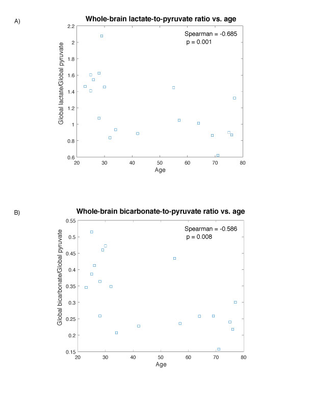
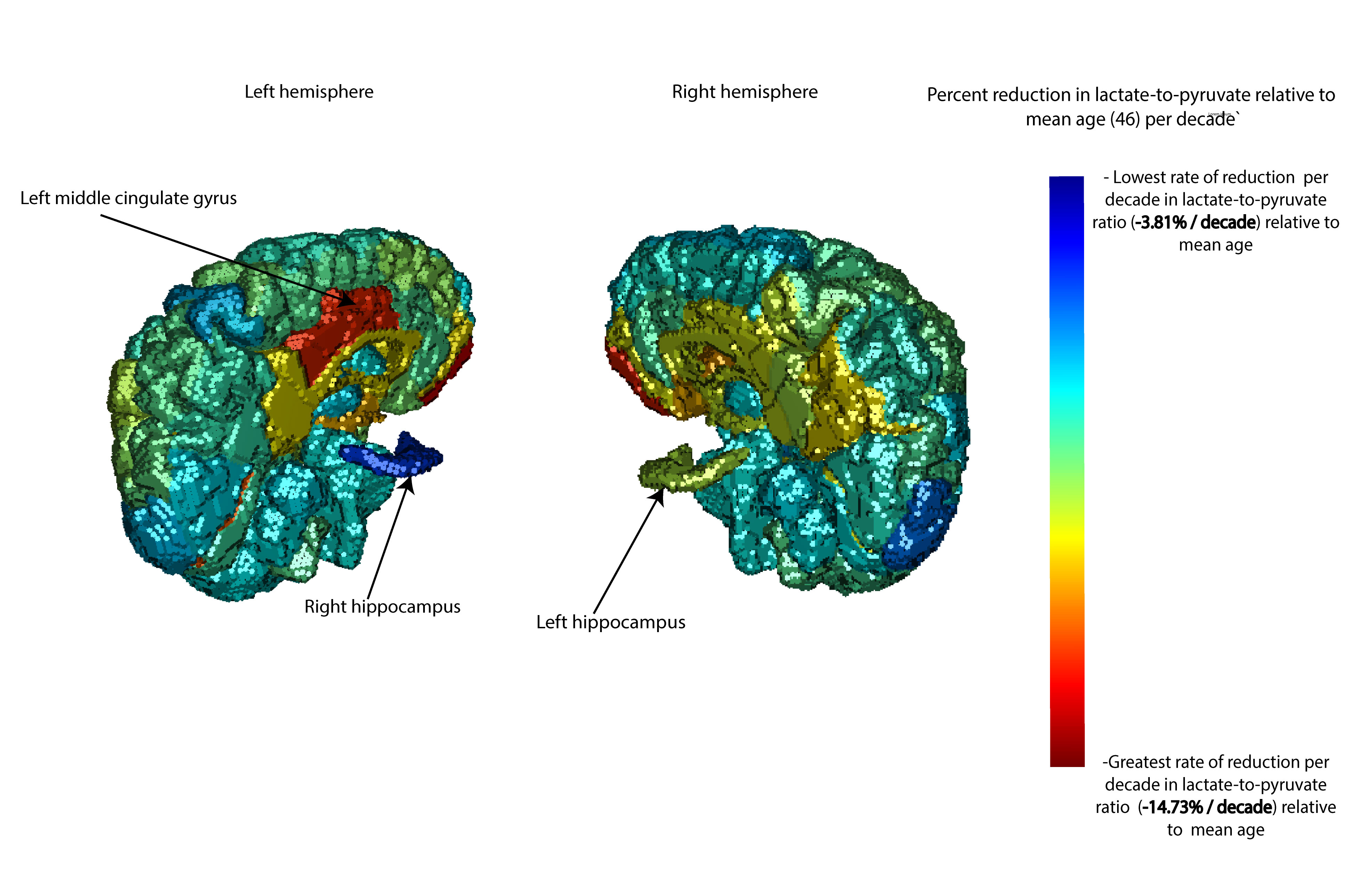
Figure 1 shows the whole brain (global) lactate-to-pyruvate ratio (LPR) and bicarbonate-to-pyruvate ratio (BPR) as a function of age for each of the 19 individuals tested. Both sets of global ratios decrease significantly with age, suggesting an age-related change in the underlying metabolic processes. To assess regional differences in lactate production, the LPR was plotted against age for each of the 126 regions resulting from the SLANT parcellation pipeline. Linear regressions for each region were used to calculate an estimated change per-decade in LPR relative to the mean age of 46. This rate of change was mapped to colours on a 3D rendered brain in Figure 2. The region with the lowest rate of change was the left hippocampus, while the left middle cingulate gyrus and bilateral gyrus rectus were among the regions with highest rates of change. Differences in the regional LPR reduction rates suggests that changes in contributory physiology, like monocarboxylate transporter expression, may be compartmentalized throughout the brain. This work lays a foundation for future clinical studies on individuals with pathological aging, as seen in Alzheimer’s disease.Conclusion
In this study, hyperpolarized 13C MRI was used to measure age-related changes to 13C-lactate and 13C-bicarbonate production in the human brain in a healthy aging population. Significant whole-brain decreases in both the LPR and BPR with age were observed, indicating age-related changes in at least one of the underlying biological factors. Parcellation was used to characterize regional differences, and multiple regions showed greater reductions in the LPR in comparison to the rest of the brain. The work presented here provides evidence of widespread decreases in the production of 13C-lactate with aging in cognitively normal individuals.Acknowledgements
Funding support from the Brain Canada and Canadian Institutes for Health Research grant PJT152928.References
[1] Carlos L´opez-Ot´ın et al. “The hallmarks of aging”. In: Cell 153.6 (2013), pp. 1194–1217.
[2] Hanzhang Lu et al. “Alterations in cerebral metabolic rate and blood supply across the adult lifespan”. In: Cerebral cortex 21.6 (2011), pp. 1426–1434.
[3] Douglas N Greve et al. “Different partial volume correction methods lead to different conclusions: an 18F-FDG-PET study of aging”. In: Neuroimage 132 (2016), pp. 334–343.
[4] Brian H Anderton. “Ageing of the brain”. In: Mechanisms of ageing and development 123.7 (2002), pp. 811–817.
[5] Zhen J Wang et al. “Hyperpolarized 13C MRI: state of the art and future directions”. In: Radiology 291.2 (2019), pp. 273–284.
[6] Casey Y Lee et al. “Lactate topography of the human brain using hyperpolarized 13C-MRI”. In: Neuroimage 204 (2020), p. 116202.
[7] Charles H Cunningham et al. “Pulse sequence for dynamic volumetric imaging of hyperpolarized metabolic products”. In: Journal of magnetic resonance 193.1 (2008), pp. 139–146.
[8] Benjamin J Geraghty et al. “Dual-echo EPI sequence for integrated distortion correction in 3D time-resolved hyperpolarized 13C MRI”. In: Magnetic Resonance in Medicine 79.2 (2018), pp. 643–653.
[9] Yuankai Huo et al. “3D whole brain segmentation using spatially localized atlas network tiles”. In: NeuroImage 194 (2019), pp. 105–119.
Figures