0631
Simultaneous pure spin-echo and gradient-echo BOLD fMRI using Echo Planar Time-resolved Imaging (EPTI) for mapping laminar fMRI responses1Athinoula A. Martinos Center for Biomedical Imaging, Massachusetts General Hospital, Charlestown, MA, United States, 2Harvard-MIT Health Sciences and Technology, MIT, Cambridge, MA, United States, 3Department of Electrical Engineering and Computer Science, MIT, Cambridge, MA, United States, 4Department of Radiology, Stanford University, Stanford, CA, United States, 5Department of Electrical Engineering, Stanford University, Stanford, CA, United States
Synopsis
We introduced a novel imaging approach SE-EPTI to address the T2’-contamination in SE-EPI for higher specificity of BOLD fMRI. EPTI resolves multi-contrast distortion/blurring-free images to simultaneously obtain: a pure SE image with minimal T2’-contamination, multiple GE images with various T2’-weightings, and conventional SE-EPI images with different levels of T2’-contamination. We demonstrated at 7T that the pure SE can significantly reduce the draining-vein-effect, and by using shorter ETLs, less T2’-contamination was introduced. A new echo-train-shifting method is also proposed for SE-EPTI to offer flexibility of achieving shorter TEs, allowing us to examine the TE dependence of the signal contribution.
Introduction
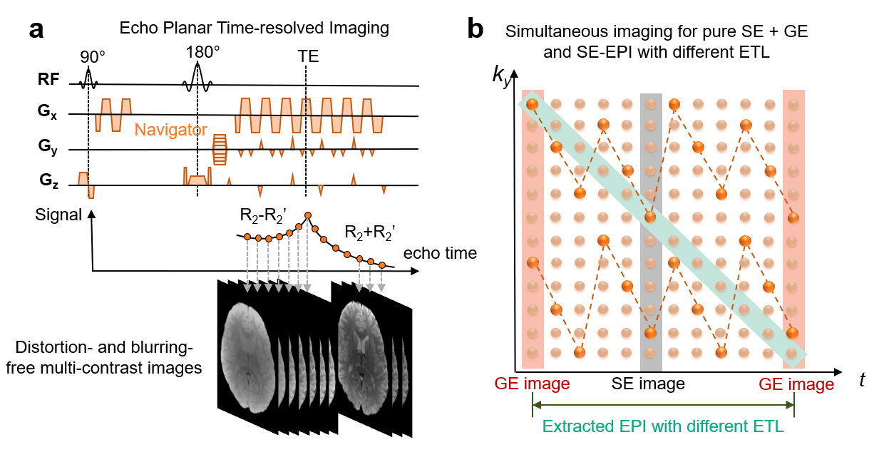
Gradient-echo (GE) BOLD fMRI is commonly used due to its high sensitivity, but suffers from the draining vein effects that limit its specificity1. Spin-echo (SE) BOLD fMRI provides higher microvascular specificity. However, it is difficult to achieve pure SE BOLD using conventional SE-EPI acquisitions because the long echo-train-lengths (ETL) will introduce T2’-contamination and therefore undesirable sensitivity to large blood vessels2-4. Alternative acquisition techniques have been developed to obtain purer T2-BOLD contrast, such as 3D GRASE5-7. Non-BOLD contrasts such as CBV8-10 and CBF11-14 have also shown promising results to achieve higher specificity.To address T2’-contamination in SE-EPI, we introduced a novel imaging approach that has been recently developed, termed echo-planar time-resolved imaging (EPTI)15-16. EPTI is able to provide full k-t data across the EPTI-readout window, enabling the generation of a time-series of distortion- and blurring-free multi-contrast images, spaced at a TE increment of an echo-spacing (~1ms) (Fig.1a). With a SE-EPTI acquisition, we can simultaneously obtain: a pure SE image with minimal T2’-contamination, multiple GE images with various T2’ weightings, and conventional SE-EPI images with different ETLs (different levels of T2’-contamination) (Fig.1b). This provides a powerful tool to investigate the effects of macro- and micro-vascular signal contributions in both GE- and SE-fMRI in a single dataset.
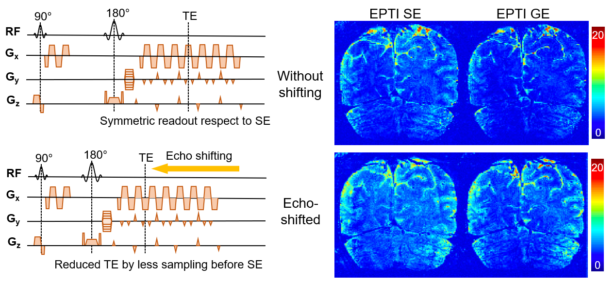
We presented preliminary SE-EPTI fMRI results at 3T last year17. This year, we extend our work to 7T to further validate that the SE signal provided by EPTI can significantly reduce the draining vein effect. We also demonstrate that by using shorter ETLs, less T2’-contamination and macro-vascular contribution will be introduced. Moreover, a new echo-train shifting method is proposed here for SE-EPTI to offer flexibility of achieving shorter TEs, to potentially examine the TE dependence of the signal contribution18. Unlike conventional partial-Fourier acquisition, the echo-train-shifting in SE-EPTI reduces TESE without reducing ky-encodings or sacrificing effective y-resolution. Rather the k-t echo-train is shifted to an earlier time-point to acquire SE data earlier in the readout window asymmetrically (Fig.4-left). Preliminary fMRI data with echo-train-shifted SE-EPTI were acquired and compared with conventional symmetric SE-EPTI.
Methods
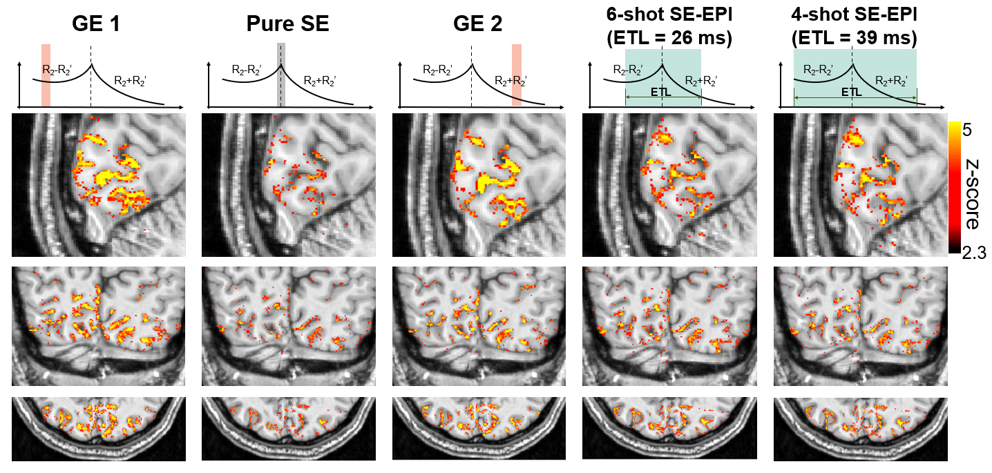
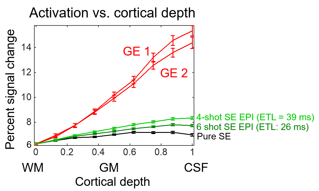
Data were acquired on a healthy volunteer with checkerboard visual stimulus on Siemens Terra 7T scanner with a custom 64-channel coil19. 3-shot SE-EPTI was designed to provide good image quality and temporal resolution for 0.9-mm isotropic resolution.First, to simultaneously obtain fMRI data for pure SE, GEs, and SEs with different ETLs, SE-EPTI without echo-train shifting was acquired with the following parameters: FOV=218×130, 0.9-mm-iso resolution, 28 slices, 45 echoes, TErange=44–88ms, TE-increment=1.09ms, volume-TR=3s×3=9s, 43 dynamics, acquisition time/run=6min27s, total 14 runs. Data were reconstructed using subspace reconstruction20-22,16, with temporal B0 drift and variation correction. After reconstruction, the pure SE (23th-echo) with TE=64ms, and two GEs that are ~17ms away from the SE were selected for GLM analysis. In addition, two conventional SE-EPIs were extracted from the reconstructed k-t data using a subset of data spanning a readout length of 26ms and 39ms respectively, corresponding to 6-shot and 4-shot EPI. B0-inhomogeneity phase evolution was removed from the k-t data prior to extracting these SE-EPI data, so that the resulting EPI images are distortion-free and geometrically-matched to the other datasets. Finally, cortical-depth analysis23 was performed using Freesurfer24,25, and 9 equi-volume26,27 layers were reconstructed.
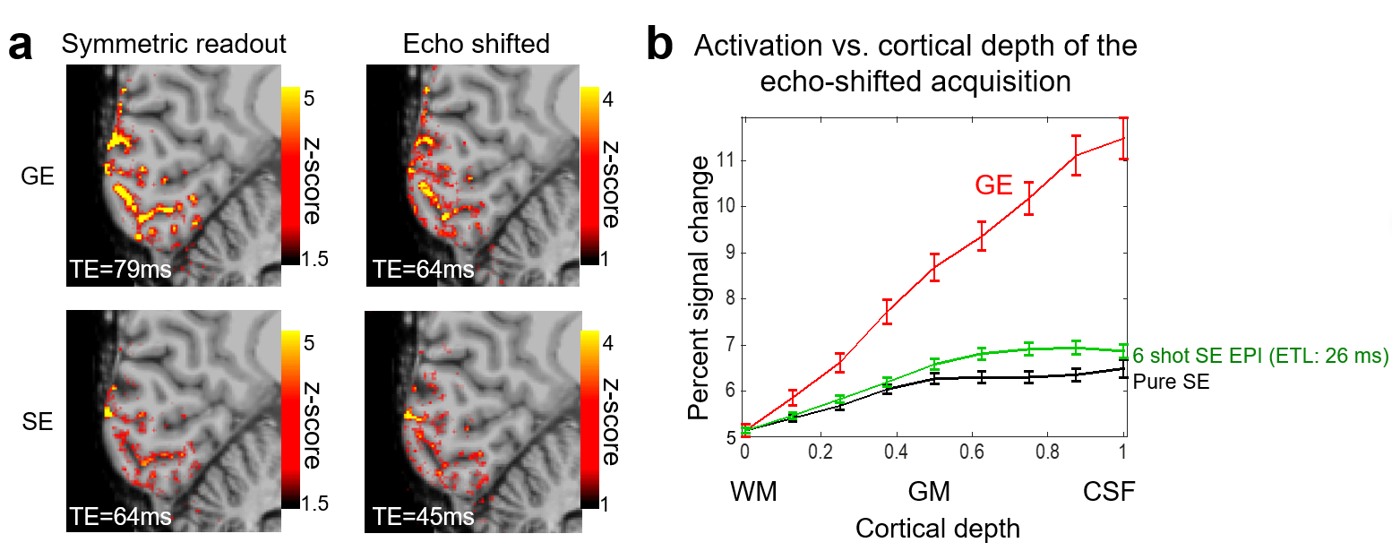
The proposed echo-train-shifted SE-EPTI was also tested that acquires 36 echoes asymmetrically, with TESE reduced from 64ms to 45ms when compared to symmetric SE-EPTI. With the shifted readout window, the volume-TR is reduced from 9s to 7.8s. The GE signal that is 17ms away from TESE was selected for comparison. Due to the reduced symmetric window length around TESE only the 6-shot SE-EPI was extracted.
Results
Figure2 shows the activation maps from the pure SE, GEs and the extracted conventional SE-EPI images provided by EPTI in a single acquisition. Higher spatial specificity was observed in the pure SE image compared to the two GEs. Although the 6-shot and 4-shot SE-EPI also present with higher spatial specificity than GE, they still show large-vessel bias and lower specificity when compared to the pure SE. This is also evident in the corresponding cortical depth profiles shown in Figure3, where GE profiles peak at the pial surface due to the expected large vessel bias, SE-EPI with shorter readout shows less bias with a less steep slope, and the pure SE with minimal T2’ contamination shows minimal bias.Figure4 demonstrates the diagram of the echo-train-shifted SE-EPTI and the improved tSNR resulted from the reduced TE for TR-matched acquisitions at 9s. Figure 5 shows preliminary results that demonstrate the feasibility of echo-shifted EPTI to provide similar activation map and cortical profiles. Nonetheless, in this data the CNR did not increase, as shown in Fig.5. The lower z-score could potentially be attributed to the shorter TR and fewer runs of the echo-shifted acquisition (~ 25% less scan time), and needs further investigation.
Conclusion
We have demonstrated on 7T that EPTI can provide simultaneous GE, pure SE and SE-EPI images, and the pure SE data can significantly reduce the large vessel bias present in GE and T2’ contaminated SE-EPI. Echo-train-shifted EPTI provides further flexibility to acquire shorter TE data to potentially help investigate TE dependence of signal contributions.Acknowledgements
This work was supported by the NIH NIBIB (R01-EB020613, R01-EB019437, R01-MH116173, R01EB016695, P41EB030006, and U01-EB025162) and the instrumentation Grants (S10-OD02363701, S10-RR023401, S10-RR023043, and S10-RR019307).References
1. Koopmans, P. J., & Yacoub, E. (2019). Strategies and prospects for cortical depth dependent T2 and T2* weighted BOLD fMRI studies. NeuroImage, 197, 668–676. https://doi.org/10.1016/j.neuroimage.2019.03.024
2. Norris, D. G. (2012). Spin-echo fMRI: The poor relation? NeuroImage, 62(2), 1109–1115. https://doi.org/10.1016/j.neuroimage.2012.01.003
3. Goense, J. B. M., & Logothetis, N. K. (2006). Laminar specificity in monkey V1 using high-resolution SE-fMRI. Magnetic Resonance Imaging, 24(4), 381–392. https://doi.org/10.1016/j.mri.2005.12.032
4. Birn R, Bandettini PA. The effect of T2’ changes on spin-echo EPI-derived brain activation maps. Proc Intl Soc Mag Reson Med. 2002;10:1324.
5. Feinberg, D., Harel, N., Ramanna, S., Ugurbil, K., & Yacoub, E. (2008). Sub-millimeter single-shot 3D GRASE with inner volume selection for T2-weighted fMRI applications at 7 Tesla. Proc Intl Soc Mag Reson Med, 16, 2373.
6. Setsompop, K., Feinberg, D. A., & Polimeni, J. R. (2016). Rapid brain MRI acquisition techniques at ultra-high fields. NMR in Biomedicine, 29(9), 1198–1221. https://doi.org/10.1002/nbm.3478
7. Kemper, V. G., De Martino, F., Vu, A. T., Poser, B. A., Feinberg, D. A., Goebel, R., & Yacoub, E. (2015). Sub-millimeter T2 weighted fMRI at 7 T: comparison of 3D-GRASE and 2D SE-EPI. Frontiers in Neuroscience, 9, 163. https://doi.org/10.3389/fnins.2015.00163
8. Huber, L., Uludağ, K., & Möller, H. E. (2019). Non-BOLD contrast for laminar fMRI in humans: CBF, CBV, and CMRO2. NeuroImage, 197, 742–760. https://doi.org/10.1016/j.neuroimage.2017.07.041
9. Huber, L., Ivanov, D., Krieger, S. N., Streicher, M. N., Mildner, T., Poser, B. A., … Turner, R. (2014). Slab-selective, BOLD-corrected VASO at 7 Tesla provides measures of cerebral blood volume reactivity with high signal-to-noise ratio. Magnetic Resonance in Medicine, 72(1), 137–148. https://doi.org/10.1002/mrm.24916
10. Jin, T., & Kim, S.-G. (2006). Spatial dependence of CBV-fMRI: a comparison between VASO and contrast agent based methods. In International Conference of the IEEE Engineering in Medicine and Biology Society (Vol. 1, pp. 25–28). IEEE. https://doi.org/10.1109/IEMBS.2006.259553
11. Wong EC, Buxton RB, Frank LR. Implementation of quantitative perfusion imaging techniques for functional brain mapping using pulsed arterial spin labeling. NMR Biomed. 1997, 10, 237-249.
12. Buxton RB. Quantifying CBF with arterial spin labeling. J Magn Reson Imaging. 2005, 22, 723-726.
13. Uludag K, Dubowitz DJ, Yoder EJ, Restom K, Liu TT, Buxton RB. Coupling of cerebral blood flow and oxygen consumption during physiological activation and deactivation measured with fMRI. Neuroimage. 2004, 23(1), 148-155.
14. Wang YI, Moeller S, Li X, Vu AT, Krasileva K, Ugurbil K, Yacoub E, Wang DJ. Simultaneous multi-slice Turbo-FLASH imaging with CAIPIRINHA for whole brain distortion-free pseudo-continuous arterial spin labeling at 3 and 7 T. Neuroimage. 2015. 113:279-88.
15. Wang, F., Dong, Z., Reese, T. G., Bilgic, B., Katherine Manhard, M., Chen, J. Setsompop, K. (2019). Echo planar time-resolved imaging (EPTI). Magnetic Resonance in Medicine, 81(6), 3599–3615. https://doi.org/10.1002/mrm.27673
16. Dong Z, Wang F, Reese TG, Bilgic B, Setsompop K. Echo planar time‐resolved imaging with subspace reconstruction and optimized spatiotemporal encoding. Magn Reson Med. 2020.
17. Wang F, Dong Z, Tian Q, Chen J, Blazejewska AI, Reese TG, Polimeni JR, Setsompop K. Cortical-depth dependence of pure T 2-weighted BOLD fMRI with minimal T 2’contamination using Echo-Planar Time-resolved Imaging (EPTI). ISMRM 2020. p1229.
18. Uludağ, K., Müller-Bierl, B., & Uğurbil, K. (2009). An integrative model for neuronal activity-induced signal changes for gradient and spin echo functional imaging. NeuroImage, 48(1), 150–165. https://doi.org/10.1016/j.neuroimage.2009.05.051.
19. Mareyam A, Kirsch JE, Chang Y, Madan G, Wald LL. A 64-Channel 7T array coil for accelerated brain MRI, ISMRM 2020, p764.
20. Liang Z-P. Spatiotemporal imagingwith partially separable functions. 2007. IEEE. p 988-991.
21. Lam F, Liang ZP. A subspace approach to high‐resolution spectroscopic imaging. Magn Reson Med. 2014;71(4):1349-1357.
22. Guo R, Zhao Y, Li Y, Wang T, Li Y, Sutton B, Liang ZP. Simultaneous QSM and metabolic imaging of the brain using SPICE: Further improvements in data acquisition and processing. Magn Reson Med. 2020;85(2):970-977.
23. Polimeni, J. R., Renvall, V., Zaretskaya, N., & Fischl, B. (2018). Analysis strategies for high-resolution UHF-fMRI data. NeuroImage, 168, 296–320. https://doi.org/10.1016/j.neuroimage.2017.04.053.
24. Fischl B. FreeSurfer. Neuroimage 2012;62(2):774-781.
25. Dale AM, Fischl B, Sereno MI. Cortical surface-based analysis: I. Segmentation and surface reconstruction. Neuroimage 1999;9(2):179-194.
26. Waehnert M, Dinse J, Weiss M, Streicher MN, Waehnert P, Geyer S, Turner R, Bazin P-L. Anatomically motivated modeling of cortical laminae. Neuroimage 2014;93:210-220.
27. Waehnert MD, Dinse J, Schäfer A, Geyer S, Bazin P-L, Turner R, Tardif CL. A subject-specific framework for in vivo myeloarchitectonic analysis using high resolution quantitative MRI. Neuroimage 2016;125:94-107.
Figures