0626
Impact of B1+-Shimming and 2-spoke pTx on 4D Angiography at 7T1Institute of Radiology, University Hospital Erlangen, Friedrich-Alexander-Universität Erlangen-Nürnberg, Erlangen, Germany, 2Physikalisch-Technische Bundesanstalt (PTB), Braunschweig und Berlin, Germany, 3Division of Medical Physics in Radiology, German Cancer Research Center (DKFZ), Heidelberg, Germany, 4Institute of Neuro-Radiology, Friedrich-Alexander Universität Erlangen-Nürnberg, Erlangen, Germany
Synopsis
4D-angiography exploiting pseudo-continuous arterial spin labeling at 7T suffers from specific absorption rate constraints, low B1+ efficiency for the labeling and B1+ inhomogeneity in the readout. In this work, we propose a B1+-phase shim trading B1+ homogeneity and transmit efficiency for the labeling combined with a dynamic transmission 2-spoke excitation readout. In volunteer measurements, the proposed approach outperformed the standard circular polarized mode by an increased vessel intensity and more vessel conspicuity.
Introduction:
4D angiography was shown to be feasible at 7T using pulsed arterial spin labeling (PASL)1,2. With pseudo-continuous arterial spin labeling (pcASL) a higher arterial signal was demonstrated at 1.5T compared to PASL3. However, at 7T, pcASL suffers from specific absorption rate (SAR) constraints, low B1+ efficiency for the labeling and B1+ inhomogeneity in the readout. This might lead to long acquisition times and poor contrast in the angiography images. To counteract high SAR values, Variable-Rate Selective Excitation (VERSE) pulses for the labeling scheme show a significant impact4. The efficiency of B1+ in the labeling could be improved by phase-only shimming5 as well as B1+ phase/amplitude shimming4. To overcome inhomogeneity in slice-selective small flip angle gradient echo sequences, multiple spokes are widely used6. In this work, we combine a B1+ phase shim trading B1+ homogeneity and transmit efficiency applied to VERSEd pcASL labeling pulses as well as 2-spoke excitation for the readout in a 4D angiography sequence at 7T. This method was compared to the standard circular polarized (CP) mode for labeling and readout.Methods:
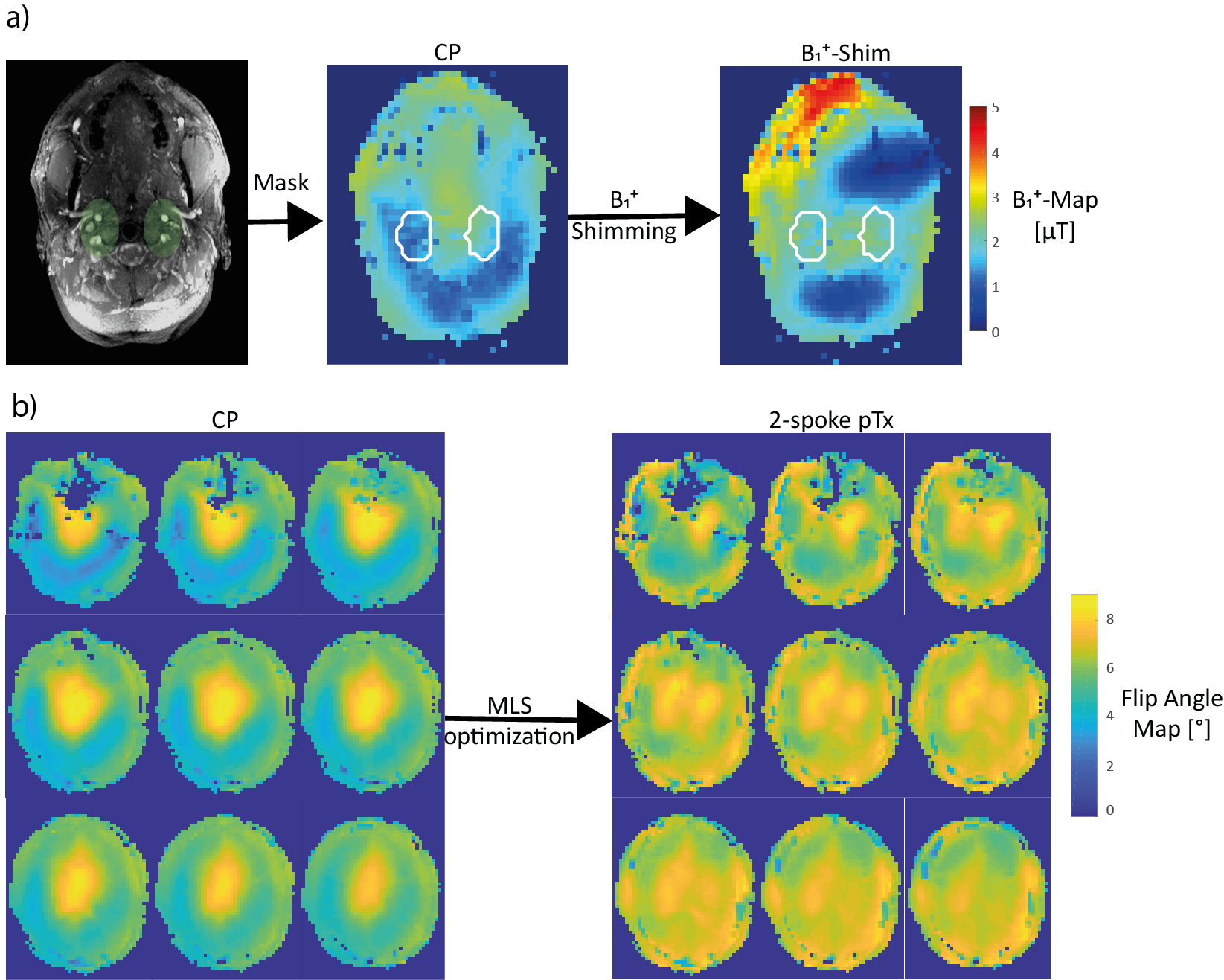
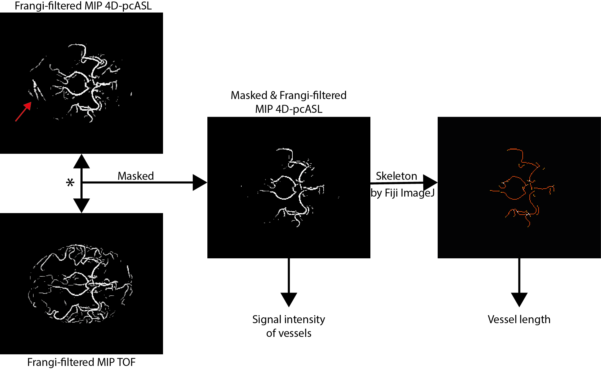
All measurements were performed on a 7T MR system (MAGNETOM Terra, Siemens Healthcare GmbH, Erlangen, Germany) using a 32-channel Rx/8Tx head-coil (Nova Medical, Wilmington, Massachusetts, USA) and carried out in accordance with the institutional guidelines and with approval of the local Ethics Committee (Friedrich-Alexander University (FAU) Erlangen-Nürnberg, Germany). An unbalanced pCASL labeling scheme (TPulse=500μs, TGap=1200μs, Gmax=4.2mT/m, Gave=0.6mT/m, labeling duration=300ms) was modified with VERSE pulses7 (50% amplitude reduction, GVERSE,max=10mT/m). For the readout a segmented spoiled turbo flash (TFL) readout was used (isotropic resolution 1mm3, 52 slices, PAT=3, partial fourier=6/8, TE=2.07ms, nominal flip angle=8°, echo spacing=4.7ms, TR=1600ms). The post labeling delay (PLD) was varied from 100:150:550ms. After each labeling 31 segments of the readout train were applied, resulting in a temporal resolution of 145.7ms and a total acquisition time of 4:40min per time-point. For the parallel transmission (pTx) pulse calculations, relative B1+ maps8 as well as B0 maps were acquired. Additional time-of-flight (TOF) images were achieved at the labeling plane for the hybrid B1+ shim and the shim values were calculated by minimizing the cost function $$$cost=0.3\cdot CoV^{2} + 0.7\cdot\eta^{2}$$$, with $$$CoV=\frac{std\left(\left|\sum_{c=1}^C B_{1,c}^{+}(r)\right|\right)}{mean\left(\left|\sum_{c=1}^C B_{1,c}^{+}(r)\right|\right)}$$$ and $$$\eta = mean\left(\frac{\left|\sum_{c=1}^C B_{1,c}^{+}(r)\right|}{\sum_{c=1}^C \left|B_{1,c}^{+}(r)\right|}\right)$$$ (compare Fig.1a). The readout pTx pulses consists of two 0.9ms long Sinc-shaped spokes placed by an iterative fourier spoke placement9. The complex weights for each spoke pulse were calculated by minimizing $$$cost=\parallel\left|Ab\right| - \left|m\right|\parallel^{2} + \lambda\parallel b \parallel^{2}$$$ with the desired magnetization pattern m, b the complex weights of each RF channel and spoke in Volt, A the system matrix containing the excitation trajectory as well as B1+, B0 maps, and the regularization factor for the RF energy $$$\parallel b \parallel^{2}$$$. A magnitude-least-squares (MLS) algorithm was used for the optimization10 (compare Fig.1b). Additionally, a 1mm3 isotropic TOF sequence was obtained at the same position (4 slabs à 20 slices, PAT=2, TE=3.57ms, flip angle=17°, TR=19ms). In total 3 subjects (mean age: 26.7±0.5years) were measured with the hybrid B1+ shim for the labeling and the 2-spokes excitation for the readout (referred as 'full pTx') compared to the sequence in the CP-mode. The 4D-pcASL images underwent postprocessing using SPM12 (Wellcome Trust Centre for Neuroimaging)11 and the FMRIB Software Library (FSL)12. To evaluate the data transversal maximum intensity projections (MIPs) of the 4D-pcASL as well as of the TOF images were created and filtered by a 2D-Frangi filter (Gaussian kernel σ=1.0, thresholds to control sensitivity α=0.4, β=0.3 and c=40). The 4D-pcASL filtered MIPs were co-registered to the TOF and masked by the filtered MIPs of the TOF. Those masked 4D-pcASL images were used for signal evaluation in the vessels and vessel length determination by processing via Fiji ImageJ as described in a previous work13 (compare Fig.2).Results:
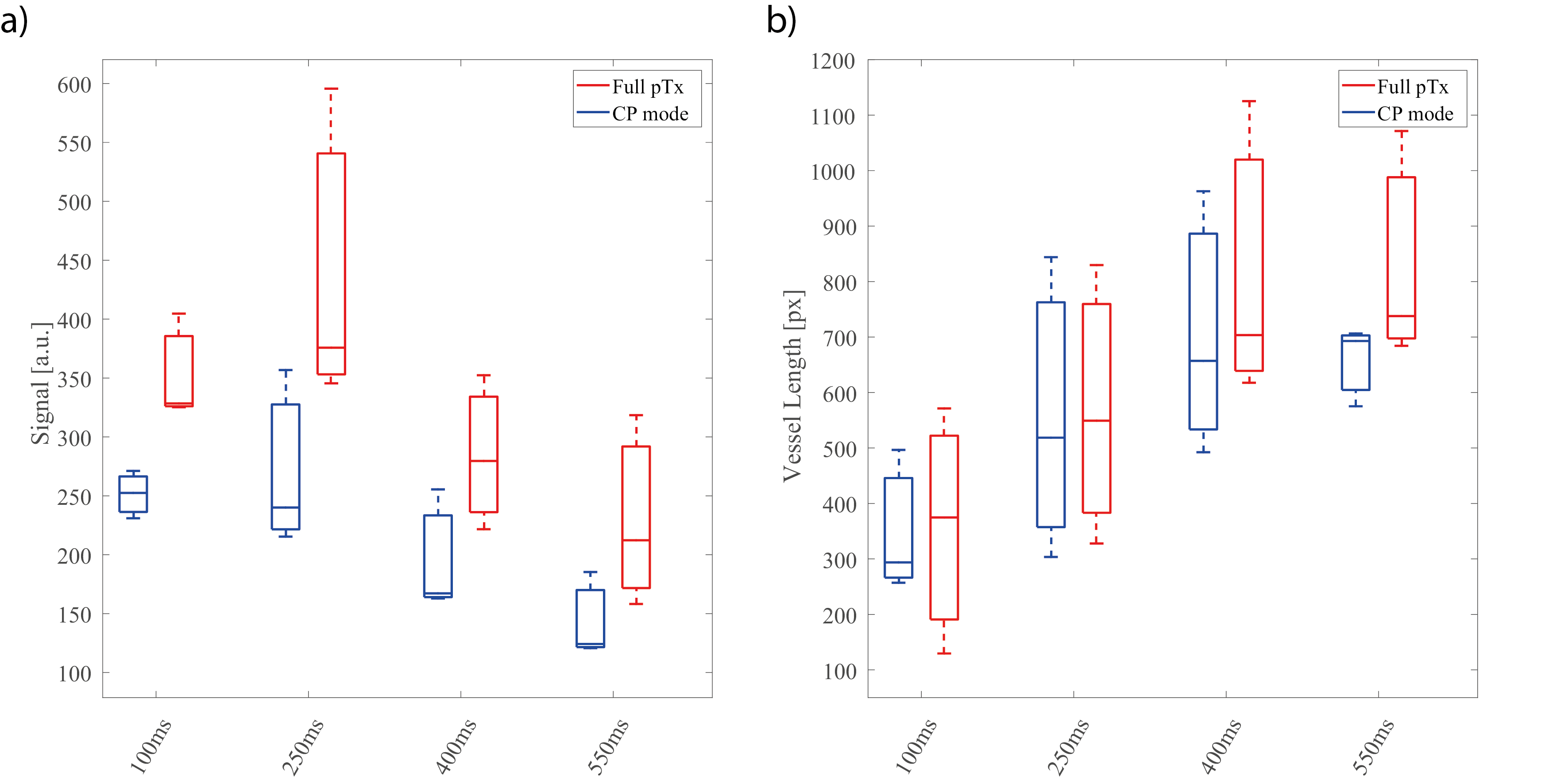
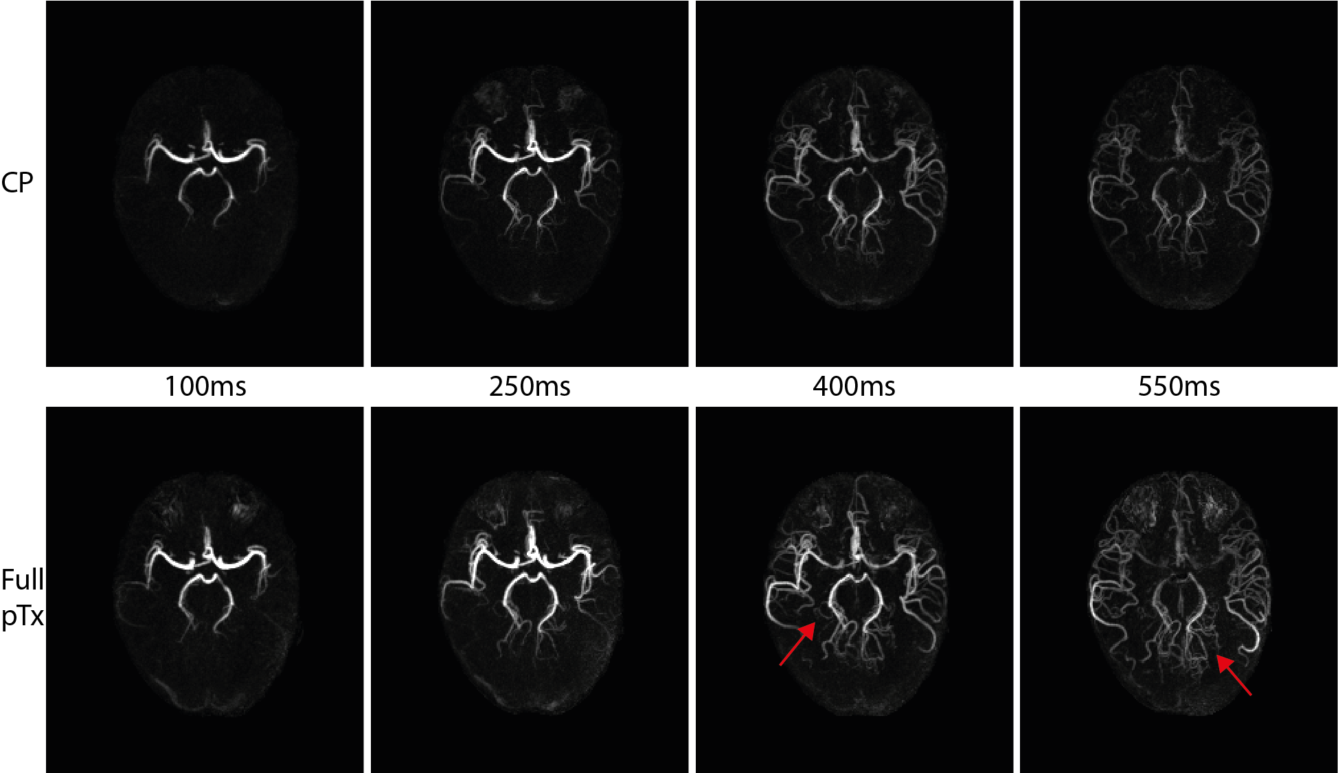
Fig.3a presents the signal intensity of the vessels at the different PLD time-points. Compared to the CP-mode, the full pTx sequence that includes the hybrid B1+ shim for the labeling and 2-spoke pTx for readout shows higher signal in the vessels at all time-points. The measured vessel length (Fig.3b) remains constant for the first three time-points (100-400ms), however with a PLD=550ms the measured vessel length for the full pTx mode increases. An example for all time-points of one subject is given in Fig.4. There, the higher signal intensity of the full pTx sequence compared to the CP-mode can also be observed as well as an increasing number of small vessels can be depicted at later time-points (red arrows).Discussion and Conclusion:
The combination of B1+-shimming for the labeling and 2-spoke dynamic pTx for the readout in 4D-pcASL MRI sequences promises great advantage over conventional CP-mode at 7T. While the higher and more homogenous distribution of the flip angles increases the vessels’ signal intensities, the B1+-shimmed labeling pulse strengthens this effect and yields even more vessel conspicuity, especially at later time-points.Acknowledgements
No acknowledgement found.References
1. Cong, F. et al. Noncontrast-enhanced time-resolved 4D dynamic intracranial MR angiography at 7T: A feasibility study. J Magn Reson Imaging 48, 111-120, doi:10.1002/jmri.25923 (2018).
2. Metzger, G. J. et al.
In Proceedings of the 21st Annual Meeting of ISMRM, Salt Lake City,
USA, 2013.
3. Koktzoglou, I., Gupta, N. & Edelman, R. R. Nonenhanced extracranial carotid MR angiography using arterial spin labeling: improved performance with pseudocontinuous tagging. J Magn Reson Imaging 34, 384-394, doi:10.1002/jmri.22628 (2011).
4. Tong, Y., Jezzard, P., Okell, T. W. & Clarke, W. T. Improving PCASL at ultra-high field using a VERSE-guided parallel transmission strategy. Magn Reson Med, doi:10.1002/mrm.28173 (2020).
5. Li X, W. D., Wu X, Van de Moortele PF, Ugurbil K, Metzger GJ. In Proceedings of the 26th Annual Meeting of ISMRM, Paris, France, 2018.
6. Padormo, F., Beqiri, A., Hajnal, J. V. & Malik, S. J. Parallel transmission for ultrahigh-field imaging. NMR Biomed 29, 1145-1161, doi:10.1002/nbm.3313 (2016).
7. Schmitter, S. et al. Contrast enhancement in TOF cerebral angiography at 7 T using saturation and MT pulses under SAR constraints: impact of VERSE and sparse pulses. Magn Reson Med 68, 188-197, doi:10.1002/mrm.23226 (2012).
8. Fautz HP, Vogel M, Gross P, Kerr A, Zhu Y. B1 mapping of coil arrays for parallel transmission. In Proceedings of the 22nd Annual Meeting of ISMRM, Toronto, Canada, 2008.
9. Saekho, S., Yip, C. Y., Noll, D. C., Boada, F. E. & Stenger, V. A. Fast-kz three-dimensional tailored radiofrequency pulse for reduced B1 inhomogeneity. Magn Reson Med 55, 719-724, doi:10.1002/mrm.20840 (2006).
10. Paige, C. C. & Saunders, M. A. LSQR: An algorithm for sparse linear equations and sparse least squares. ACM Transactions on Mathematical Software 8, 43-71 (1982).
11. Wang, Z. et al. Empirical optimization of ASL data analysis using an ASL data processing toolbox: ASLtbx. Magnetic resonance imaging 26, 261-269, doi:10.1016/j.mri.2007.07.003 (2008). 12 Jenkinson, M., Beckmann, C. F., Behrens, T. E., Woolrich, M. W. & Smith, S. M. Fsl. NeuroImage 62, 782-790, doi:10.1016/j.neuroimage.2011.09.015 (2012).
13. Meixner, C. R. et al. High resolution time-of-flight MR-angiography at 7T exploiting VERSE saturation, compressed sensing and segmentation. Magnetic resonance imaging, doi:10.1016/j.mri.2019.08.014 (2019).
Figures