0598
CEST Imaging vs. Diffusion-Weighted Imaging vs. FDG-PET/CT vs. Combined Method: Prediction Capability for Recurrence in NSCLC Patients1Radiology, Fujita Health University School of Medicine, Toyoake, Japan, 2Joint Research Laboratory of Advanced Medical Imaging, Fujita Health University School of Medicine, Toyoake, Japan, 3Division of Functional and Diagnostic Imaging Research, Department of Radiology, Kobe University Graduate School of Medicine, Kobe, Japan, 4Canon Medical Systems Corporation, Otawara, Japan, 5Diagnostic Radiology, Hyogo Cancer Center, Akashi, Japan
Synopsis
No major reports have been reported the comparison of capability for differentiating recurrence from non-recurrence groups in candidates for surgical resection due to lung cancer among CEST imaging, DWI and PET/CT. We hypothesized that CEST imaging, which was determined as APT-weighted (APTw) imaging at 3.5 ppm, had equal or better potential for prediction of postoperative recurrence prediction in postoperative lung cancer patients, when compared with DWI and FDG-PET/CT. The purpose of this study was to compare the prediction capability of among single- and multi-parametric approaches by APTw imaging, DWI, and FDG-PET/CT in NSCLC patients.
Introduction
Prediction of recurrence group from non-recurrence group in postoperative non-small cell lung cancer (NSCLC) patients are essential for radiological examination in routine clinical practice. Currently, CT and MR imaging including diffusion-weighted imaging (DWI) as well as dynamic contrast-enhanced MR imaging have been applied for morphological evaluation, although FDG-PET and PET/CT are applicable molecular imaging technique in various clinical and academic interest. As compared with FDG-PET or PET/CT, chemical exchange saturation transfer (CEST) imaging at 3.5 ppm (APT-weighted imaging: APTw imaging) has been suggested as the new technique for MR-based molecular imaging and reported as having the potential for diagnosis of thoracic lesions as well as pulmonary nodules (1, 2). However, no major reports have been reported the comparison of capability for differentiating recurrence from non-recurrence groups in candidates for surgical resection due to lung cancer among APTw imaging, DWI and PET/CT. We hypothesized that APTw imaging had equal or better potential for prediction of postoperative recurrence prediction in postoperative lung cancer patients, when compared with DWI and FDG-PET/CT. In addition, multiparametric approach among all three methods (i.e. combined method) had better potential than single-parametric approach on each method in this setting. The purpose of this study was to compare the prediction capability of among single- and multi-parametric approaches by APTw imaging, DWI, and FDG-PET/CT in NSCLC patients.Materials and Methods
Sixty-seven consecutive NSCLC (38 men, 29 women; mean age 71 years) patients prospectively underwent CEST imaging and DWI at 3T MR system (Vantage Titan 3T, Canon Medical Systems Corporation), FDG-PET/CT, surgical resection and/ or more than 2 years follow-up examinations. Based on the results of follow up examination, 67 operated patients were divided as follows: non-recurrence group (n=52) and recurrence group (n=15). To obtain CEST data in each subject, respiratory-synchronized FASE imaging was conducted following a series of magnetization transfer (MT) pulses. Then, magnetization transfer ratio asymmetry (MTRasym) was calculated from z-spectra in each pixel, and MTRasym map was computationally generated. To obtain radiological indexes on CEST imaging, DWI and PET/CT, ROIs were placed over each lesion, and determined MTRasym, apparent diffusion coefficient (ADC) and maximum standard uptake value (SUVmax). Then, Student’s t-test was performed to determine the MTRasym, ADC and SUVmax between recurrence and non-recurrence groups in operated patients. Multivariate logistic regression analysis was performed to investigate the discriminating factors of recurrence from non-recurrence groups. In addition, ROC analyses were also performed to differentiating two groups. Finally, sensitivity, specificity and accuracy were compared each other by means of McNemar’s test. A p value less than 0.05 was considered as significant in this study.Results
Representative cases are shown in Figures 1. MTRasym, ADC and SUVmax had significant difference between recurrence and non-recurrence groups (p<0.05). Results of multivariate regression analysis are shown in Figure 2. Multivariate regression analyses identified MTRasym (Odds ratio [OR]: 3.71, p=0.0008) and SUVmax (OR: 5.8, p=0.009) as significant differentiators. Results of ROC analyses and diagnostic performance comparison are shown in Figure 3. For differentiating recurrence from non-recurrence group, specificity (SP) or accuracy (AC) of combined indexes (SP: 96.2 [50/52]%, AC: 94.0 [63/67] %) were significantly higher than those of MTRasym (SP: 82.7 [43/52]%, p<0.05; AC: 83.6 [58/67] %, p<0.05) and SUVmax (SP: 84.6 [44/52]%, p<0.05).Conclusion
MTRasym and SUVmax were determined as significant predictors for distinguishing recurrence from non-recurrence groups. Multiparametric approaches of MRI and PET/CT have better potential than PET/CT alone in these settings.Acknowledgements
This study was technically and financially supported by Canon Medical Systems Corporation.References
- Ohno Y, Yui M, Koyama H, et al. Chemical Exchange Saturation Transfer MR Imaging: Preliminary Results for Differentiation of Malignant and Benign Thoracic Lesions. Radiology. 2016; 279(2): 578-589.
- Ohno Y, Kishida Y, Seki S, et al. Amide proton transfer-weighted imaging to differentiate malignant from benign pulmonary lesions: Comparison with diffusion-weighted imaging and FDG-PET/CT. J Magn Reson Imaging. 2018; 47(4): 1013-1021.
Figures
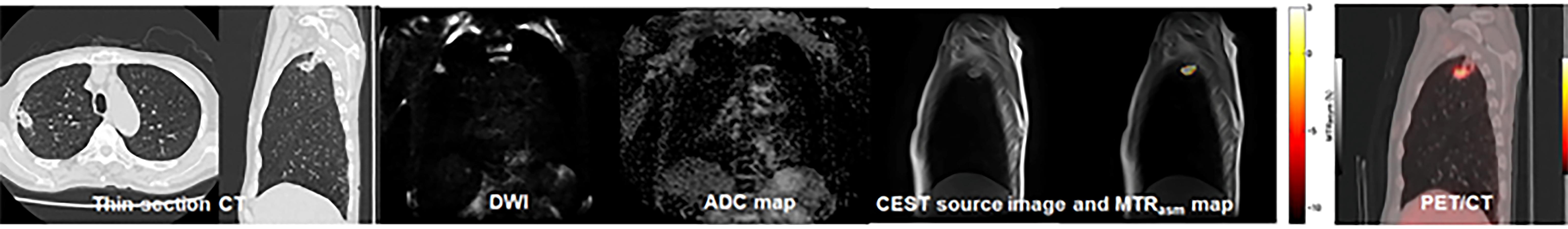
Figure 1. 62-year-old female with invasive adenocarcinoma (L to R: thin-section CT, DWI, ADC map, MTRasym map fused with T2WI, and SUVmax map fused with CT) and determined as recurrence group.
Thin-section CT demonstrates a nodule in the right upper lobe. ADC of this nodule was 1.05×10⁻³mm2/s. SUVmax was 2.6. APTw image shows low MTRasym with the value of 1.25. This case was false-negative on PET/CT, and true-positive on DWI and APTw image. When combined both indexes, PET/CT with APTw image was diagnosed as recurrence group and determined as true-positive case.
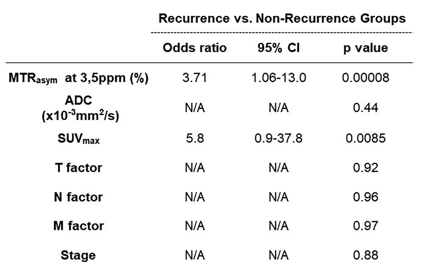
Figure 2. Results of multivariate regression analysis for distinguishing recurrence from non-recurrence groups in NSCLC patients.
MTRasym at 3.5ppm and SUVmax were determined as significant predictor for distinguishing recurrence from non-recurrence groups (p<0.05).
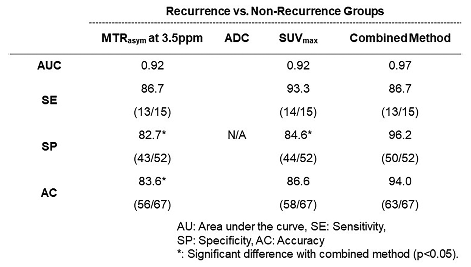
Figure 3. Comparison of diagnostic performance among MTRasym at 3.5ppm, ADC and SUVmax for distinguishing recurrence from non-recurrence groups in NSCLC patients.
Specificity and accuracy of combined indexes were significantly higher than those of MTRasym and SUVmax on prediction of postoperative recurrence in lung cancer patients (p<0.05).