0572
Resting-state fMRI Predicts Task Activation Patterns Using a Graph Convolutional Network1GE Healthcare, MR Research China, Beijing, China, 2Center for Biomedical Imaging Research, Department of Biomedical Engineering, School of Medicine, Tsinghua University, Beijing, China, 3Department of Psychiatry, University of Connecticut School of Medicine, Farmington, MI, United States, 4Department of Psychiatry and Behavioral Sciences, Stanford University, Stanford, CA, United States
Synopsis
Resting-state fMRI has the clinical potential as diagnostic and prognostic markers because of its easy implementation/standardization in data acquisitions, and its ability to parcellate functionally connected neural networks. It is of importance to examine whether the task-free spontaneous activity could be used to predict individuals’ task-induced activation. Here we proposed a graph convolutional network-based framework which utilized the information of the brain connections for the convolution step, and showed the ability of using resting-state fMRI to predict individual differences in activations of tasks from human connectome project. This framework could be extended to other resting-state fMRI researches.
Introduction
Resting-state fMRI has a great potential in translating fMRI researches into clinical realm, because of its easy implementation and standardization in terms of data acquisitions across research sites, and its ability to parcellate spontaneous brain networks without external stimulations. However, it is still obscure whether the intrinsic task-free brain activities could also be used to predict individuals’ task-induced activation, ultimately by only acquiring resting-state that would shorten the total scanning time and reduce the dependence of data quality on subjects’ task performance. Convolution neural network has been enormously applied to image processing. To better take advantage of the brain functional organizations from resting-state functional connectivity (FC), we proposed a graph convolutional network (GCN) 1 based framework and aimed to use resting-state fMRI to predict individuals’ activation patterns evoked by tasks from different behavioral domains.Methods
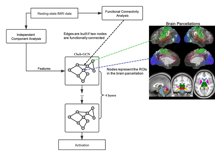
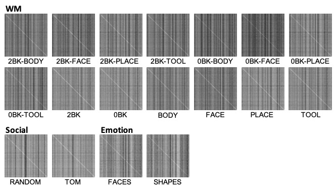
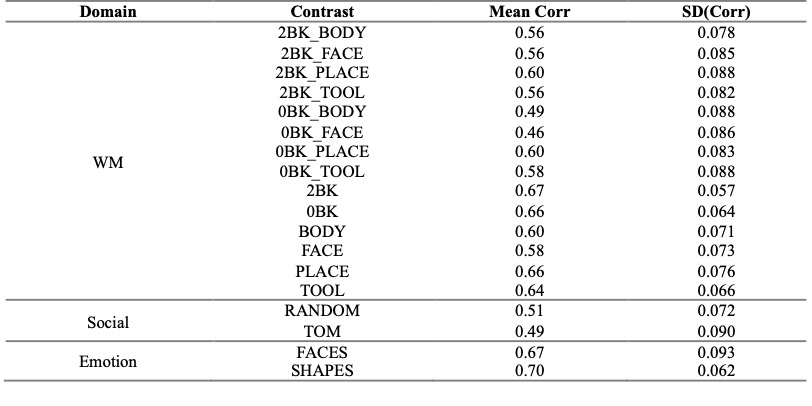
A subgroup of 547 subjects from Human Connectome Project (www.humanconnectome.org) with a complete set of 4 fifteen-minute resting-state and 7 task-related fMRI data were included in this study 2. Task fMRI data included 86 contrasts from 7 task domains: working memory (WM), gambling, motor, language, social, relational, and emotion. fMRI data underwent the minimal preprocessing, while resting-state fMRI data was additionally FIX-cleaned to remove artifacts from different sources 3. All data was resampled onto the set of 91282 “grayordinates” in standard space, including the two cortical hemispheres represented in a surface-based space and sub-cortical grey matters and the cerebellum in a voxel-based space. Group independent component analysis (gICA) 4 was implemented to parcellate data of all subjects into 50 shared brain networks, individuals’ connectivity maps for each network were derived following the dual regression procedure 4 and were entered as features of the GCN.To generate the graph for GCN modeling, a customized brain parcellation of 399 ROIs (360 cortical regions 5 + 39 subcortical structures) were applied as graph nodes, individual FC matrix of those ROIs was calculated as the edges (Fig. 1) 5. To ignore weak connections between nodes, a threshold of FC > (0.3 * FCmax) was used on the FC maps for each subject, notably that edges retained after thresholding might vary across subjects. Fig. 1 shows the overview of the graph convolutional network for this study (Fig. 1). Four Chebyshev spectral graph convolutional layers (Cheb-GCN) 6 were used, instance normalization was added for each of the first three layers to reduce over-fitting. Data from 447, 50 and 50 subjects were used for training, validation, and testing respectively. To reduce the computation complexity while to still predict task-evoked activations in a voxel-wise way, we first extracted connectivity values of the 50 gICA connectivity maps for the 399 ROIs and generated a 399*50 matrix as input features. During the loop across voxels per task, values of all 50 gICA maps for the ROI (i.e. node K) to which the current voxel belongs were replaced by the values of the current voxel. Using this way, the voxel difference in activation level even within same ROIs could still be maintained. Each task was trained separately, outputs of each network were the actual activation levels of each voxel for all conditions under a same task, thus the dimension of the output was determined by the number of contrasts in each task fMRI. The mean-squared-error between the predicted and actual activation value of node K was used as the loss function. The network was trained and evaluated using pyTorch 7. Pearson correlations of the predicted activation map and actual activation map were calculated, here results of 18 contrasts from 3 tasks (WM, social and emotion) are shown as examples.
Results and Discussion
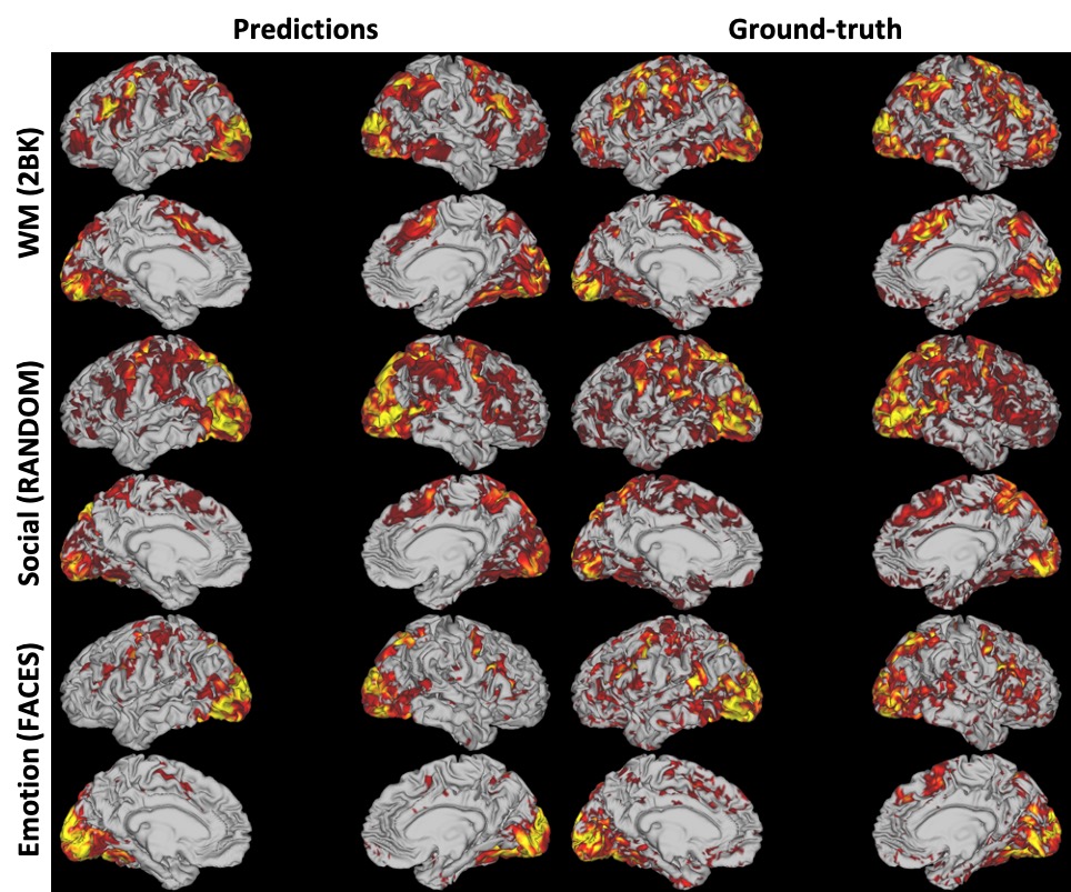
As indicated in Fig. 2, the predicted task activation maps resemble the actual activation patterns across the WM (2BK), social (RANDOM), emotion (FACES) behavioral domains. Full results of all 18 contrasts of the 50 testing subjects could be found in Table 1, overall the correlation coefficients are comparable with the prediction using general linear model incorporated not only fMRI but also structural MRI features 8, especially the correlations of the emotion task are higher with the proposed method. To evaluate the performance of the proposed method in predicting activation at individual level, the between-subjects cross-correlation matrices of predicted versus actual activation maps for the task contrasts listed in Table 1 were calculated. The bright diagonals indicate that for each subject the predicted map mostly resembles the subject’s actual map (paired two sample t-test, p < 0.0001) (Fig. 3), and the model predicts individual variations in response to tasks instead of the group average.Conclusion
The current study showed the ability of predicting individual difference of task-evoked activation patterns from different behavioral domains by resting-state fMRI with GCN, and we believe the performance of the current model could be improved by fine-tuning of the network structures. We proposed a GCN based framework by taking account of the functional organizations in the brain, which could be applied to other scenarios in resting-state fMRI research such as prediction of personal traits and symptoms of psychiatric disorders.Acknowledgements
No acknowledgement found.References
1. Wu Z, Pan S, Chen F, Long G, Zhang C, Philip SY. A comprehensive survey on graph neural networks. IEEE Transactions on Neural Networks and Learning Systems 2020.
2. Barch DM, Burgess GC, Harms MP, Petersen SE, Schlaggar BL, Corbetta M, Glasser MF, Curtiss S, Dixit S, Feldt C. Function in the human connectome: task-fMRI and individual differences in behavior. NeuroImage 2013;80:169-189.
3. Glasser MF, Sotiropoulos SN, Wilson JA, Coalson TS, Fischl B, Andersson JL, Xu J, Jbabdi S, Webster M, Polimeni JR. The minimal preprocessing pipelines for the Human Connectome Project. NeuroImage 2013;80:105-124.
4. Beckmann CF, Mackay CE, Filippini N, Smith SM. Group comparison of resting-state FMRI data using multi-subject ICA and dual regression. NeuroImage 2009;47(Suppl 1):S148.
5. Glasser MF, Coalson TS, Robinson EC, Hacker CD, Harwell J, Yacoub E, Ugurbil K, Andersson J, Beckmann CF, Jenkinson M. A multi-modal parcellation of human cerebral cortex. Nature 2016;536(7615):171-178.
6. Defferrard M, Bresson X, Vandergheynst P. Convolutional neural networks on graphs with fast localized spectral filtering. arXiv preprint arXiv:160609375 2016.
7. Paszke A, Gross S, Massa F, Lerer A, Bradbury J, Chanan G, Killeen T, Lin Z, Gimelshein N, Antiga L. Pytorch: An imperative style, high-performance deep learning library. 2019. p 8026-8037.
8. Tavor I, Jones OP, Mars R, Smith S, Behrens T, Jbabdi S. Task-free MRI predicts individual differences in brain activity during task performance. Science 2016;352(6282):216-220.
Figures