0564
In vivo Two-photon Magnetic Resonance Imaging of Human Brain at 3T1Electrical Engineering and Computer Sciences, University of California, Berkeley, Berkeley, CA, United States, 2Helen Wills Neuroscience Institute, University of California, Berkeley, Berkeley, CA, United States
Synopsis
We are exploring in vivo brain imaging with two-photon excitation on a 3T scanner. To do that, we built a customized coil that is large enough to fit a standard birdcage transceiver coil inside of it. The resulting two-photon gradient-echo brain images excited using a 25.5 kHz z-axis RF were overall similar to standard single-photon images with equivalent parameters. Interestingly, we observed some slight differences in tissue contrast whose cause are being investigated.
Introduction:
Multiphoton excitation is an innovative MRI scanning technique. Despite using frequencies far from the Larmor frequency, spins can still be excited if conservation of energy and angular momentum are followed. For example, if the traditional xy-plane RF field is shifted from the Larmor frequency by 25.5 kHz, no excitation is expected. However, if a z-direction RF field, called $$$B_{1z}$$$, parallel to the $$$B_0$$$ field, at 25.5 kHz is applied along with the 25.5 kHz-offset xy-plane RF field, called $$$B_{1xy}$$$, then two-photon excitation occurs. One z-photon is absorbed together with one xy-photon to excite each spin. In imaging, it has been shown that we can use extra z-photons to turn slice selective xy-RF pulses (e.g. Sincgauss pulses) into scaled versions of their single-photon counterparts when the offset in frequency for the xy-pulse is set to meet the two-photon resonance condition1. Using an extra, uniform coil that produces a magnetic field in the z-direction, we can perform gradient echo scans with all single-photon excitations replaced by two-photon counterparts. This idea has been previously applied on an Aspect 1T wrist scanner2 and for the human hand, it was found that two-photon images looked similar to their single photon counterparts. However, there was still much to be desired in terms of SNR and $$$B_{1z}$$$ field homogeneity due to the small bore size. In this project, we built a new version of the $$$B_{1z}$$$ coil and associated setup for use on a clinical whole-body 3T scanner to resolve the imperfections of previous work. As result, we successfully acquired the first two-photon brain image.Methods:
We designed and built a large size solenoid coil to produce an uniform $$$B_{1z}$$$ field over the human brain, parallel to the $$$B_0$$$ field and perpendicular to the $$$B_{1xy}$$$ field. For this coil we used a large PVC pipe with an 18-inch diameter and 20-inch length. Two parallel strands of 14 AWG wire were used to make a helix with 20 windings on the outer surface of pipe. We added another 20 windings on the inner surface of cylinder. The inner helix is mirrored with the outer helix and is joined at the bottom. Both windings meet at the top of the pipe and connect to the output of an amplifier. To stabilize the windings on the pipe, we applied epoxy on both inner and outer surfaces of the pipe. Lastly, we wrapped the coil with a UL94 V-0 rated flame-retardant sound-dampening mat for the safety of the human experiment (Figure 1).In this project, we targeted 25.5KHz for the frequency of the $$$B_{1z}$$$ field. In order to achieve a large $$$B_{1z}$$$ field strength, the coil was tuned and matched to 3 ohms for use with a 500W audio power amplifier. When scanning, the power amplifier amplifies a signal from a waveform generator in a gated burst mode, which is synchronized to the scanner with a Schmitt Trigger circuit to ensure that our $$$B_{1z}$$$ field will be off during the receive period. For the cable that connects the coil to the amplifier, we added 15 baluns each separated by a Larmor frequency quarter wavelength to reduce the EMI effects of the long cable and improve the safety of the design (Figure 2).
The setup was implemented on a GE 750W 3T scanner (GE Healthcare, Waukesha, WI). To compare single-photon ($$$B_{1xy}$$$ at the Larmor frequency) and two-photon ($$$B_{1xy}$$$ at 25.5 kHz away from the Larmor frequency) images, we calibrated the flip angles based on 1D projections of signal magnitude in manual pre-scans in gradient echo sequences. Signals were measured to ensure that single- and two-photon scans with proper $$$B_{1xy}$$$ strengths produced similar amounts of signal and all other parameters were kept equivalent. We ensured that the frequency offset $$$B_{1xy}$$$ fields did not produce any excitation without the $$$B_{1z}$$$ fields.
Results:
We successfully obtained 2D GRE two-photon images with several TRs and FAs from a healthy volunteer. During scanning, no appreciable vibration nor increased acoustic noise were observed. Figure 3 shows the same slice with both single- and two-photon excitation. The overall SNR and image appearances appear to be comparable. Interestingly, the gray/white matter contrast of the two-photon image appears to be slightly enhanced compared to the single-photon image. In addition, we also explored various TRs while fixing flip angles (Figure 4), and various flip angles while fixing the TR (Figure 5). In all comparisons, the single- and two-photon images share overall similarities for the most part but some small differences also exist throughout the brain.Discussion and Conclusion:
We have demonstrated that brain images can be obtained by using two-photon excitation on a clinical 3T scanner. The preliminary results suggest that two-photon excitation may provide some promise for creating different tissue contrasts. However, there are still many un-answered questions that need to be further explored, such as where these differences come from and what happens at different offset frequencies and $$$B_{1z}$$$ amplitudes. To help answer these questions, we plan to improve the SNR by using arrays of surface coil receivers instead of the birdcage coil. Furthermore, more work needs to be done with pulse sequence and hardware design to streamline the process of setting up two-photon scans.Acknowledgements
No acknowledgement found.References
1. Han, V, Liu, C. Multiphoton magnetic resonance in imaging: A classical description and implementation. Magn Reson Med. 2020; 84: 1184– 1197.
2. Chi, J, Han, V, Liu, C. In vivo Two-photon Magnetic Resonance Imaging of Human Hand at 1T. In: ISMRM Annual Scientific Meeting & Exhibition 2020.
Figures
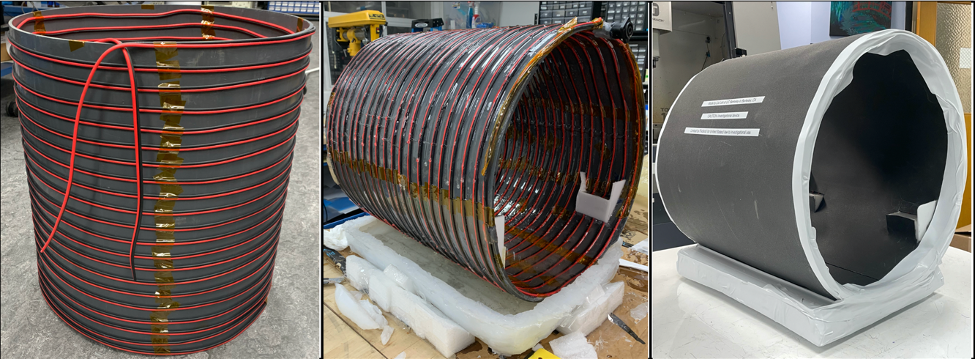
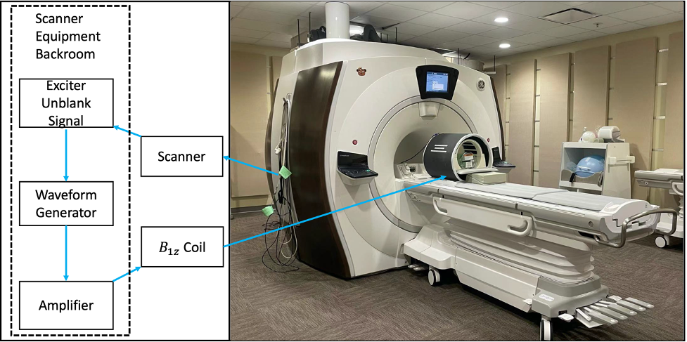
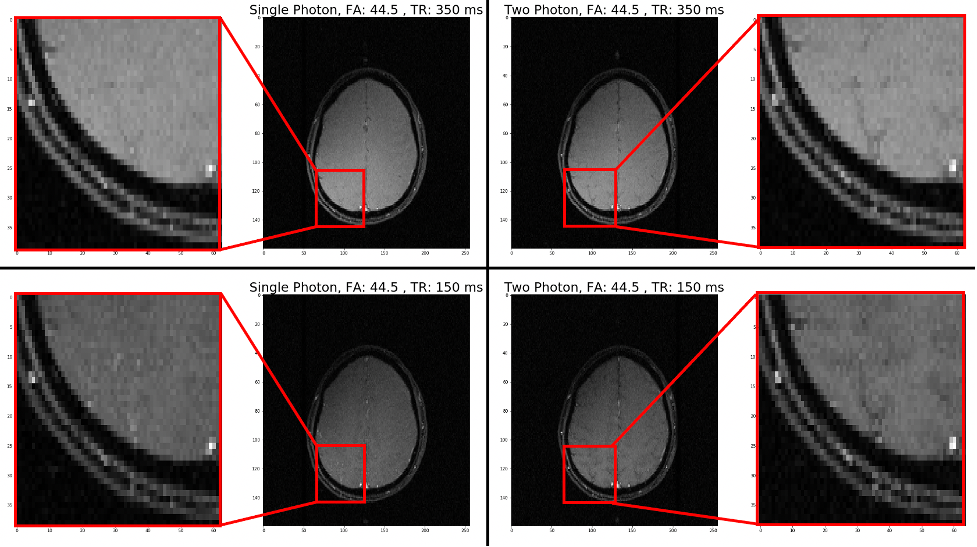
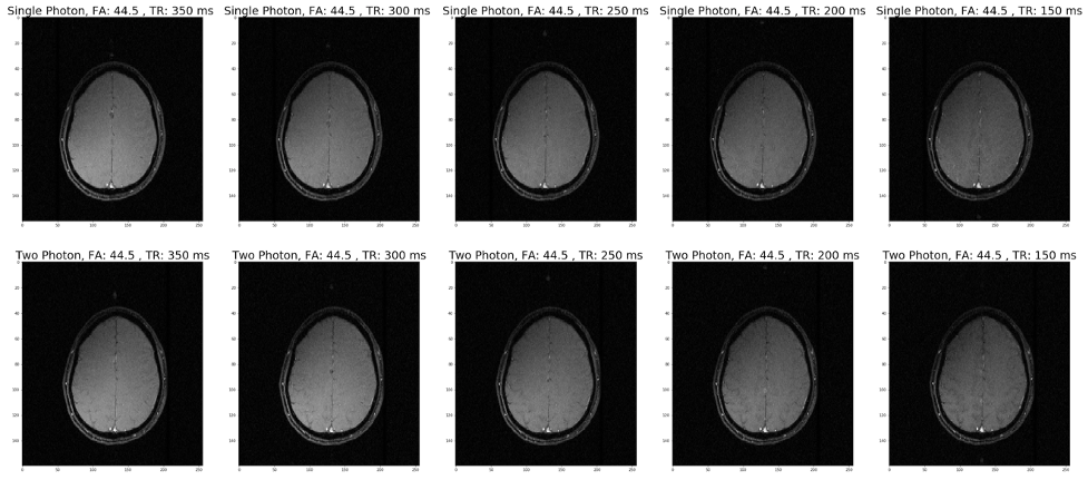
Figure 4: Single-photon (top row) vs. two-photon (bottom row) images of human brain with fixed flip anlges (FA) and various TR. Each column is a pair of single- and two-photon images with matched equivalent flip angle. Parameters: FOV 27.6 cm, slice thickness 3 mm, matrix 256x160, TE 10ms, flip angle 44.5o, TR (from 150 ms to 350 ms with step of 50 ms).
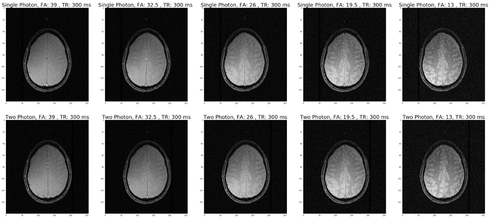
Figure 5: Single-photon (top row) vs. two-photon (bottom row) images of human brain with fixed TR and various FA. Each column is a pair of single- and two-photon images with matched equivalent flip angle. Parameters: FOV 27.6 cm, slice thickness 3 mm, matrix 256x160, TE 10ms, TR 300ms, flip angles (13o, 19.5o, 26o, 32.5o and 39o).