0560
Simultaneous imaging of hard and soft biological tissues in a low-field MRI scanner1I3M, CSIC, Valencia, Spain, 2Tesoro Imaging, Valencia, Spain
Synopsis
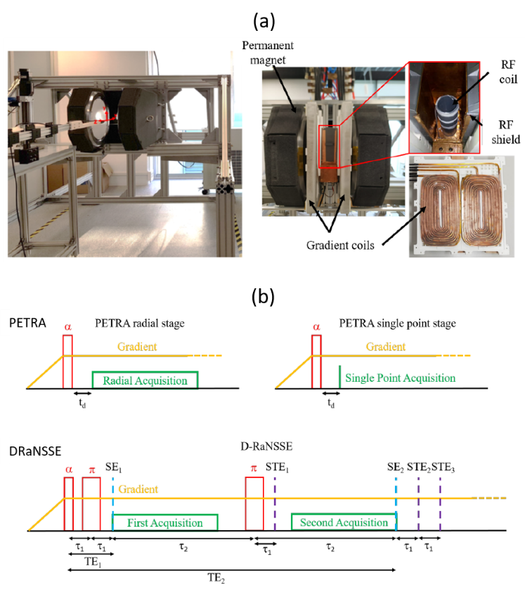
Here we present the first demonstration of dental imaging in a low cost MRI setup at sub-Tesla fields (260 mT). We show 3D reconstructions of a rabbit head and human teeth acquired in “DentMRI – Gen I” (Fig. 1(a)), a home-made special-purpose MRI scanner designed with the goal of demonstrating dental imaging at low field strengths. We use two variations of zero echo time (ZTE) pulse sequences (Fig. 1(b)): standard PETRA , and Double Radial Non Stop Spin Echo (DRaNSSE), which we have devised to address limitations we find with PETRA. We reconstruct images by Algebraic Reconstruction Techniques (ART).
Introduction
Here we present the first demonstration of dental imaging in a low cost MRI setup at sub-Tesla fields (260 mT) [1]. We show 3D reconstructions of a rabbit head and human teeth acquired in “DentMRI – Gen I” (Fig. 1(a), [1]), a home-made special-purpose MRI scanner designed with the goal of demonstrating dental imaging at low field strengths. We use two variations of zero echo time (ZTE) pulse sequences (Fig. 1(b)): standard PETRA [2,3], and Double Radial Non Stop Spin Echo (DRaNSSE), which we have devised to address limitations we find with PETRA. We reconstruct images by Algebraic Reconstruction Techniques (ART, [4]).Methods
“DentMRI – Gen I” operates with a “C”-shaped permanent NdFeB magnet that provides 260 mT over a spherical region of 150 mm in diameter. The system is equipped with a gradient system capable of reaching strengths > 0.4 T/m along any spatial direction, and a TxRx RF solenoid coil able to induce a flip angle of 90 degrees in a few microseconds. We drive the solenoid with an rf power amplifier fed by a direct digital synthesizer on our field-programmable gate-array (FPGA), where we also digitize, down-convert and filter the previously amplified MR signal.We acquired all images with two variations of standard ZTE pulse sequences: Pointwise Encoding Time Reduction with Radial Acquisition (PETRA [2,3]); and a sequence we have devised to overcome specific k-space coverage and contrast limitations, Double Radial Non-Stop Spin Echo (DRaNSSE [1]).
In PETRA a hard rf pulse homogeneously excites the sample after the onset of the encoding gradient fields. Data acquisition starts next, after a dead time usually set by the TxRx switch and the rf coil ring-down. Every acquisition follows a radial direction (spoke) in a 3-dimensional k-space. Due to the finite dead time, the center of k-space is filled in a pointwise manner on a Cartesian grid.
In DRaNSSE, after the initial rf pulse, two refocusing π-pulses produce two different spin echoes. The first one (SE1) is induced at an echo time (TE1) as short as possible to include contributions from both hard and soft tissues. The second echo (SE2) occurs at a later time (TE2), when the short-lived signal from hard tissues has already faded away. Images are reconstructed by Algebraic Reconstruction Techniques [4], since we found that conventional regridding and Fourier reconstruction results in highly non-stationary noise (due to the non-Cartesian acquisition) and a worse SNR compared to ART [1].
Results
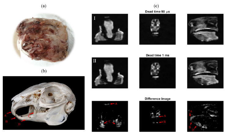
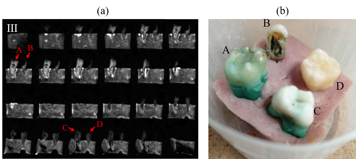
Figure 2(c) contains selected slices from the full 3D ART reconstruction of a rabbit head employing a PETRA sequence with 0.5 mm isotropic resolution. We present two images: one with a short (90 µs) dead time (Fig. 2(c) top) with a scan time of 61 minutes; and one with a long (1 ms) dead time (Fig. 2(c) middle) with a scan time of 31 minutes. The bottom image in Fig. 2(c) shows difference between both images to highlight hard tissues.Figure 3(a) shows 2 dimensional slices obtained from 3D ART reconstruction of four human teeth embedded into a piece of ham (emulating the gum) employing PETRA with 0.5 mm isotropic resolution. The image was acquired with 100 µs dead time in a total scan time of 65 minutes.
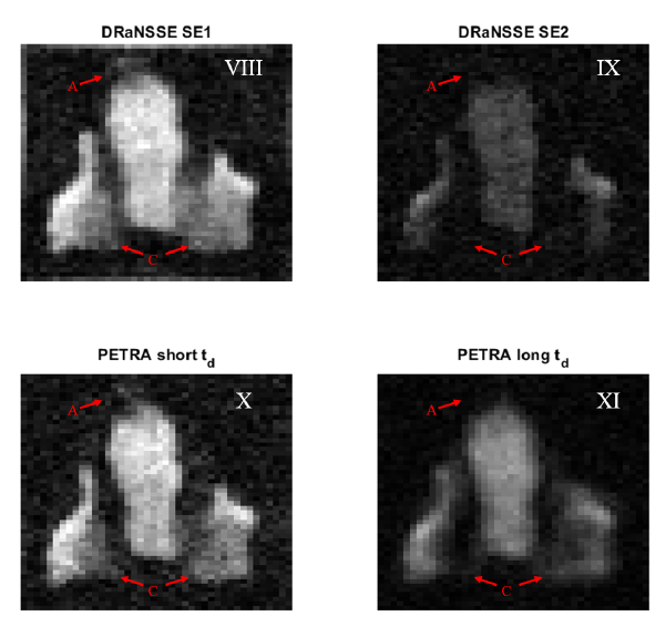
Figure 4 shows slice reconstructions from DRaNSSE (top) and PETRA (bottom) acquisitions. These images were acquired with 1 mm isotropic resolution with a total scan time of 30 (15) minutes for DRaNSSE (PETRA). Due to the long echo time for the second echo (10 ms), tissue contrast is higher with DRaNSSE.
Discussion/Conclusion
In the present work we have demonstrated the capability of our new low-cost “DentMRI – Gen I” scanner to simultaneously image hard and soft biological tissues; we show that human teeth can be imaged with high resolution at low magnetic fields; and we have devised a new pulse sequence (DRaNSSE) that, compared to standard sequences such as PETRA, yields higher SNR images and enhanced tissue contrast.Using this as a starting point, we are currently working on a prototype for clinical dental imaging. The main remaining challenge is to reduce scan times. To that end we will explore four major upgrades: i) we will explore balanced steady-state free precession protocols; ii) we will use quantum dynamical decoupling techniques, which can prolong the lifetime of the magnetic resonance signal of hard tissues; iii) we will perform dual species MRI on protons and 31P nuclei, since the latter are more abundant in hard tissues and they provide complementary information; and iv) we will study the possibility of slice selection with zero-echo time sequences for fast 2D imaging [5].
Acknowledgements
We thank anonymous donors for their tooth samples, Andrew Webb and Thomas O’Reilly (LUMC) for discussions on hardware and pulse sequences, and Antonio Tristán (UVa) for information on reconstruction techniques. This work was supported by the European Commission under Grants 737180 (FET-OPEN: HISTO- MRI) and 481 (ATT RAC T: DentMRI). Action co-financed by the European Union through the Programa Operativo del Fondo Europeo de Desarrollo Regional (FEDER) of the Comunitat Valenciana 2014-2020 (IDIFEDER/2018/022).References
[1] Algarín, J. M., Díaz-Caballero, E., Borreguero, J., Galve, F., Grau-Ruiz, D., Rigla, J. P., Bosch, R., González, J. M., Pallás, E., Corberán, M., Gramage, C., Aja-Fernández, S., Ríos, A., Benlloch, J. M., & Alonso, J. “Simultaneous imaging of hard and soft biological tissues in a low-field dental MRI scanner”. Scientific Reports, 10, 21470, dec 2020. https://doi.org/https://doi.org/10.1038/s41598-020-78456-2
[2] M. Weiger, K. P. Pruessmann, A.-K. Bracher, S. Köhler, V. Lehmann, U. Wolfram, F. Hennel, and V. Rasche, “High-resolution ZTE imaging of human teeth,” NMR in Biomedicine, vol. 25, no. 10, pp. 1144–1151, oct 2012. [Online]. Available: http://doi.wiley.com/10.1002/nbm.2783
[3] D. M. Grodzki, P. M. Jakob, and B. Heismann, “Ultrashort echo time imaging using pointwise encoding time reduction with radial acquisition (PETRA),” Magnetic Resonance in Medicine, vol. 67, no. 2, pp. 510–518, feb 2012.
[4] R. M. Gower and P. Richtarik, “Randomized iterative methods for linear systems,” SIAM Journal on Matrix Analysis and Applications, vol. 36, no. 4, pp. 1660–1690, 2015.
[5] Galve, F., Alonso, J., Algarín, J. M. & Benlloch, J. M. Magnetic resonance imaging method with zero echo time and slice selection. Patent application ESP202030504 (2020).
Figures