0556
Quantifying Brain Iron Deposition in patients with Parkinson’s Disease Using MRI-R2*: A new specific approach developed from a multicenter study
Laila khedher1, Jean Marie Bonny2, Ana Marques3, Marie Vidailhet4, Frédéric Torny5, Luc Defebvre6, Stéphane Thobois7, Elena Moro8, Philippe Remy9, Christian Geny10, Wassilios Meissner11, Solène Frismand12, Anne Doe de Maindreville13, Jean-Luc Houeto14, Olivier Rascol15, and Franck Durif1,3
1University Clermont Auvergne, Clermont Ferrand, France, 2INRA, UR370 Qualité des Produits Animaux, Saint Genès Champanelle, France, 3CHU Clermont Ferrand, Clermont Ferrand, France, 4Fédération des maladies du système nerveux GH La Pitié Salpêtrière, Paris, France, 5CHU Dupuytren, Service de Neurologie, Limoges, France, 6Hopital Roger Salengro, Service de Neurologie et Pathologie du Mouvement, Lille, France, 7Hopital Pierre Wertheimer, Neurologie C, Lyon, France, 8CHU de Grenoble, Service de Neurologie, Grenoble, France, 9Hopital Henri Mondor, Service de Neurologie, Creteil, France, 10CHRU Montpellier, Service de Neurologie, Montpellier, France, 11CHU Bordeaux, Service de Neurologie, Bordeaux, France, 12Hopital Central-CHU Nancy, Service de Neurologie, Nancy, France, 13Pole Neurologie-Gériatrie, Reims, France, 14CHU de Poitiers, Poitiers, France, 15Centre d’Investigation Clinique CIC 1436, CHU PURPAN-Place du Dr Baylac, Hopital Pierre Paul Riquet, Toulouse, France
1University Clermont Auvergne, Clermont Ferrand, France, 2INRA, UR370 Qualité des Produits Animaux, Saint Genès Champanelle, France, 3CHU Clermont Ferrand, Clermont Ferrand, France, 4Fédération des maladies du système nerveux GH La Pitié Salpêtrière, Paris, France, 5CHU Dupuytren, Service de Neurologie, Limoges, France, 6Hopital Roger Salengro, Service de Neurologie et Pathologie du Mouvement, Lille, France, 7Hopital Pierre Wertheimer, Neurologie C, Lyon, France, 8CHU de Grenoble, Service de Neurologie, Grenoble, France, 9Hopital Henri Mondor, Service de Neurologie, Creteil, France, 10CHRU Montpellier, Service de Neurologie, Montpellier, France, 11CHU Bordeaux, Service de Neurologie, Bordeaux, France, 12Hopital Central-CHU Nancy, Service de Neurologie, Nancy, France, 13Pole Neurologie-Gériatrie, Reims, France, 14CHU de Poitiers, Poitiers, France, 15Centre d’Investigation Clinique CIC 1436, CHU PURPAN-Place du Dr Baylac, Hopital Pierre Paul Riquet, Toulouse, France
Synopsis
Several postmortem studies have shown an accumulation of iron in the substantia nigra (SN) in Parkinson’s disease (PD). The iron concentration can be estimated by MRI via the MRI-R2* mapping. 1, 2 In order to assess the changes in R2* occurring in PD patients compared to healthy controls, a multicenter transversal prospective study was carried out in a large cohort of PD patients (n = 98) going from the early to the late stage of the disease and matched controls (n = 66).
Objectives
Several postmortem studies have shown an accumulation of iron in the substantia nigra (SN) in Parkinson’s disease (PD). The iron concentration can be estimated by MRI via the MRI-R2* mapping. 1, 2 In order to assess the changes in R2* occurring in PD patients compared to healthy controls, a multicenter transversal prospective study was carried out in a large cohort of PD patients (n = 98) going from the early to the late stage of the disease and matched controls (n = 66).Materials and Methods
The images acquired at 3Tesla 3 in thirteen different clinical sites were registered, then normalized to the Montreal Neurological Institute neuroanatomic (MNI) space by DARTEL approach 4 for level group magnetic resonance imaging (MRI) analyses. After parametric reconstruction, R2* was measured in different subcortical regions of interest (the substantia nigra (SN); the red nucleus (RN); the striatum (STR); the globus pallidus external (GPe) and the globus pallidus internal (GPi)) segmented from an automatic atlas (available from https://www.nitric.org/projects/atag). R2* values measured separately in the left and the right parts in each brain structure. In this study, the observed inter-individual variability was significantly higher than the disease effect. For this raison, an original strategy (intrasubject subcortical quantitative referencing, ISQR) was developed using the measurement of R2* in the red nucleus (RN) as an intra-individual reference. This structure is not directly involved in the PD circuit but exhibits non-specific variability equivalent to that presented in the subcortical structures of interest. ISQR is based on the calculation of the difference between a structure of interest and the RN: R2*(normalized) = [R2*(structure) – R2*(RN)]Results
an increased R2* value was only achieved in the SN of all PD patients compared to healthy controls (p(SNL) =0.0008, p(SNR) =0.0015). After stratification into 4 subgroups according to the duration of the disease (PD - G1 <5 years; PD - G2, between 5 and 10 years; PD - G3, between 10 and 15 years and PD - G4> 15 years), no significant R2* difference was found in all regions of interest by comparing PD subgroups with each other (figure1). Our data did not reveal a significant correlation between R2* in all target regions and the clinical features. These results can be explained by the high interindividual variability in all subcortical regions of R2* parameter. Applying our ISQR developed strategy, increased R2* normalized values were obtained in the left and in the right part of the SN when comparing ; all PD to controls (p(SNR) and p(SNL) <10-4), each PD subgroup to controls (p(SNR)≤0.0002, p(SNL)≤0.00001), and between each PD subgroup (p(SNR) = 0.03, p(SNL) = 0.01) (figure 2) with an overall significant effect of the increase in normalized R2* in both SNR and SNL structures, and also with a significant increase in the effect size between the PD subgroups (effect size(G1.vs.G2) = 0.9 s-1, effect size(G1.vs.G3) = 3 s-1, effect size(G1.vs.G4) = 5.6 s-1, effect size(G2.vs.G3) = 1.9 s-1, effect size(G2.vs.G4) = 4.7 s-1 and effect size(G3.vs.G4) = 2.9 s-1). Moreover, a significant difference were obtained when compared PD with Hoehn and Yahr scale (HY) < 2 and > 2 (p(SNR)=0.03 and p(SNL)=0.04). In addition, a significant correlation between R2* normalized and clinical features (disease duration, H&Y stages, MDS UPDRS III) was found.Conclusion
These results determine that MRI-R2 * mapping is a technique for detecting subtle iron-related variations in subcortical regions of PD patients. A necessary condition is to take into account the variations that are not directly related to the PD. These variations, rarely reported in the literature, could explain the contradictory results obtained by quantitative MRI - R2* from the literature. Our ISQR strategy improves its specificity and makes R2* a credible biomarker to monitor the evolution of PD.Acknowledgements
This work was supported by the France Parkinson association, the Federation for Brain Research, the Clinical Research Hospital Program, the NS-PARK Network and the University Hospital of Clermont-Ferrand.References
1. Ghassaban K, et al. (2019). Regional High Iron in the Substancia Nigra Differentiates Parkinson's Disease Patients From Healthy Controls. Front Aging Neurosci ; 11 :106. 2. Ulla, Miguel et al. “Is R2* a new MRI biomarker for the progression of Parkinson's disease? A longitudinal follow-up.” PloS one vol. 8,3 (2013): e57904. 3. David Gay, et al. A standardised clinical multicentric whole brain T2* mapping protocol at 3T. ISMRM Annual Meeting Montréal, 2016, Singapour. 4. J. Ashburner. A fast diffeomorphic image registration algorithm NeuroImage, 38 (2007), pp. 95-113Figures
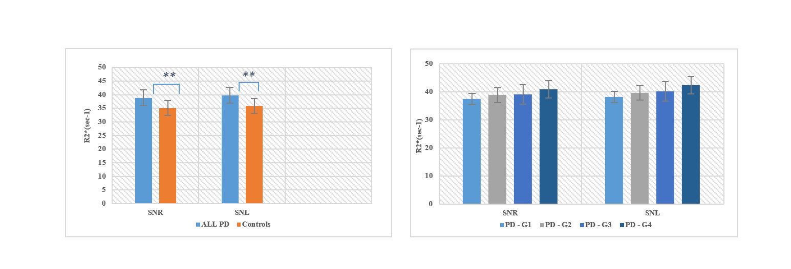
Figure 1. R2* (sec-1) in each region
of interest ; in PD patients and controls and between PD sub-groups (based
on disease duration ; disease (PD - G1 <5 years; PD - G2, between 5 and
10 years; PD - G3, between 10 and 15 years and PD - G4> 15 years). The p-values
were calculated using two samples t-tests. ***p≤0.0001 **p≤0.005, *p≤0.05.
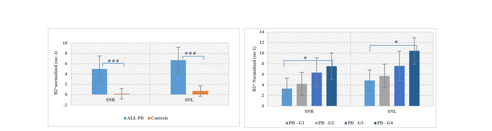
Figure 2. R2*
normalized (sec-1); in PD patients and controls and between PD sub-groups (based
on disease duration ; disease (PD - G1 <5 years; PD - G2, between 5 and
10 years; PD - G3, between 10 and 15 years and PD - G4> 15 years). The p-values
were calculated using two samples t-tests. ***p≤0.0001 **p≤0.005, *p≤0.05.