0540
Characterizing white matter microstructural alterations in de novo Parkinson's disease using diffusion MRI1PERFORM Centre, Concordia University, Montreal, QC, Canada, 2Computer Science and Software Engineering, Concordia University, Montreal, QC, Canada, 3Robarts Research Institute, Western University, London, ON, Canada, 4Department of Medical Biophysics, Western University, London, ON, Canada, 5School of Biomedical Engineering, Western University, London, ON, Canada, 6The Brain and Mind Institute, Western University, Lonon, ON, Canada
Synopsis
Understanding microstructural alterations in white matter can be instrumental in revealing pathogenesis and devising effective plans to treat Parkinson’s disease (PD). Diffusion MRI can be used to characterize the status of white matter integrity. With voxel-based analysis using DTI measures and fixel-based analysis (FBA), we demonstrated for the first time strengthened and weakened white matter integrity, which is subject to laterality of motor symptoms in de novo Parkinson’s disease. The findings suggest that the disease gives rise to both functional degeneration and the creation of compensatory networks.
Introduction
Parkinson’s disease (PD) is a progressive neurodegenerative disorder characterized by the loss of dopaminergic neurons in the midbrain. The disease primarily affects the patient’s motor functions, but as a result of alterations in brain connectivity, can also induce a range of non-motor symptoms. Knowledge regarding the disease-related changes in white matter pathways can effectively help improve the understanding of the disease and its treatment strategy. Microstructural imaging techniques, including diffusion tensor imaging (DTI), allows inspection of white matter integrity to study the progression of neurological conditions. Previous voxel-based analyses with DTI measures, such as fractional anisotropy (FA) and mean diffusivity (MD) have demonstrated regions that are correlated with PD, but the results have been inconsistent [1], partially due to smaller patient cohorts. Using diffusion MRI, fixel-based analysis (FBA) [2] is a relatively recent technique that offers tract-specific insights regarding white matter integrity. So far, very few studies have investigated whiter matter changes related to PD using FBA, as well as the comparison between DTI measures and FBA metrics in a large PD cohort. We present a study that reveals alterations in white matter tissues that are subject to symptom laterality in a large de novo PD cohort using both DTI and FBA metrics.Methods
In total, 144 de novo PD patients (age = 61±9y, 52 females) and 62 healthy subjects (age = 61±10y, 22 females) were collected from the Parkinson’s Progression Markers Initiative (PPMI) database (www.ppmi-info.org/data) in preparation of this article. For up-to-date information on the study, visit www.ppmi-info.org. All patients were in early stages of PD (H&Y Scale =1~2). Except for three patients, the motor functions of the PD cohort were affected more severely unilaterally (62 on the left). Multi-centre 3T brain MRI data were acquired for the collected subjects, including T1w MRI (1x1x1mm3), T2w MRI (1x1x2mm3), and DWI scans (64 directions, b=1000 s/mm2). Structural images were pre-processed with non-uniformity correction and skull stripping. Image distortion of DWI scans was corrected using nonlinear registration between B0 and T2w images. DTI metrics, such as fractional anisotropy and mean diffusivity were derived from the preprocessed DWI scans. Fixel measures, including fiber-cross-section (FC), fiber density (FD), and a joint measure of fiber density and cross-section (FDC) were obtained for all subjects using the single-shell 3-Tissue CST method in MRtrix3. For the analysis, scans of PD patients that were more affected on the left side in their motor functions were mirrored to the right side, while the scans were kept the same for the healthy subjects and patients who experience motor malfunction equally bilaterally. Statistical analysis was performed to compare PD and healthy subjects for the DTI and fixel-based measures. For the study, the subjects were nonlinearly registered to a common template space using fixel-based group-wise registration at 1.3x1.3x1.3mm3 resolution, and the effects of age and sex were controlled for all statistical tests. Family-wise error correction was used to reveal regions and fiber tracts with statistical significance (p<0.05).Results
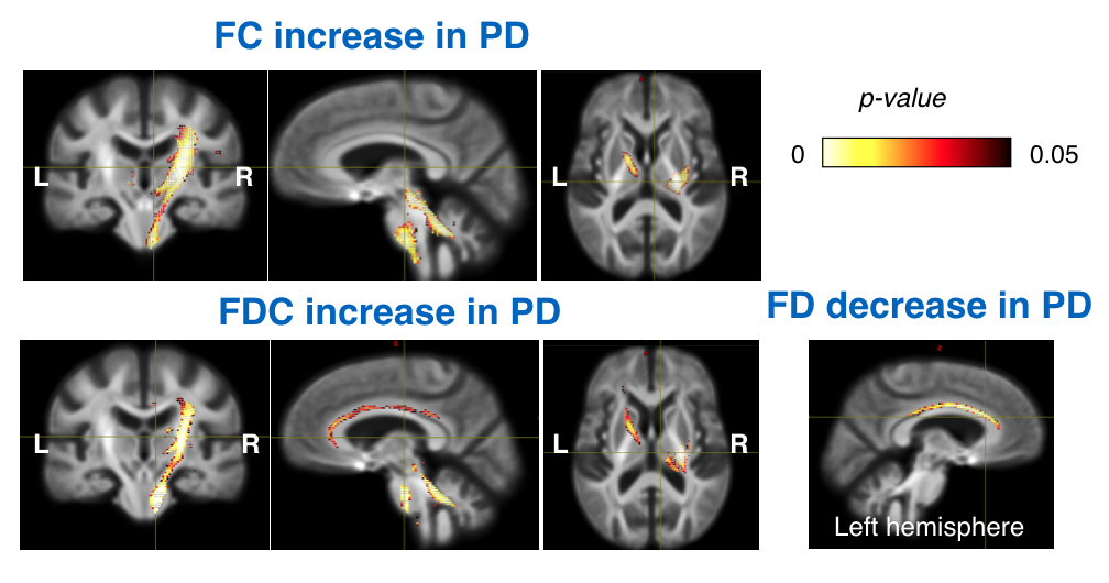
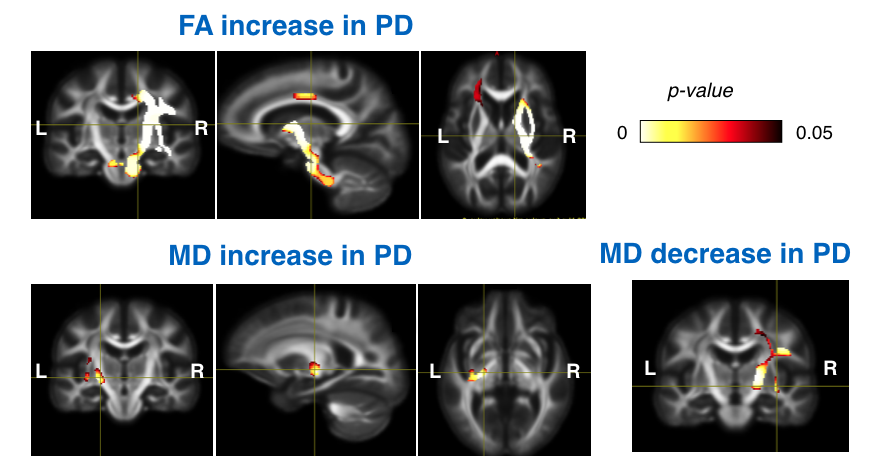
The results of FBA and DTI-metric comparison are shown in Figs. 1 & 2, respectively. From FBA, in the PD population, we found increased FC and FDC in the posterior limb of internal capsule, cingulum, and the dentatorubrothalamic (DRT) tract in the right hemisphere. In addition, FD in the left cingulum was significantly lower for PD patients. In terms of DTI measures, compared to healthy subjects, PD patients had widespread FA increase in the right internal capsule, external capsule, superior corona radiata, cingulum, and the middle cerebellar peduncle. Reduction of MD in the patients also occurred in the right internal capsule and superior corona radiata. On the other hand, MD increase in PD could be found in small regions within the left posterior limb of internal capsule and external capsule.Discussion
The higher FC and FDC in the white matter tracts signify greater white matter integrity, implying that the brain is performing a contralateral compensation for the motor function deterioration. The same trend is also replicated in the elevated FA and decreased MD in the same hemisphere. Degeneration of white matter is shown in both FBA and DTI metrics in the hemisphere that corresponds to more severe motor symptoms, but the regions between the analyses don’t overlap with each other, since FBA emphasizes on tract-specific measures and DTI metrics focus on volumetric regions. As the internal capsule and DRT play important roles in motor control, the discovered patterns can potentially be important in devising effective treatment plans in early stages of the disease. This study aligns the hemisphere with more severe motor symptoms for the patients since we hypothesize that early PD affects the hemispheres differently, as confirmed by the results.Conclusion
Using DTI measures and FBA, we have demonstrated patterns of white matter alterations in a large de novo PD cohort. The findings suggest both functional degeneration and compensation of the brain in the early stage of disease, with the impacts being subject to the laterality of the motor symptoms. Future longitudinal studies will help fully characterize the trajectory of disease-related white matter changes as a potential biomarker for improved prognosis and treatments.Acknowledgements
This work was supported by CIHR, NSERC, the Canada First Research Excellence Fund.
PPMI—a public-private partnership—is funded by the Michael J. Fox Foundation for Parkinson's Research and funding partners, including AbbVie, Avid, Biogen, Bristol-Myers Squibb, Covance, GE Healthcare, Genentech, GlaxoSmithKline, Lilly, Lundbeck, Merck, Meso Scale Discovery, Pfizer, Piramal, Roche, Sanofi Genzyme, Servier, Teva, and UCB.
References
1. Atkinon-Clement et al., “Diffusion tensor imaging in Parkinson’s disease: Review and meta-analysis,” Neuroimage Clin, vol 16, pp 98-110, 2017.
2. Raffelt et al., “Investigating white matter fibre density and morphology using fixel-based analysis,” Neuroimage, vol 144, pp 58-73, 2017.
Figures