0519
Predicting delayed union in osteoporotic vertebral fractures in the acute phase with intravoxel incoherent motion1Division of Radiology and Nuclear Medicine, Sapporo Medical University Hospital, Sapporo, Japan, 2Department of Orthopaedic Surgery, Sapporo Medical University School of Medicine, Sapporo, Japan, 3Department of Orthopaedic Surgery, Sapporo Maruyama Orthopedic Hospital, Sapporo, Japan
Synopsis
Previous studies have reported that the disorder of intramedullary perfusion in the vertebral fracture (VF) delays the bone union process. However, few reports evaluating VF using intravoxel incoherent motion (IVIM) exist. The IVIM parameters between favorable and unfavorable VF prognosis were compared, and we investigated whether possible to evaluate for the VF prognosis. ADC, D, D*, and f in VF as IVIM parameters were measured, and the IVIM parameters between the favorable and unfavorable prognosis groups was compared. The IVIM parameters were significantly different between groups. Therefore, it is concluded that the IVIM analysis enables the prediction of VF prognosis.
Background and Purpose
Vertebral fractures (VF) is one of the most frequent and serious osteoporotic fractures. Osteoporotic VFs (OVF) have been managed conservatively with rest or corset use, and the outcomes have generally been favorable. However, recent studies have revealed that some patients develop nonunion or collapse of the vertebral body, leading to unfavorable outcomes, with residual pain, decreased activities of daily living, and decreased quality of life.1 Performing therapeutic interventions such as vertebroplasty and surgical treatment from an early stage will be possible if the occurrence of such nonunion or collapse can be predicted at the initial treatment stage. Some reports exist that the blood flow in the VF part was evaluated by using a contrast medium because the intramedullary perfusion disorder delays the bone union process.2 Meanwhile, the intravoxel incoherent motion (IVIM) has been demonstrated to be an attractive approach for assessing tissue water diffusivity and microcapillary perfusion.3 However, few reports evaluating OVF using IVIM are available, and it is unclear whether IVIM parameters enables the prediction of VF prognosis. This study aimed to compare IVIM parameters between favorable and unfavorable VF prognosis.Materials and Methods
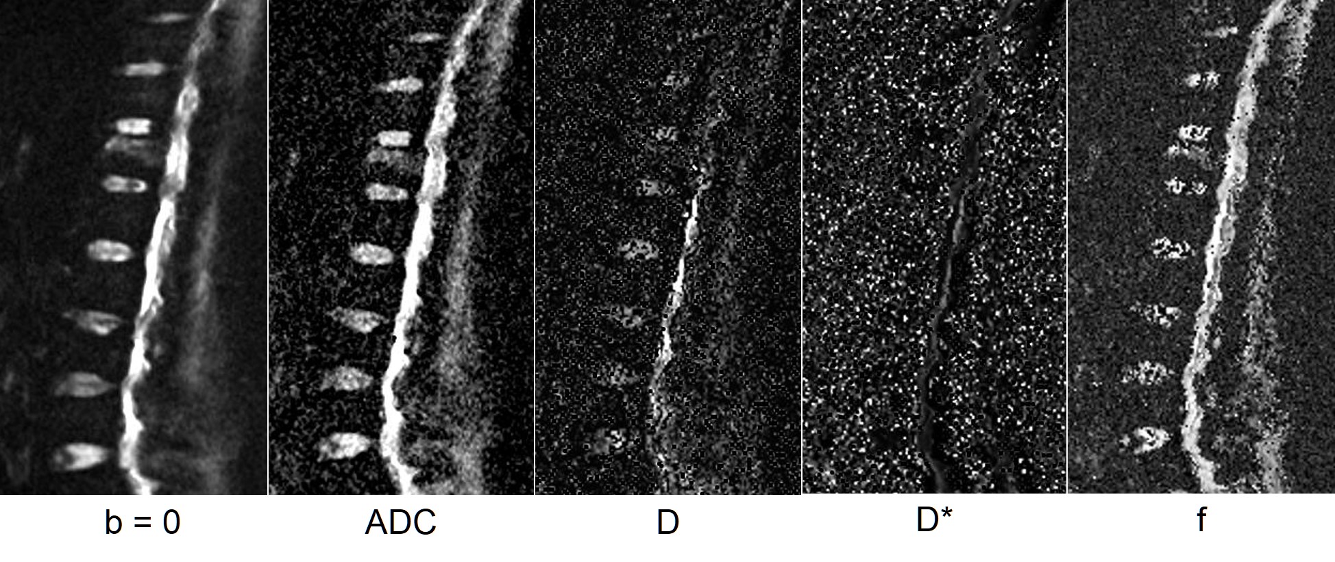
This study enrolled 30 patients who showed high-signal change of VF on STIR or T2-weighted sagittal images. In addition, plain X-rays of the thoracolumbar were performed at the time of enrollment and 3-month follow-up. VFs indicating cleft sign or collapse were defined as VF >25% on plain X-rays as the unfavorable prognosis group. Moreover, magnetic resonance imaging was performed using a GE Signa Creator 1.5T (GE Healthcare, Wauwatosa, WI, USA). ADC, D, D*, and f map were created from IVIM (TR, 3,300 ms; TE, 85 ms; b value = 0, 10, 20, 30, 50, 80, 120, 200, 300, 500, and 800 s/mm2) at the time of enrollment (Fig. 1). ADC, D, D*, and f in VF were measured and the favorable and unfavorable prognosis groups were compared.Results
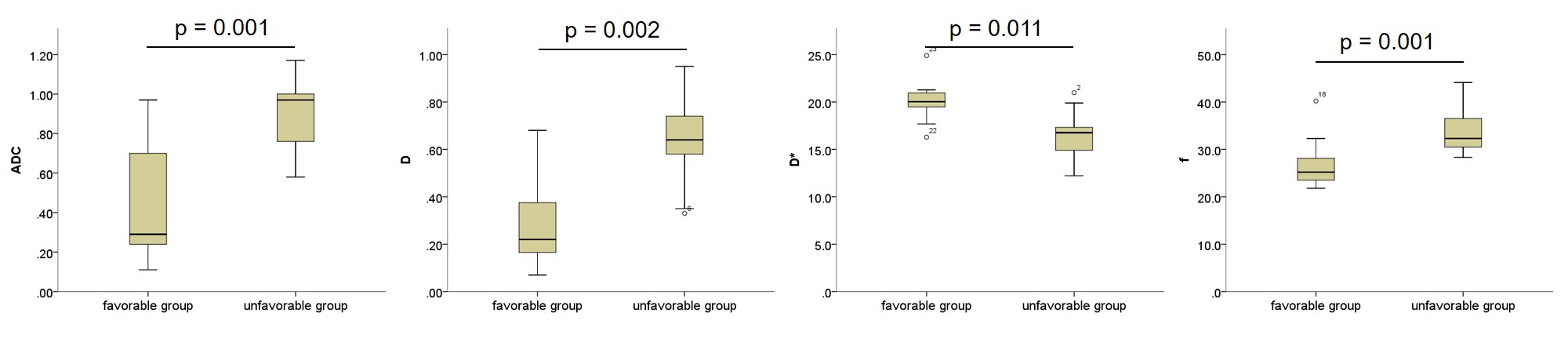
This study had 14 favorable (11 females and 3 males) and 16 unfavorable prognosis group (13 females and 3 males). The ADC of the favorable and unfavorable groups were 0.45 ± 0.32 and 0.89 ± 0.18, respectively. Similarly, D, D*, and f were 0.29 ± 0.20 and 0.63 ± 0.18, 20.1 ± 2.17 and 16.5 ± 2.32, and 27.0 ± 5.37 and 33.7 ± 4.32, respectively (Fig. 2). All IVIM parameters were significantly different (p < 0.05).Conclusion
IVIM analysis enables the prediction of VF prognosis.Acknowledgements
I am grateful to MR room staff, especially Mitsuhiro Nakanishi and Nobuyasu Yoshinaka for useful discussions and their help regarding patient treatment. This study is supported by grant of Japan osteoporosis foundation.References
1. Hoshino M, Nakamura H, Terai H. et al. Factors affecting neurological deficits and intractable back pain in patients with insufficient bone union following osteoporotic vertebral fracture. Eur Spine J. 2009; 18 (9): 1279-1286.
2. Kanchiku T, Taguchi T, Toyoda K. et al. Dynamic contrast-enhanced magnetic resonance imaging of osteoporotic vertebral fracture. Spine (Phila Pa 1976). 2003; 28 (22): 2522-2526.
3. Le Bihan D. Intravoxel incoherent motion perfusion MR imaging: a wake-up call. Radiology. 2008; 249 (3); 748-752.
Figures