0515
A rapid in vivo method for mapping cortical connections of primate amygdala with infrared neural stimulation and 7T fMRI1Interdisciplinary Institute of Neuroscience and Technology, Zhejiang University, Hangzhou, China, 2Dept of Neuroscience, University of Rochester School of Medicine, Rochester, NY, United States, 3University of Arizona, Dept of Physiology, Tucson, AZ, United States
Synopsis
We have previously shown that INS-fMRI is a rapid method for mapping mesoscale brain networks in the macaque monkey brain. Here, we extend this capability by stimulating deep brain sites. We test this new method by stimulating the basal nucleus of amygdala in the macaque monkey. The connections we identified are consistent with dye-tracing studies. In conclusion, our results indicate that INS-fMRI is a promising novel method for mapping connections of deep brain structures at high spatial resolution.
Introduction
One recent development in Non-Human Primate (NHP) MRI technology is a method for studying brain connections termed INS-fMRI (Xu et al. 2019). Infrared neural stimulation (INS), delivered via optic fibers, induces neuronal firing via optically induced heat transients (Shapiro et al. 2012). When INS is used to stimulate focal (submillimeter) sites on surface cortex, connected sites are activated and can be imaged using BOLD functional MRI in ultrahigh field; this results in a map of a brain-wide network connected to a single site.Here, we extend this capability by stimulating deep brain sites. To archive precise targeting, we combine MR imaging techniques with grid implantation and a fiber pushing system. We test this new method by stimulating the basal nucleus of amygdala in the macaque monkey, because it is considered one of the most interconnected subcortical hubs of the primate brain (Bickart, Dickerson, and Barrett 2014; Tomasi and Volkow 2011). The basal nucleus of the amygdala is a region characterized by a diversity of neuronal responses and exhibits strong connections with major limbic as well as sensory association cortical areas (Barbas 2007; Pitkänen and Amaral 1991). We demonstrate that this deep brain stimulation with INS-fMRI can reveal known cortical connections of the basal nucleus of the amygdala.
Methods
The experiments were conducted in 2 rhesus monkeys. Animals were anesthetized with sufentanil and isoflurane. A guiding grid filled with gadolinium-based contrast agent was installed on the monkey skull and imaged in the scanner. After MR imaging, one of the grid holes was chosen for craniotomy of a 1-mm burr hole in skull. An optical fiber of diameter 200 micrometer was then inserted into brain through this burr hole and reached the target area with a syringe-based pushing system. Laser stimulation was delivered to the fiber tip. Stimuli consisted of a train of laser pulses (wavelength 1875nm, 250µs) at 200Hz for 0.5 seconds. Radiant exposures were from 0.1 to 1.0 J/cm2 per pulse.Data was acquired in a 7-Tesla MR scanner (Siemens, Erlangen, Germany) with an 18-cm transmit coil and a customized 6-channel receive coil with a 6-7 cm diameter. We obtained functional images with a single-shot echo planar imaging (EPI) sequence (TE 25 ms; TR 2000 ms; matrix size 86 × 72; flip angle 90°). The raw images were preprocessed with slice-timing and motion correction. The preprocessed data were then used for identifying significant voxels. The time-courses of all voxels were fitted with the expected time-course given stimulus onsets and canonical hemodynamic response in generalized linear model (GLM). Voxels with significant T-test p-values (under Benjamini-Hochberg FDR control) in GLM were considered as voxels with significant response to INS.
Results
We found that our method can precisely reach target sites. Since the optical fiber appeared completely dark on T1 images. We could determine the fiber tip with an error as small as the size of a single voxel. In our case, it was 0.3 mm.With this method, we stimulated the left amygdala of monkey M and right amygdala of monkey Y. In the example on Figure 1A and B, the fiber tip was identified in the basal nucleus of amygdala. The stimulation of this site resulted in the activation of a distinct cluster of voxels (E-H: p<0.0001, FDR 5.5%) in the insula (representative schematics of insula shown in 1C and D), as well as small patches of cortex in the surrounding lateral sulcal regions. Figures 1E-H show the pattern of activation in four different coronal sections along the anterior-posterior axis of the brain. The activated voxels included the retroinsular region and the granular and dysgranular insula (Id) (1E through H), auditory areas CM, RM and R, as well as patches of the secondary somatosensory cortex SII.
We then examined the timecourses of the BOLD activations at these presumed connected sites. The average time course of the fMRI signal in these voxels (shown in Figs 5I-L) showed increased fMRI signals at the expected latency after the stimulus triplet (vertical red lines) and with a magnitude consistent with the BOLD response. We also show that an event related stimulation paradigm could achieve similar results (the right amygdala in monkey Y, Figure 1 M-P). This also resulted in ipsilateral activation sites in the insula and in somatosensory cortex.
Discussion and Conclusion
Our results show that we can precisely stimulate basal nucleus of the amygdala. The connected sites identified are consistent with known connections from dye-tracing studies. Adding INS-fMRI to the existing connection mapping methods has several advantages. The primary advantage of this method is the focality of the energy delivery. Compared to other methods such as electrical stimulation used in conjunction with MRI, the INS stimulus is relatively small and focal.In conclusion, our results indicate that INS-fMRI is a promising novel method for mapping connections of deep brain structures at high spatial resolution. When used systematically on large scale, it will lead to a columnar connectome. Furthermore, the ability to stimulate in deep tissue offers a potentially new approach for therapeutic efforts.
Acknowledgements
This work was supported by: National Key R&D Program of China 2018YFA0701400 (to A.W.R.), Chinese NSF Instrumentation Grant No. 31627802 (A.W.R.), Key Research and Development Program of Zhejiang Province 2020C03004 (to A.W.R.), Fundamental Research Funds for the Central Universities 2019XZZX003-20 (to A.W.R.), and China-US grant (NSFC 8191101288 to A.W.R., 1R01MH121706 to K.M.G.). We acknowledge the following for their roles in experimental data collection: Gang Chen, Hsin-yi Lai, Meizhen Qian, Shuxian Qu, Zhiyan Quan, Toru Takahata, Jianbao Wang, Pingyi Wang, Wang Xi, Bin Xu, Songping Yao, Hong Yin, Jichao Yu, Xiongjie Yu, Yuying Zhai, Xiaotong Zhang, Ying Zhang, Dengfeng Zhou, Liang Zhu.References
Barbas, Helen. 2007. 'Flow of information for emotions through temporal and orbitofrontal pathways', Journal of anatomy, 211: 237-49.
Bickart, Kevin C, Bradford C Dickerson, and Lisa Feldman Barrett. 2014. 'The amygdala as a hub in brain networks that support social life', Neuropsychologia, 63: 235-48.
Pitkänen, A, and David G Amaral. 1991. 'Demonstration of projections from the lateral nucleus to the basal nucleus of the amygdala: a PHA-L study in the monkey', Experimental brain research, 83: 465-70.
Shapiro, Mikhail G, Kazuaki Homma, Sebastian Villarreal, Claus-Peter Richter, and Francisco Bezanilla. 2012. 'Infrared light excites cells by changing their electrical capacitance', Nature communications, 3: 1-11.
Tomasi, Dardo, and Nora D Volkow. 2011. 'Association between functional connectivity hubs and brain networks', Cerebral cortex, 21: 2003-13.
Xu, Augix Guohua, Meizhen Qian, Feiyan Tian, Bin Xu, Robert M Friedman, Jianbao Wang, Xuemei Song, Yi Sun, Mykyta M Chernov, and Jonathan M Cayce. 2019. 'Focal infrared neural stimulation with high-field functional MRI: A rapid way to map mesoscale brain connectomes', Science advances, 5: eaau7046.
Figures
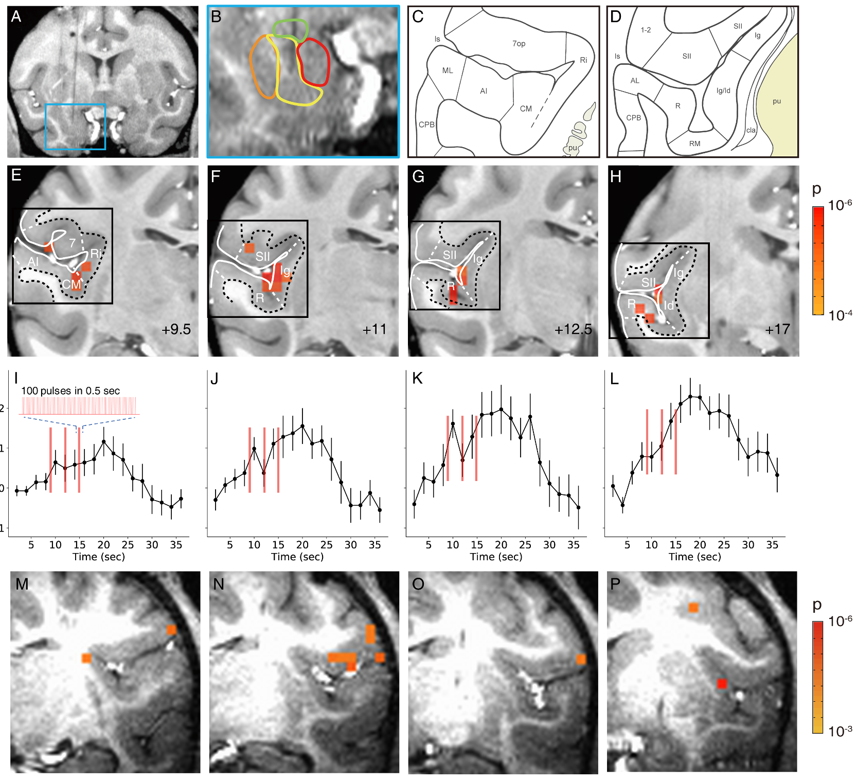
Figure 1. Activation in insula and lateral sulcal areas elicited by stimulation of the basal nucleus of the amygdala.
A-L. Monkey M-P. Monkey Y. A-B. Stimulation site in the basal nucleus. C-D. Schematics of cortical areas in the lateral sulcus. E-H. Activations within the lateral sulcus. Voxels: p<0.0001 (FDR 5.5%). I-L: Mean time courses of significant voxels in E-H (mean of significant voxels). Inset: enlarged schematic of each stimulus train (red line). Stimulation: block design. Error bars: SEM. M-P. Activations within the lateral sulcus are seen in monkey Y. Voxels: p<0.001.