0469
Regulating labeling efficiency in arterial spin labeling using a multi-coil B0 shim array: Application to territory mapping1Massachusetts General Hospital, Charlestown, MA, United States, 2Siemens Medical Solutions USA, Inc., Malvern, PA, United States, 3Qbio Inc, San Carlos, CA, United States, 4Dept. of Electrical Engineering, Massachusetts Institute of Technology, Cambridge, MA, United States, 5Harvard Medical School, Boston, MA, United States, 6Biomedical Engineering, University of Michigan, Ann Arbor, MI, United States, 7Siemens, S.A., Madrid, Spain, 8Neurology, University of Pennsylvania, Philadelphia, PA, United States, 9Radiology, University of Pennsylvania, Philadelphia, PA, United States, 10A.A. Martinos Center for Biomedical Imaging, Department of Radiology, Massachusetts General Hospital, Charlestown, MA, United States
Synopsis
We apply dynamic local B0 field control with a multi-coil (MC) shim array to improve ASL labeling. Labeling efficiency can be regulated by dynamically shimming the labeling plane during a pCASL tagging pulse train. We demonstrate this capability through territory mapping with an unspecialized MC head shim array – Through shimming, we shift target arteries relative to the labeling pulse bandwidth. Implementation takes less than 10 minutes during a scan, without previous subject information. We also simulate the improvement possible with a specialized MC neck shim array, which enables regulation in more inferior labeling planes and higher field control efficiency.
Introduction
Arterial spin labeling (ASL) MRI is a noninvasive technique for measuring cerebral blood flow (CBF). Pseudo-continuous ASL (pCASL) is the most widely used ASL implementation because it affords high labeling efficiency1,2. The label is achieved with a long train of short, Hanning-windowed block pulses. However, the labeling efficiency of pCASL depends on several subject-specific factors, including off-resonance of the B0 field3,4,5. The challenge of B0 off-resonance is exacerbated in pCASL because the scanner shim usually prioritizes the field homogeneity of the imaging volume in the brain over that of the labeling plane. Here we evaluate the addition of a secondary shim system focused on the field homogeneity local to the labeling plane to improve inversion efficiency and provide higher sensitivity and resilience to artifactual hypoperfusion in regions of low labeling efficiency. We also show that local control of the B0 field at the labeling plane allows selection of perfusion flow territories without modification to the pulse sequence. We control the B0 field at the labeling plane using a local multi-coil (MC) B0 shim array during the labeling step and demonstrate its effect on modulating pCASL labeling efficiency. We also obtain flow territory maps of CBF by specifically controlling the B0 fields near specific arteries using the MC B0 shim array. We further simulate the performance of a dedicated ASL MC shim array with 7-turn loops designed for improved field control efficiency and flexibility in ASL labeling region.Methods
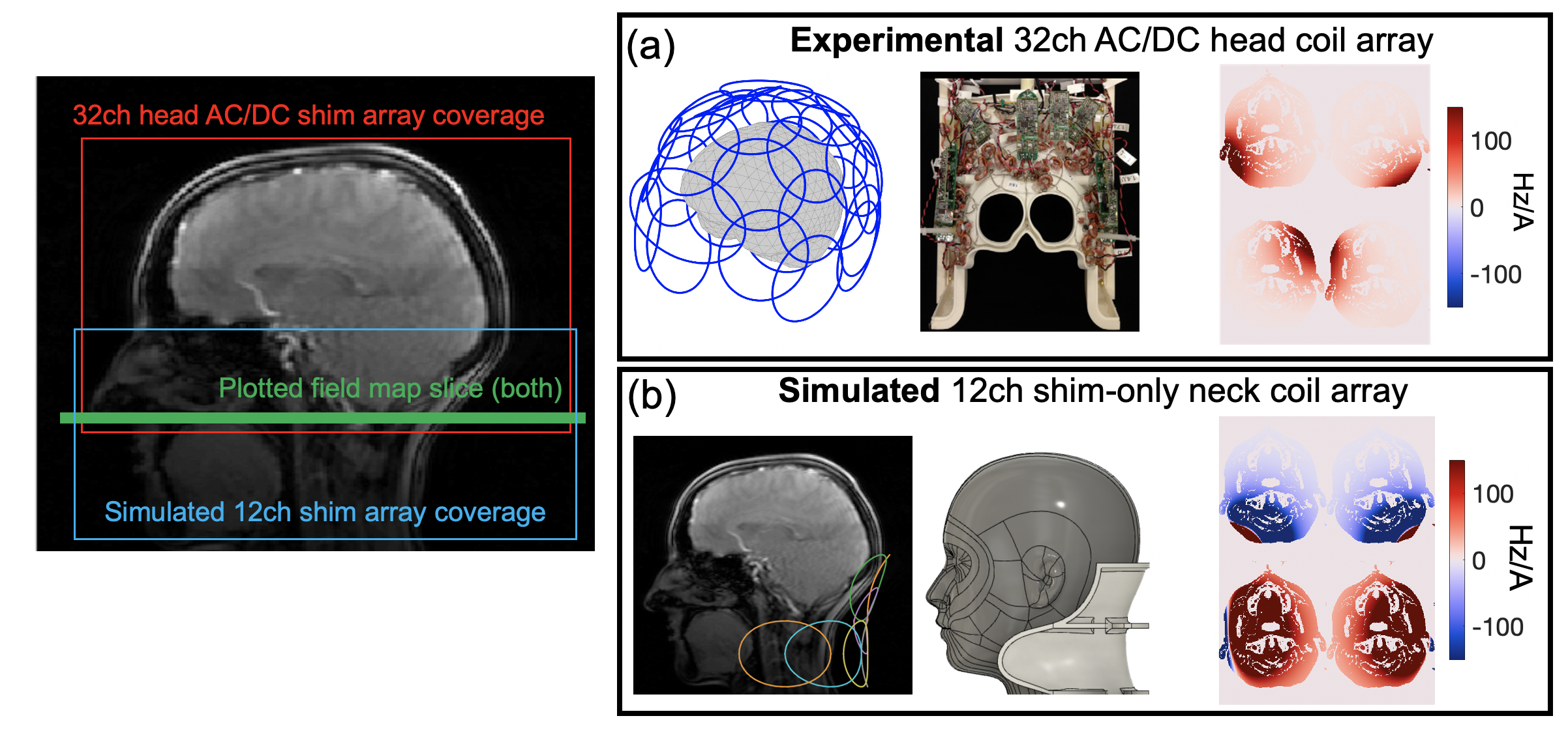
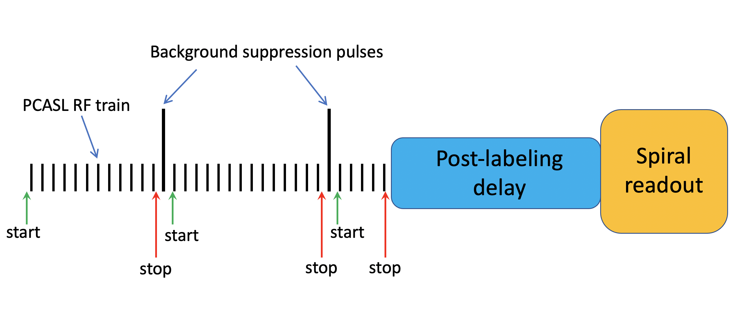
MC B0 shim arrays use an array of independently driven loops to generate nonlinear, rapidly-switchable B0 offsets in the body. While originally designed for improving B0 homogeneity to reduce artifacts in imaging and spectroscopy, we examine the extension of MC shim arrays to ASL (labeling efficiency, robustness, and territory mapping). In proof-of-concept experiments, we employed a 32-channel “AC/DC” array with single-turn loops used both for RF receive and for carrying DC currents6 (See Fig. 1b). pCASL experiments were performed on a MAGNETOM Prisma 3T MRI scanner (Siemens Healthcare, Erlangen) using the scanner body coil for transmit. Figure 1 shows a block diagram of the labeling, saturation, and spatial encoding blocks of the pCASL sequence, indicating the points in time when the MC shim field is switched on and off.The acquisition used a prototype pCASL sequence with 1.8s labeling duration, 1.8s post-labeling delay, 4-pulse background suppression, single-shot 3D stack-of-spirals readout with 2 spiral interleaves for each partition, spatial resolution of 3.75mm isotropic, TR/TE=5000/10.3 ms, 8 control-label pairs, and scan time of 1 min 44 sec7. Time of flight (TOF) images were acquired and segmented immediately prior to the ASL acquisition to obtain vessel masks for the two carotid and two vertebral arteries. For territory mapping, one carotid is marked as “desired” while the other three vessels are masked as “reject”.
Previous work has shown that pCASL labeling efficiency falls off rapidly for B0 shifts over ~100 Hz off-resonance3. To shift spins outside the pCASL “passband”, a convex objective function is used with a fast solver (Mosek, Copenhagen, Denmark) which uses the available degrees of freedom to shift the “reject” voxels while holding the “desired” voxels on-resonance8. The pipeline required 1 min 20 sec for TOF image acquisition, 2 minutes for data transfer and masking, and 10 seconds for the field solver.
Results
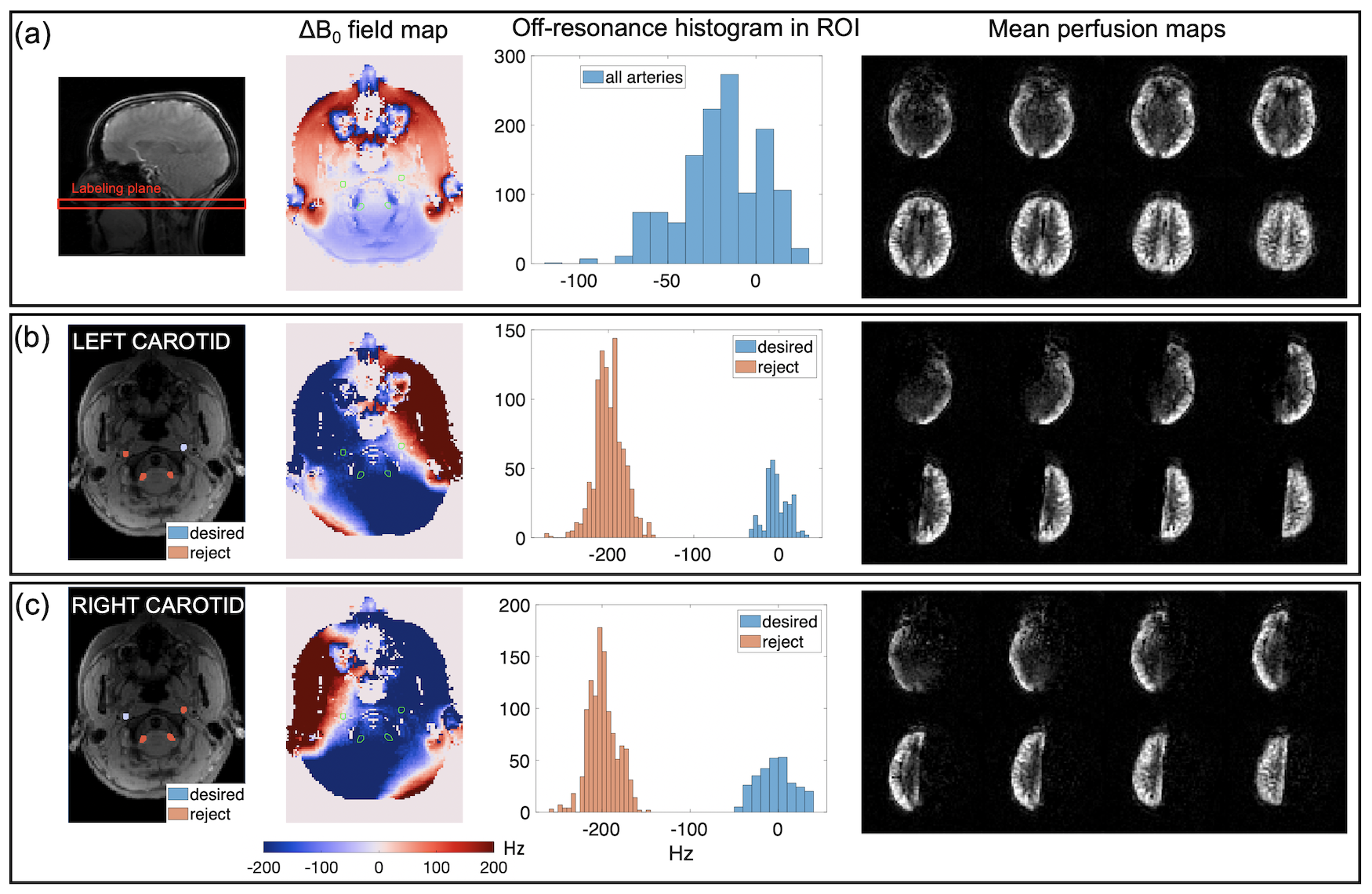
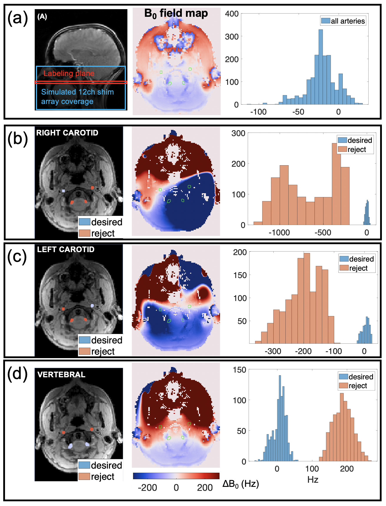
Figure 3 shows experimental territory mapping in vivo. The B0 field maps and histograms show spectral separation which shifts the “reject” arteries outside the pCASL passband.Figure 4 shows simulations of similar territory mapping methodology with the proposed 12-ch ASL shim array design.
Discussion
The 32-ch MC shim array provided sufficient B0 control to map the left and right anterior cerebral circulation but was unable to successfully target the vertebral arteries. By contrast, simulations suggest that the dedicated 12-ch ASL shim array, with its stronger fields at the level of labeling, should successfully separate all three target arteries. The 12-ch array will also allow for labeling at more inferior labeling planes as compared to the 32-ch brain array, which is advantageous for some applications5.In comparison to other pCASL flow territory mapping techniques that involve either dedicated RF labeling coils9, the use of RF phase modulation to acquire images with different labeling efficiency distributions10, or rotating in-plane gradients during labeling1,11, the current approach does not require modifications to the standard pCASL imaging sequence or advanced postprocessing strategies. Further, the ability to shim B0 homogeneity at the labeling plane is expected to improve the labeling efficiency of non-selective pCASL as well as the labeled arteries in selective pCASL. We note that these approaches can be implemented with any sufficiently rich basis set of B0 shim fields, allowing for implementations that synergize with other specialized coil arrays. Finally, this approach will have a particular impact for 7T pCASL, where off-resonance poses a severe obstacle to reproducible ASL4.
Acknowledgements
Funding support from NIH NIBIB U24EB028984 and R01EB028797.References
[1] Dai W, Garcia D, de Bazelaire C, Alsop DC. Continuous flow-driven inversion for arterial spin labeling using pulsed radio frequency and gradient fields. Magn Reson Med 2008;60:1488–1497
[2] Alsop DC, Detre JA, Golay X, et al. Recommended implementation of arterial spin-labeled perfusion MRI for clinical applications: a consensus of the ISMRM perfusion study group and the European consortium for ASL in dementia. Magn Reson Med. 2015;73:102-116
[3] Jahanian H, Noll DC, Hernandez-Garcia L. B0 field inhomogeneity considerations in pseudo-continuous arterial spin labeling (pCASL): effects on tagging efficiency and correction strategy. NMR Biomed 2011;24:1202–1209.
[4] Luh WM, Talagala SL, Li TQ, Bandettini PA. Pseudo-continuous arterial spin labeling at 7 T for human brain: estimation and correction for off-resonance effects using a Prescan. Magn Reson Med 2013;69:402–410.
[5] Zhao L, Vidorreta M, Soman S, Detre JA, Alsop DC. Improving the Robustness of Pseudo-Continuous Arterial Spin Labeling to Off-Resonance and Pulsatile Flow Velocity, Magn Reson Med 2017;78:1342-1351
[6] Stockmann JP, Witzel T, Keil B, Polimeni JR, Mareyam A, LaPierre C, Setsompop K, Wald LL. A 32-channel combined RF and B0 shim array for 3T brain imaging. Magn Reson Med. 2016 Jan;75(1):441-51
[7] Vidorreta M, Wang Z, Chang YV, Wolk DA, Fernandez-Seara MA, Detre JA. Whole-brain background-suppressed pCASL MRI with 1D-accelerated 3D RARE Stack-of-Spirals readout. PloS ONE, 2017;12:e0183762
[8] Arango N, Stockmann J, Strasser B, Gagoski B, Andronesi O, Wald L, White J, Adalsteinsson E. Dynamically switched B0 field control for dual optimization of tailored volume lipid suppression and B0 homogeneity for brain chemical shift imaging at 3T using local multi-coil-shim-array. In: Int. Soc. Magn. Res. Med. ; 2018, p.1062.
[9] Zaharchuk, G., Ledden, P.J., Kwong, K.K., Reese, T.G., Rosen, B.R. and Wald, L.L. (1999), Multislice perfusion and perfusion territory imaging in humans with separate label and image coils. Magn. Reson. Med., 41: 1093-1098. https://doi.org/10.1002/(SICI)1522-2594(199906)41:6<1093::AID-MRM4>3.0.CO;2-0
[10] van Osch, M. J., Teeuwisse, W. M., Chen, Z., Suzuki, Y., Helle, M., & Schmid, S. Advances in arterial spin labelling MRI methods for measuring perfusion and collateral flow. Journal of Cerebral Blood Flow & Metabolism, 38(9);2018:1461-1480.
[11] Helle, M., Norris, D.G., Rüfer, S., Alfke, K., Jansen, O. and van Osch, M.J.P. (2010), Superselective pseudocontinuous arterial spin labeling. Magn. Reson. Med., 64: 777-786. https://doi.org/10.1002/mrm.22451.
Figures