0461
Combined active and passive shimming of the temporal lobes using graphite-silicone earplugs and a multi-coil B0 shim array1A. A. Martinos Center for Biomedical Imaging, Massachusetts General Hospital, Charlestown, MA, United States, 2Dept. of Biomedical Engineering, Stony Brook University, Stony Brook, NY, United States, 3Asbury Carbons, Asbury, NJ, United States, 4Harvard Medical School, Boston, MA, United States, 5Dept. of Electrical Engineering, Stanford University, Stanford, CA, United States
Synopsis
Neuroimaging of the human brain temporal lobes has long been impeded by severe B0 inhomogeneity arising from air-tissue susceptibility interfaces around the ear canals and temporal bone. Here, we propose localized passive shimming using graphite-embedded silicone ear plugs, which replace standard ear plugs used during MRI. We further synergistically combine the passive shims with active multi-coil B0 shimming to boost shim performance. Our results show improvements in B0 homogeneity as assessed using field maps and signal loss and distortion in gradient-echo EPI slices.
Introduction
MR imaging and spectroscopy applications in the temporal lobes are limited by severe B0 inhomogeneity in this area. Focal inhomogeneity near the ear canals and air-filled temporal bone remain difficult to fully correct with sources placed outside the head [1] despite recent advances in higher-order spherical harmonic shimming [2] and multi-coil (MC) B0 shim arrays [3]. To clarify, the sources of the inhomogeneity reside inside the head, and Laplace’s equation no longer holds. Therefore, B0 perturbations cannot be readily nulled using currents outside the head. We hypothesize that a more effective remedy is to place a shim source inside the ear canal, near the focal inhomogeneity. Since actively-driven shim coils inside the ear canal present technical challenges, we opted for passive shimming using ear plugs made from diamagnetic materials. We complement the passive shims with active multi-coil B0 shimming using a 32-channel 3 Tesla “AC/DC” array with high-spatial order B0 shimming capability [4].Passive B0 shimming has previously been realized using graphite and/or bismuth placed near the face or inside the mouth to compensate inhomogeneity in the frontal lobes [5] and neck [6]; these authors further suggested introducing graphite foam plugs into the ear canal. We opted to mix pyrolytic graphite into a silicone putty, which readily molds itself to the ear canal and allows for a higher density of graphite.
Methods
We designed ear shims to replace conventional foam ear plugs used in MRI scans at our site (3M, St. Paul, MN). Moldable silicone putty (Mack’s silicone putty, McKeon Products, Warren, MI) was mixed with vein graphite. This design provided the required malleability while maintaining adequate sound attenuation. The vendor provided noise reduction rating for silicone plugs is 22 dB compared to 29 dB for foam plugs.Vein graphite is a naturally occurring form of “pyrolytic” graphite, which is deposited directly from a high temperature, carbonaceous geologic fluid. Pyrolytic graphite possesses the highest diamagnetic susceptibility of any readily available material due to its carbon benzene structure at the atomic scale [7]. Vein graphite samples used were prepared by mechanically sieving a commercial vein graphite product (Asbury Carbons, Asbury, NJ).
We kneaded graphite particles into silicon putty until a uniform consistency and desired malleability was achieved, which we found to be approximately 50% graphite flake by mass. We also tested the efficacy of adding spherical shim pieces to the ear lobe; a mixture of 35% silicon, 55% graphite, and 10% bismuth powder was used (100 mesh, 99.9% Bismuth, Sigma-Aldrich, St. Louis, MO). Malleability was a lesser concern outside of the ear canal, so we incorporated bismuth as additional diamagnetic material [8].
Temperature mapping was performed by placing ear shims on a phantom and running a pulse sequence at SAR and gradient PNS limits for 10 minutes each, acquiring infrared images before and after.
To assess shim performance, we acquired B0 field maps using a vendor-provided two-echo sequence (2mm iso.) on four healthy volunteers after standard 2nd-order spherical-harmonic shimming (2SH). We also acquired gradient-echo echo planar imaging (EPI) slices with both anterior-posterior (AP) and posterior-anterior (PA) phase encode directions to better assess distortion impact (2 mm iso., echo-spacing=0.76ms). We acquired EPI slices with dynamic, slice-optimized multi-coil active shimming, with and without ear shims.
Results
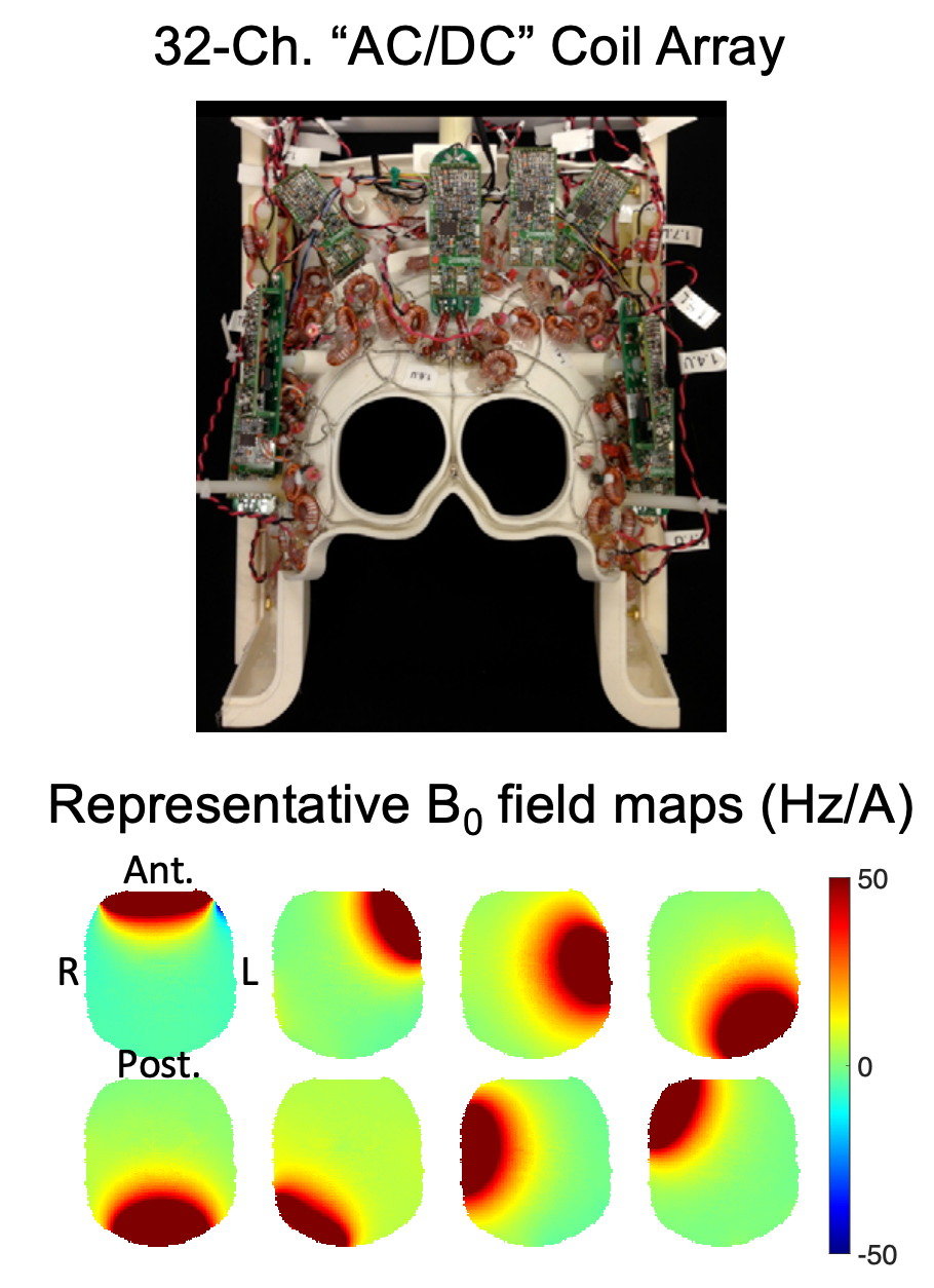
Figure 1 shows the graphite ear shim and lobe shim as worn by a subject. Subjects reported comparable comfort using the silicone ear plugs as compared to conventional ear plugs. Infrared images revealed no significant change in temperature during scanning.Figure 2 shows the 32-ch AC/DC multi-coil shim array used to perform active shimming and representative B0field maps at the level of the temporal lobes.
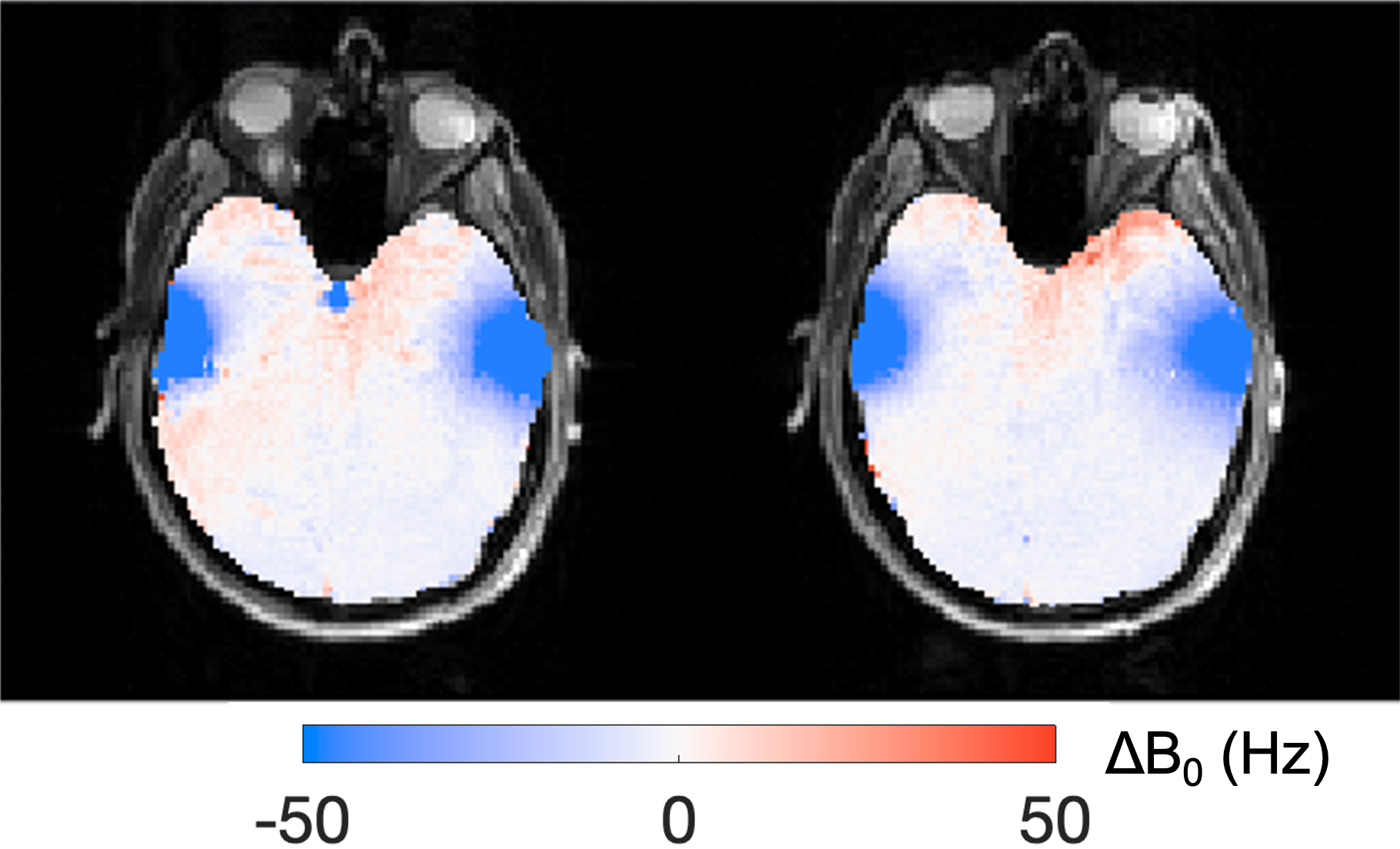
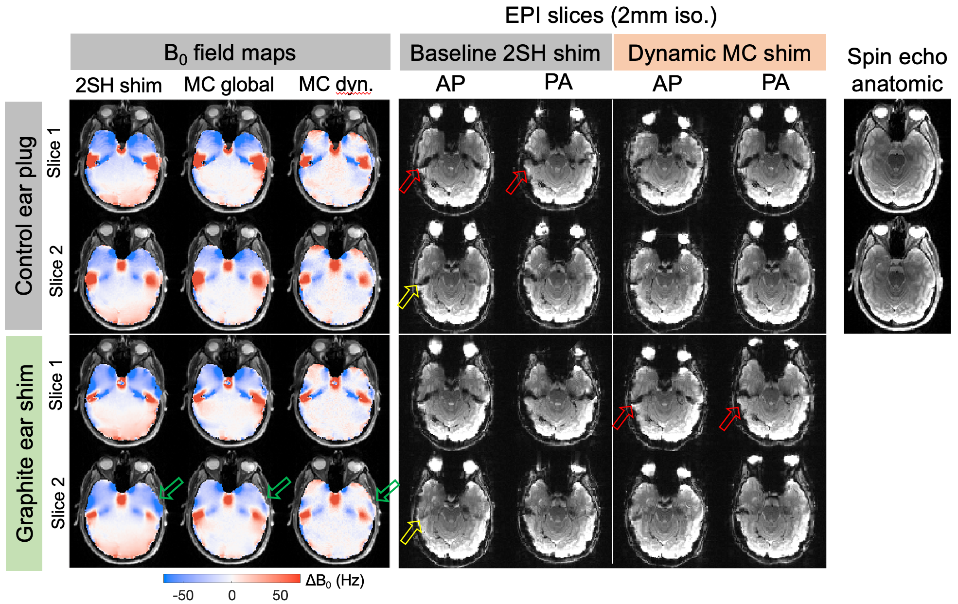
The difference B0 field map in figure 3 shows the highly-focal nature of the B0 field offset generated by the ear shim in the temporal lobes. Figure 4 shows improvements in B0 field homogeneity in one subject provided by the ear shims, active MC shims, and the combined approach. EPI slices show both reductions in geometric distortion as well as modest signal recovery.
We found that some adaptation of the ear shim size to subject-specific anatomy provided better B0 shim performance. The final ear shim plugs measured ~3cm length and had total weight of 2.6g for males and 2.0g for females. Lobe shims measured ~.75 cm3.
Discussion
We show a safe and non-toxic ear shim that replaces standard MRI ear plugs. Passive ear shims provide greater improvements in temporal lobe field homogeneity than those achieved by MC shimming alone, since it does not have high enough spatial-order fields to null the focal spot above the ear canals. These spots show significant improvement when ear shims are used either alone or in conjunction with active shimming.In some regions, the ear shim overcompensates the baseline B0 offset; however, this is largely nulled by global or dynamic MC shimming, further demonstrating the synergy between the active and passive shims.
Differences in ear shim performance across subjects may be attributed to variations in ear canal geometry between individuals [9]. In future iterations, we will use segmented structural images to tailor the shim design and optimize net diamagnetic moment for nulling the average off-resonance pattern.
Acknowledgements
Funding support from NIH NIBIB R01EB028797References
1. de Graaf, R.A. and C. Juchem, CHAPTER 4 B0 Shimming Technology, in Magnetic Resonance Technology: Hardware and System Component Design. 2016, The Royal Society of Chemistry. p. 166-207.
2. Kim, T., et al., Gradient-echo EPI using a high-degree shim insert coil at 7 T: Implications for BOLD fMRI. Magnetic Resonance in Medicine, 2017. 78(5): p. 1734-1745.
3. Juchem, C., et al., Dynamic multi-coil shimming of the human brain at 7 T. J Magn Reson, 2011. 212(2): p. 280-8.
4. Stockmann, J.P., et al., A 32-channel combined RF and B0 shim array for 3T brain imaging. Magn Reson Med, 2016. 75(1): p. 441-51.
5. Wilson, J.L., M. Jenkinson, and P. Jezzard, Optimization of static field homogeneity in human brain using diamagnetic passive shims. Magn Reson Med, 2002. 48(5): p. 906-14.
6. Lee, G.C., et al., Pyrolytic graphite foam: a passive magnetic susceptibility matching material. J Magn Reson Imaging, 2010. 32(3): p. 684-91.
7. Fischbach, D.B., Diamagnetic Susceptibility of Pyrolytic Graphite. Physical Review, 1961. 123(5): p. 1613-1614.
8. Koch, K.M., et al., Sample-specific diamagnetic and paramagnetic passive shimming. J Magn Reson, 2006. 182(1): p. 66-74.
9. Stinson, M.R. and B.W. Lawton, Specification of the geometry of the human ear canal for the prediction of sound‐pressure level distribution. The Journal of the Acoustical Society of America, 1989. 85(6): p. 2492-2503.
Figures
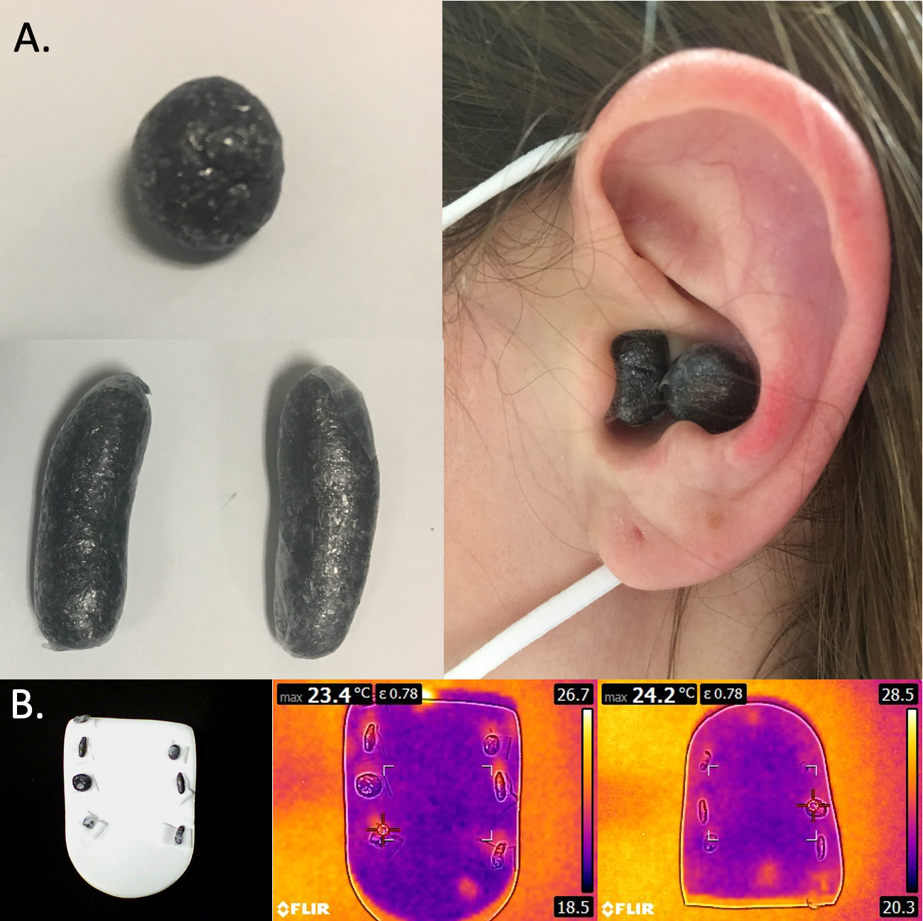
Figure 1. Panel A: Diamagnetic pyrolytic graphite was blended into a silicon putty to create a passive earplug shim. Spherical pieces were placed in the lobe adjacent to the elongated shim, which was inserted into the ear canal.
Panel B: Thermal images were obtained before and after scanning ear shim material in a receive array head coil. We conducted imaging of the shims at baseline and after running a turbo spin echo at 99% of the SAR limit for 10 minutes and gradient slew test with maximum slew rate EPI for 10 minutes. No significant change in temperature was observed.