0458
Extreme Looping Star: Quiet fMRI at high spatiotemporal resolution1Department of Radiology and Biomedical Imaging, University of California San Francisco, San Francisco, CA, United States, 2UC Berkeley - UC San Francisco Joint Graduate Program in Bioengineering, Berkeley and San Francisco, CA, United States, 3King's College London, London, United Kingdom, 4GE Healthcare, Munich, Germany, 5GE Healthcare, Paris, France, 6GE Healthcare, Menlo Park, CA, United States
Synopsis
The Looping Star pulse sequence was recently introduced as an acoustically silent alternative to EPI-based fMRI pulse sequences. In this abstract, we present improvements to the spatiotemporal resolution of Looping Star using the “extreme MRI” approach, without sacrificing functional sensitivity. We demonstrate the application of silent fMRI with increased temporal resolution and increased spatial resolution using extreme Looping Star, in comparison with standard Looping Star, on a motor task and a visual task across two sites.
Introduction
Recently, we presented Looping Star as a new method for 3D quiet multi-gradient echo structural and functional BOLD1,2 imaging . Here we present modifications of Looping Star to boost spatiotemporal resolution, which we call “Extreme Looping Star”. Pulse sequence optimizations resolves echo-in and echo-out overlap by separating the RF excitations in time. To exploit spatiotemporal image sparsity, we randomized the 3D center-out radial sampling and used Extreme MRI reconstruction based on multi-scale low-rank decomposition3. For high resolution Looping Star the data can be flexibly reconstructed to provide both 1) dynamic BOLD information and 2) high-spatial resolution images for optional concurrent anatomical referencing.New Looping Star
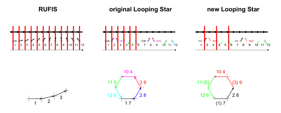
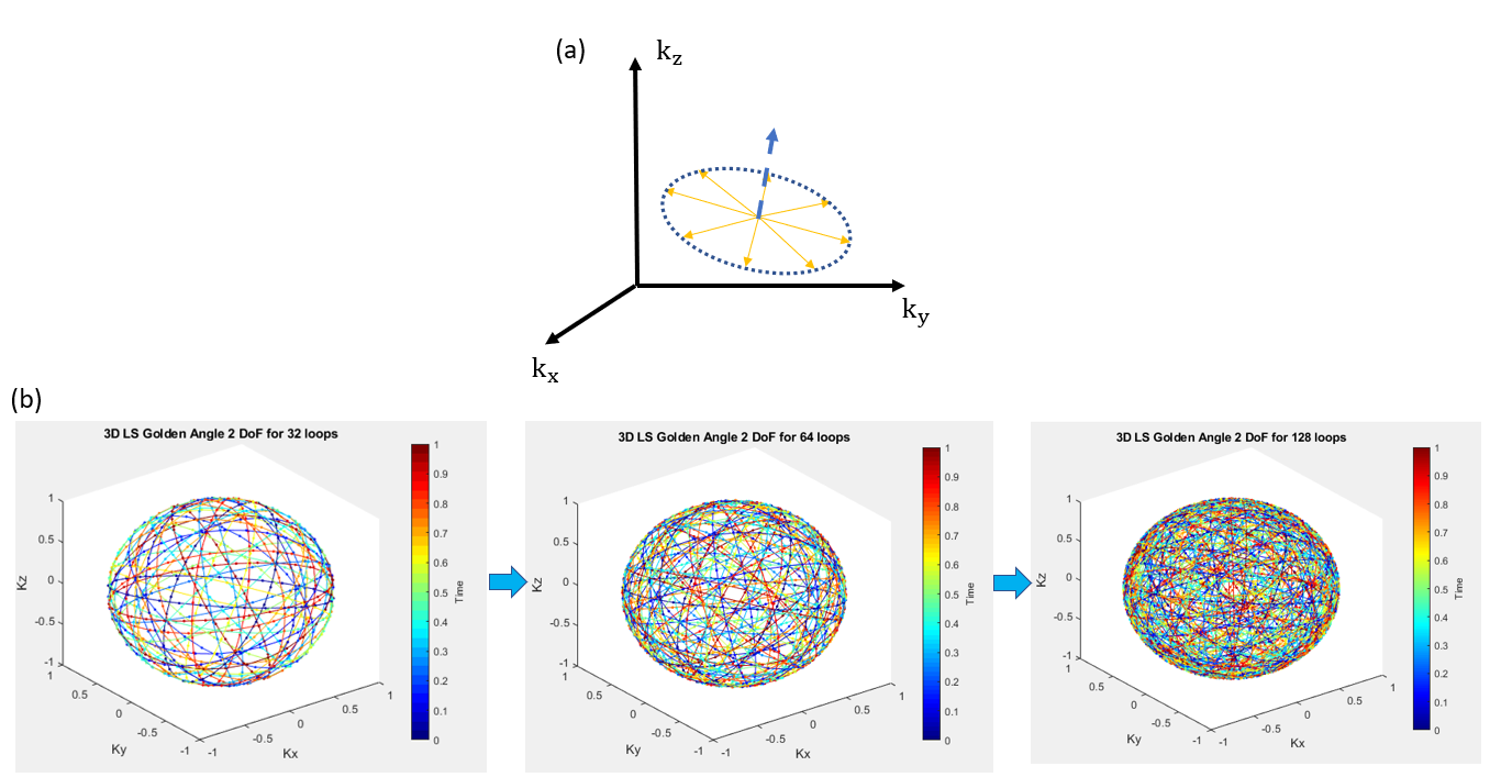
The original Looping Star approach is described in detail at 1. Rather than exciting at every spoke, the new Looping Star Scheme (Fig. 1) addresses the echo in/out overlap problem by exciting only every second spoke during the excitation phase.For 3D spatial encoding, the loops are rotated according to the 3D golden-angle radial trajectory ordering4 to achieve uniform k-space coverage and incoherent spatiotemporal sampling (Fig. 2). Rather than rotating each spoke, the normal vector of a group of coplanar spokes (a loop) are distributed in a golden angle fashion.
Extreme Looping Star with Modified Multi-scale Low Rank Model
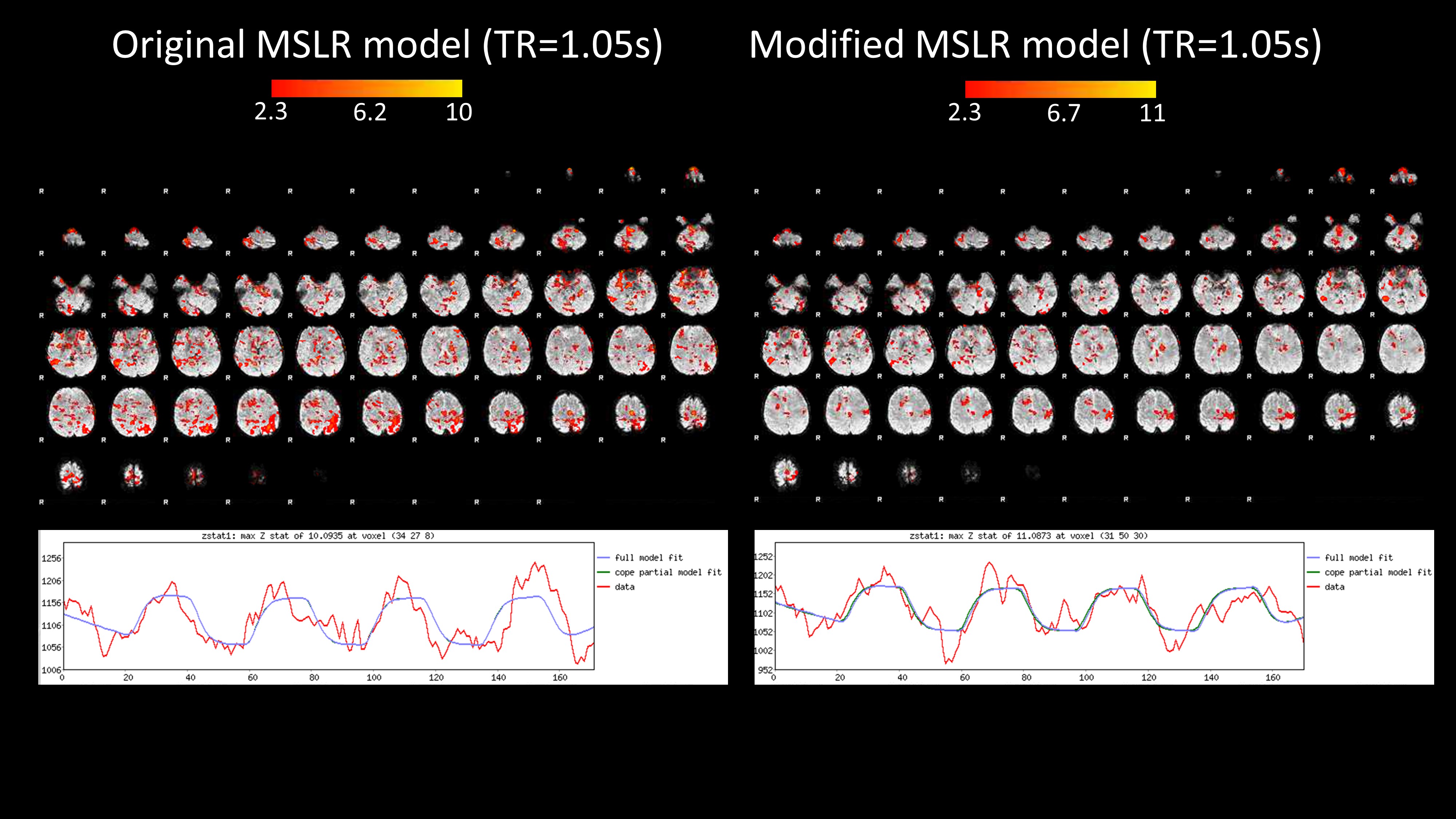
The “Extreme MRI” multi-scale low-rank (MSLR) model3 representation works extremely well for reconstructing images under cases of bulk motion such as for pediatric MRI or lung MRI when incoherent k-space sampling is utilized. In contrast, typical fMRI imaging paradigms require the subject to hold their position throughout the scan. Thus, we can precondition the MSLR model as follows:$$\textbf{X}=\textbf{X}_{static}+\sum_{j=1}^{J}\sum_{b=1}^{B_j}\textbf{M}_{jb}\textbf{L}_{jb}\textbf{R}^H_{(jb)}$$
where J is the number of scales for MSLR, and $$$B_j$$$ is the number of blocks for each scale, and $$$\textbf{X}_{static}$$$ is a static volumetric image reconstructed using JSENSE5 with all the k-space samples from the whole scan. $$$\textbf{X}_{static}$$$ is effectively an incoherent but uniformly-oversampled volume. Since $$$\textbf{X}_{static}$$$ is treated as a constant under this modified reconstruction model, we can utilize the same stochastic optimization and update equations as extreme MRI by focusing only on the spatiotemporal difference image ($$$\textbf{X}-\textbf{X}_{static} $$$).
fMRI Acquisitions
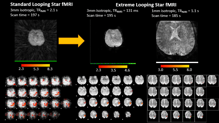
Two fMRI acquisitions were performed on a 3T MR750 scanner (GE Healthcare, Chicago, IL) at two sites.Motor task: A motor fMRI paradigm involved the participant tapping the fingers of their right hand with 20s duration and 20s break between blocks and was acquired with an 8-ch brain coil. Acquisition parameters were: 24 spokes per loop, 2 echoes, BW=±31.25kHz, FOV=(19.2cm)^3, resolution=(3mm)^3, FA=2, TE=[0ms, 26.88ms] for high temporal resolution, and 16 spokes per loop, 2 echoes, BW=±50kHz, FOV=(16.0cm)^3, resolution=(1mm)^3 resolution, FA=2, TE=[0ms, 12ms] for high spatial resolution. Original Looping Star mirrored the acquisition parameters of the high temporal resolution extreme Looping Star but with TR=2.1s and a sub-Nyquist sampling factor per volume of 0.25. Extreme reconstruction parameters were set to produce TR=0.131s and 1488 volumes for high temporal resolution with an effective sub-Nyquist sampling factor per volume of 0.01, and TR = 1.1s and 172 volumes for high spatial resolution with an effective sub-Nyquist sampling factor per volume of 0.006.
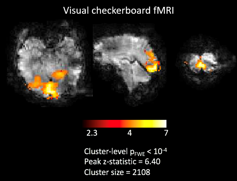
Visual task: A visual fMRI paradigm involved an 8Hz visual checkerboard with 10s duration and 20s break between blocks and was acquired with a 32-channel Nova Medical brain coil. Acquisition parameters were: 24 spokes per loop, 2 echoes, BW=±31.25kHz, FOV=(19.2cm)^3, resolution=(3mm)^3, FA=2, TE=[0ms, 26.88ms]. Extreme reconstruction parameters were set to produce TR = 0.155s and 1280 volumes for an effective sub-Nyquist sampling factor per volume of 0.025.
fMRI Pre-processing & Analysis
Steady-state signal stabilization was corrected for by removing the first volumes from the dataset. This dataset was pre-processed with FSL in native space, including 3mm FWHM smoothing and FILM pre-whitening. Motion correction was not used. A high pass filter of 40s was used. The first level general linear model design matrix included gamma convolution with temporal derivatives and temporal filtering. The whole brain was thresholded at z > 2.3 and corrected cluster significance threshold p=0.05.Results
Figure 3 shows the results for the fMRI finger-tapping motor task experiment with standard Looping Star with the new approach and Extreme Looping Star. Motor activity for the right hand was clearly shown in both approaches. However, Extreme Looping Star can be performed under finer temporal resolution or finer spatial resolution that preserves anatomic detail and motor activation. The 1mm Extreme Looping Star data can also be combined and reconstructed into a single high-resolution image for perfectly concurrent anatomical referencing. Using the modified MSLR allows for improved SNR of task activation maps (Fig. 4).Figure 5 shows the results for the fMRI visual task experiment. Like the motor task with high temporal resolution, visual activity was clearly shown.
Conclusion
By adapting the Looping Star pulse sequence for Extreme MRI reconstruction, we developed a quiet fMRI method that provides high spatiotemporal dynamic BOLD information. Moving forward, we now intend to evaluate this Extreme Looping Star fMRI for studies of hyperacoustic patients who are sensitive to the loud acoustic MRI scanner noise.Acknowledgements
This presentation represents independent research supported by the National Institute for Health Research (NIHR) Biomedical Research Centre at South London and Maudsley NHS Foundation Trust and King’s College London. Nikou is in receipt of a PhD studentship funded by the NIHR Maudsley Biomedical Research Centre. The views expressed are those of the author(s) and not necessarily those of the NHS, the NIHR or the Department of Health and Social Care.References
1. Wiesinger F, Menini A, Solana AB. Looping Star. Magn Reson Med. 2019;81(1):57-68. doi:https://doi.org/10.1002/mrm.27440
2. Dionisio‐Parra B, Wiesinger F, Sämann PG, Czisch M, Solana AB. Looping Star fMRI in Cognitive Tasks and Resting State. J Magn Reson Imaging. 2020;52(3):739-751. doi:https://doi.org/10.1002/jmri.27073
3. Ong F, Zhu X, Cheng JY, et al. Extreme MRI: Large-scale volumetric dynamic imaging from continuous non-gated acquisitions. Magn Reson Med. 2020;84(4):1763-1780. doi:https://doi.org/10.1002/mrm.28235
4. Chan RW, Ramsay EA, Cunningham CH, Plewes DB. Temporal stability of adaptive 3D radial MRI using multidimensional golden means. Magn Reson Med. 2009;61(2):354-363. doi:10.1002/mrm.21837
5. Ying L, Sheng J. Joint image reconstruction and sensitivity estimation in SENSE (JSENSE). Magn Reson Med. 2007;57(6):1196-1202. doi:https://doi.org/10.1002/mrm.21245
Figures