0439
Denoising of Hyperpolarized 13C MR Images of the Human Brain Using Patch-based Higher-order Singular Value Decomposition1Department of Radiology and Biomedical Imaging, University of California, San Francisco, CA, United States, 2Department of Neurological Surgery, University of California, San Francisco, CA, United States, 3Center for Cancer Research, National Cancer Institute, National Institutes of Health, Bethesda, MD, United States
Synopsis
Quantifying metabolism in hyperpolarized (HP) 13C MRI can be challenging because of low signal-to-noise ratio for downstream metabolites. To overcome this limitation, we investigated a new patch-based singular value decomposition method to denoise dynamic imaging data and tested it in numerical simulations and on 6 HP [1-13C]pyruvate EPI human brain datasets. The sensitivity enhancement provided by denoising significantly improved quantification of metabolite dynamics. With denoising, [1-13C]pyruvate and its metabolites [1-13C]lactate and [13C]bicarbonate had ≥5-fold sensitivity gain, improving the number of quantifiable voxels for mapping pyruvate-to-bicarbonate conversion rates (kPB) by 2-fold, and providing whole-brain coverage for mapping pyruvate-to-lactate conversion rates (kPL).
Introduction
Hyperpolarized (HP) 13C MRI is an emerging MR molecular imaging technique to detect disease and early treatment response by measuring metabolic conversions of HP substrates to downstream metabolites, utilizing a MR signal enhancement of >104 – 105 via hyperpolarization.1,2 Despite the enormous gain in MR signal, the spatial resolution of this dynamic imaging method is limited by the signal-to-noise ratio (SNR) for metabolic products such as [13C]bicarbonate in HP-13C MR studies with [1-13C]pyruvate.Image denoising approaches have recently been shown to improve the sensitivity of HP-13C studies. The higher-order singular value decomposition (HOSVD) technique3 has been employed for denoising multidimensional data and has shown particularly powerful results for denoising dynamic spectroscopic imaging.4-6 The HOSVD can also be performed at the patch level by taking account of similarity between patches in nearby regions in the image and has shown superior performance over the global HOSVD method when applied to T1-, T2- and diffusion-weighted 1H MR images.7 In this study, we investigated the feasibility of using a patch-based HOSVD method to denoise dynamic 13C metabolic images and applied this approach to dynamic HP-13C EPI data from healthy volunteer brain studies acquired with single- and multi-channel arrays.
Methods
The patch-based HOSVD denoising algorithm involved pre-filtering noisy data by applying the HOSVD transform globally to the whole dynamic data, followed by grouping similar patches in the pre-filtered images into stacks and performing HOSVD on each stack. By thresholding the SVD transform coefficients, the denoised stacks were estimated, and the multiple estimates at each voxel were aggregated to obtain final denoised images (Figure 1). The MATLAB code for this global-local HOSVD (GL-HOSVD) denoising method was modified based on existing scripts.7 For simulation, metabolic data were generated using a brain model accessible from the BrainWeb Simulated Brain Database8 and the hyperpolarized-mri-toolbox9 for kinetic modeling of pyruvate and lactate dynamics in gray and white matter. The image quality, accuracy, and reliability of quantitative analyses with denoising were first evaluated using the simulated [1-13C]pyruvate and [1-13C]lactate dynamics at different noise levels. The results were compared to those from a global HOSVD method (tensor rank truncation-image enhancement (TRI))4. The GL-HOSVD method was further applied to 6 sets of HP [1-13C]pyruvate multi-slice EPI data of the human brain acquired with a multichannel array (n = 5) or a single channel volume coil (n = 1). The SNR improvement was quantified, and voxel-wise kinetic modeling was performed on both non-denoised and GL-HOSVD denoised data to compare the number of voxels quantifiable based on SNRauc (SNR of temporally summed metabolic signals) <3 for pyruvate and lactate/bicarbonate and fitting error (<30% relative variability) criteria.Results
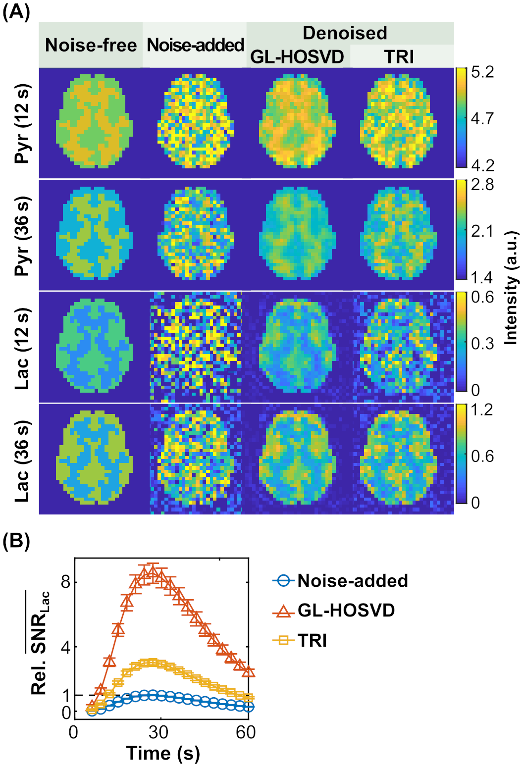
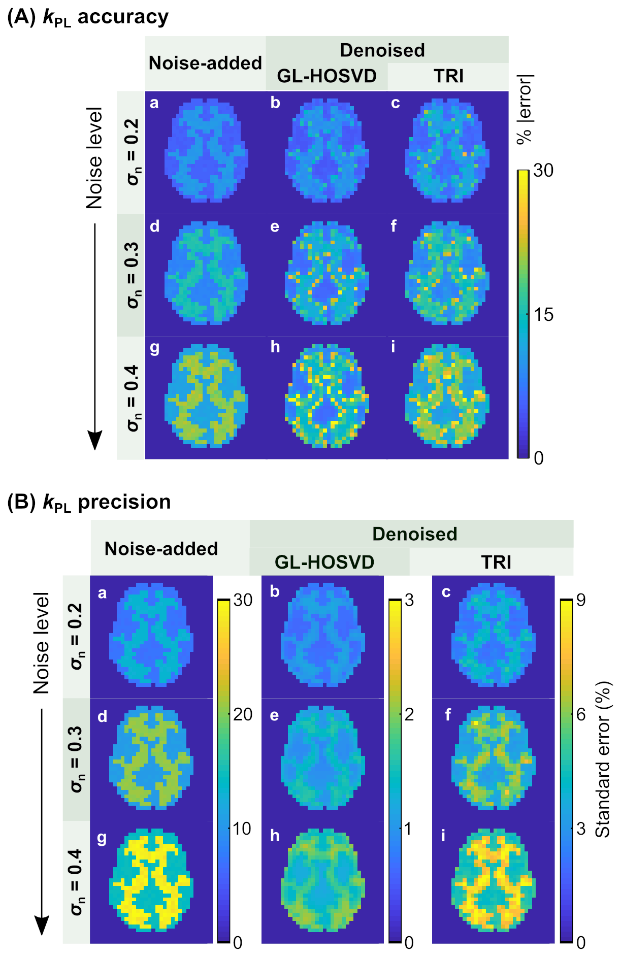
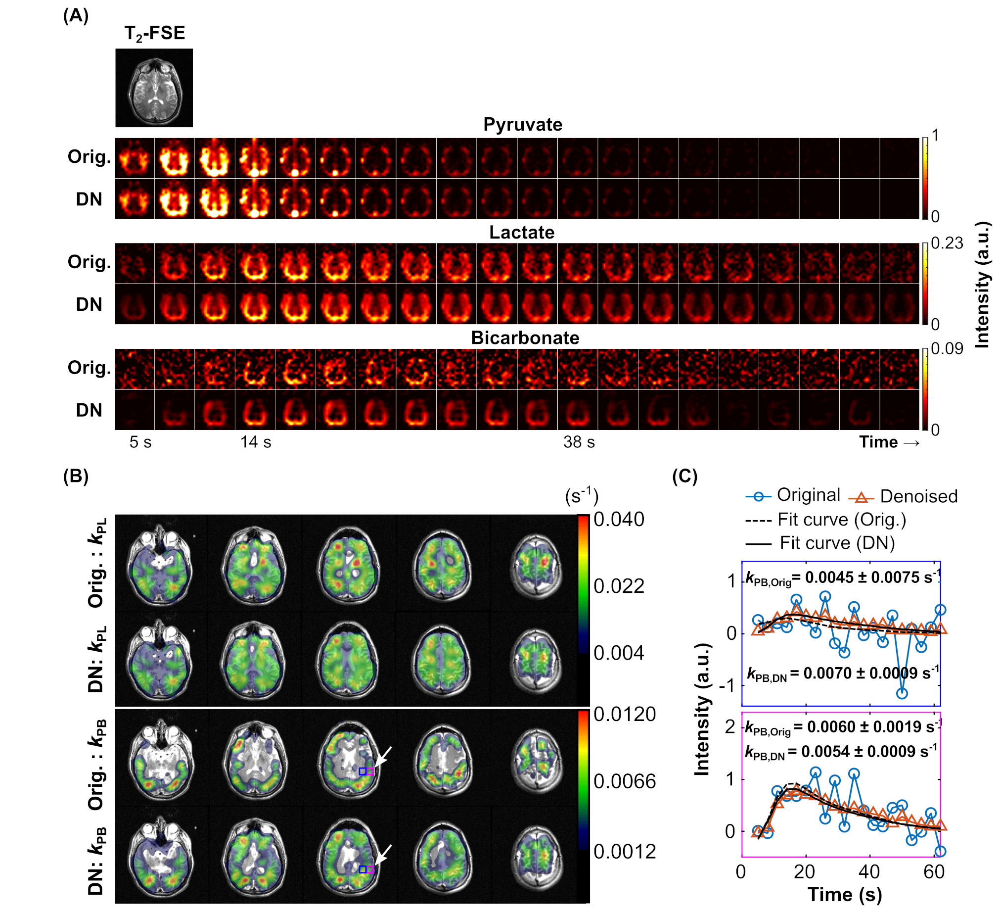
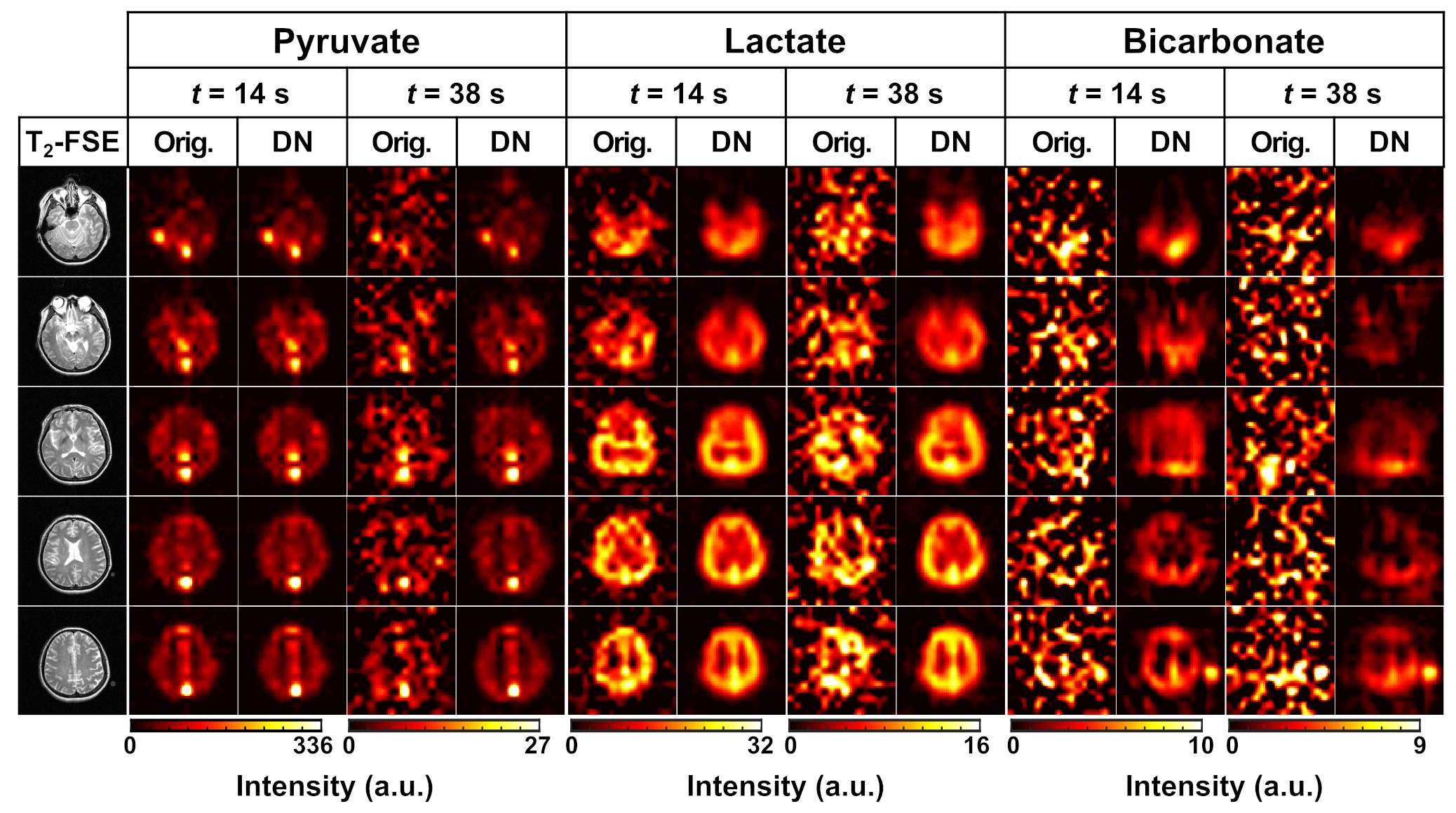
Simulation results demonstrated over 8-fold increases in the apparent SNR of [1-13C]pyruvate and [1-13C]lactate with the GL-HOSVD denoising, which was almost 3 times greater than that achieved with TRI (Figure 2). From 500 replicate simulations of the voxel-wise quantification of kPL (pyruvate-to-lactate conversion rate), smaller kPL bias and improved precision were obtained from GL-HOSVD, though elevated error at the gray/white matter boundary was observed due to the binary mask used for this numerical simulation (Figure 3A). Compared to the non-denoised data, the mean standard error in the kPL fit decreased by 9-fold for GL-HOSVD (just 3-fold for TRI), indicating substantial improvement in kPL precision (Figure 3B). Figure 4A displays the denoising results using GL-HOSVD for the HP [1-13C]pyruvate EPI data acquired from a healthy volunteer using the multichannel array. With denoising, a 5+ fold sensitivity gain was achieved for all metabolites, which was most apparent for [13C]bicarbonate and late-phase [1-13C]lactate. GL-HOSVD denoising provided nearly whole-brain coverage for voxelwise kPL mapping satisfying the SNR and error criteria. Also, the number of voxels satisfying SNR and error criteria for mapping of kPB increased by 2-fold (Figure 4B). The outstanding benefit of GL-HOSVD at even higher noise levels was also illustrated in the denoising of the volunteer dataset acquired using the volume coil (Figure 5). The denoised images demonstrated significantly improved sensitivity for all metabolites. The computation time for denoising one EPI dataset (3 metabolites x 8 slices, matrix size of 16x16) using a patch size of 5x5 was on average 7.75 seconds.Discussion and Conclusion
This study demonstrated that the patch-based denoising method of GL-HOSVD can greatly improve image quality and quantification in dynamic HP-13C MR studies of the human brain. With regard to improving SNR of image data and the accuracy of quantified kinetic rates, the GL-HOSVD method was superior to the global denoising approach (TRI). By denoising HP-13C EPI in vivo datasets using GL-HOSVD, low SNR metabolites were better visualized, and more voxels met kPL and kPB fitting criteria. This great improvement in the image quality and quantification of metabolite dynamics by the patch-based denoising supports the possibility of acquiring metabolic data at higher spatial resolution in future clinical studies, thereby minimizing partial volume effects and facilitating improved extraction of metabolic information for improved clinical management. It could be beneficial to other image-based 13C studies using HP [2-13C]pyruvate and [13C]urea, which may exhibit even lower SNR compared to [1-13C]pyruvate due to shorter T1 or lower polarization.Acknowledgements
This research was supported by NIH (P41EB013598, P01CA118816, T32CA151022, P50CA097257) and the UCSF NICO project.References
1. Wang ZJ, Ohliger MA, Larson PEZ, et al. Hyperpolarized 13C MRI: State of the Art and Future Directions. Radiology 2019;291:273–284.
2. Le Page LM, Guglielmetti C, Taglang C, Chaumeil MM. Imaging Brain Metabolism Using Hyperpolarized 13C Magnetic Resonance Spectroscopy. Trends in Neurosciences 2020;43:343–354.
3. De Lathauwer L, De Moor B, Vandewalle J. A Multilinear Singular Value Decomposition. SIAM J. Matrix Anal. & Appl. 2000;21:1253–1278.
4. Chen H, Autry AW, Brender JR, et al. Tensor image enhancement and optimal multichannel receiver combination analyses for human hyperpolarized 13C MRSI. Magn Reson Med 2020;84:3351–3365.
5. Brender JR, Kishimoto S, Merkle H, et al. Dynamic Imaging of Glucose and Lactate Metabolism by 13C-MRS without Hyperpolarization. Sci Rep 2019;9:3410.
6. Nguyen HM, Peng X, Do MN, Liang Z-P. Denoising MR Spectroscopic Imaging Data With Low-Rank Approximations. IEEE Trans. Biomed. Eng. 2013:60:78-89.
7. Zhang X, Peng J, Xu M, et al. Denoise diffusion-weighted images using higher-order singular value decomposition. NeuroImage 2017;156:128–145.
8. Collins DL, Zijdenbos AP, Kollokian V, et al. Design and construction of a realistic digital brain phantom. IEEE Trans. Med. Imaging 1998;17:463–468.
9. Larson PEZ, Chen H-Y, Gordon JW, et al. Investigation of analysis methods for hyperpolarized 13C-pyruvate metabolic MRI in prostate cancer patients: Hyperpolarized Pyruvate Prostate Cancer Analysis Methods. NMR in Biomedicine 2018;31:e3997
Figures