0375
Application of APTw imaging in prediction of lymph node metastasis in rectal cancer1The First Affiliated Hospital of Dalian Medical University, Dalian, China, Dalian, China, 2Philips Healthcare,Beijing,China, Beijing, China
Synopsis
The purpose of this study was to evaluate the value of amide proton transfer weighted (APTw) imaging in the identification of lymph node metastasis in rectal cancer. Results showed that, for patients with rectal cancer, the APTw value in group with lymph node metastasis was significantly lower than those in group without lymph node metastasis. APTw imaging can be used as a promising non-invasive method to predict the lymph node metastasis in rectal cancer.
Synopsis
The purpose of this study was to evaluate the value of amide proton transfer weighted (APTw) imaging in the identification of lymph node metastasis in rectal cancer. Results showed that, for patients with rectal cancer, the APTw value in group with lymph node metastasis was significantly lower than those in group without lymph node metastasis. APTw imaging can be used as a promising non-invasive method to predict the lymph node metastasis in rectal cancer.(AUC:0.770;sensitivity :70% specificity :80% )Introduction
Rectal cancer is one of the most common malignant tumors in the digestive tract. Whether there is lymph node metastasis in patients with rectal cancer has an important impact on the decision-making of treatment plan and the prognosis of patients. Therefore, the accurate determination of lymph node metastasis is an important step in the treatment of rectal cancer. In this study, the amide-proton-transfer weighted (APTw) imaging on the primary foci of rectal cancer was evaluated for prediction of lymph node metastasis.[1-3]Materials and Methods
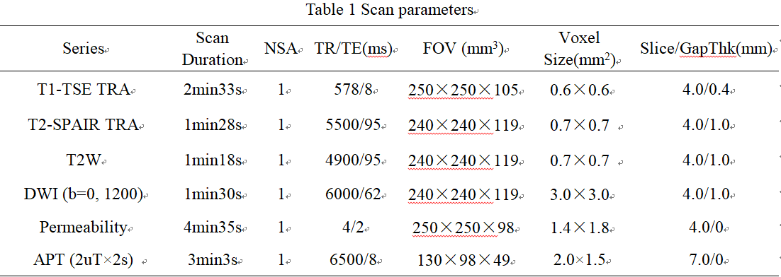
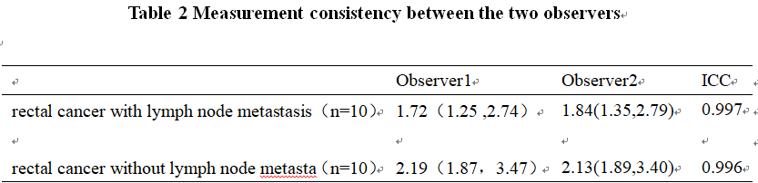
This study has been approved by the local IRB. A total of 20 cases with informed consent were scanned for a retrospective study from March 2019 to December 2020.10 patients with lymph node metastasis (group A,7males,3 females) and 10 patients without lymph node metastasis (group B 7males,3 females )were enrolled(age range from 28 to 77 years 62.1±11.3). All patients underwent 3D APTw imaging and routine MR examinations on a 3.0T MR scanner (Ingenia CX, Philips Healthcare). APTw imaging was performed using a saturation pulse with duration of 2s and strength of 2μT. Three regions of interest (ROIs) were placed on the image of rectal cancer. All ROIs were placed within the interested tissue by avoiding blood vessels. A ROI was manually placed by two radiologists (8years and 2years of clinical experience) on the same location of the lesion according to the APTw image, and its APT value was measured (Fig.1). The ICC (Inter-class correlation coefficient) was used to test the measurement consistency between the two observers. Mann-Whitney U test was used to compare the difference of APT value between group A and group B. If there is a significant difference, the diagnostic efficiency would be test by the ROC curve analysis.Results
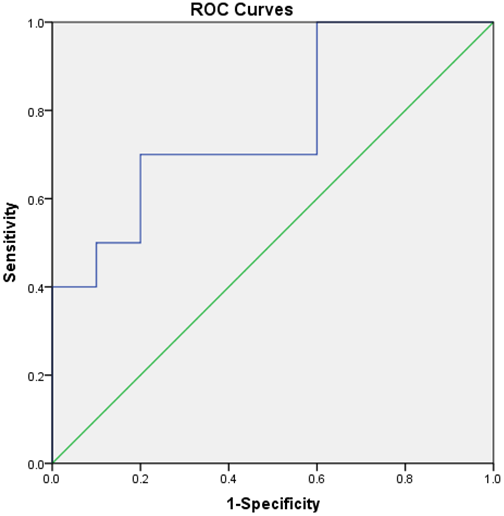
APT values measured by the two doctors were well consistent (ICC > 0.75). The APT values measured for the primary foci of the rectal cancer in group A (1.75[1.67, 2.75]%) were significantly lower than those in group B (2.20[1.87, 3.50]%) (Fig. 2). The area under the ROC curve of APT value to distinguish the lesions of group A and B was 0.770, and the diagnostic sensitivity and specificity were 70% and 80% . (Fig. 3).Discussion and Conclusion
The APT values of rectal cancer with lymph node metastasis were significantly lower than those of rectal cancer without lymph node metastasis. Therefore, APTw imaging can be sensitive in prediction of the lymphatic metastasis in rectal cancer.Acknowledgements
No acknowledgement found.
References
[1] Zhou Jinyuan, HeoHye-Young, Knutsson Linda, et al. APT-weighted MRI: Techniques, current neuro applications, andchallenging issues.Magn Reson Imaging, 2019, 50: 347-364.
[2] Takayama Y, Nishie A, Sugimoto M, et al. Amide proton transfer (APT) magnetic resonance imaging of prostate cancer: comparison with Gleason scores. MAGMA 2016; 29: 671-679.87.
[3] Zhou J, Payen JF, Wilson DA, et al. Using the amide proton signals of intracellular proteins to detect PH effects in MRI[J]. Nat Med, 2003,9:1085-1090.
Figures
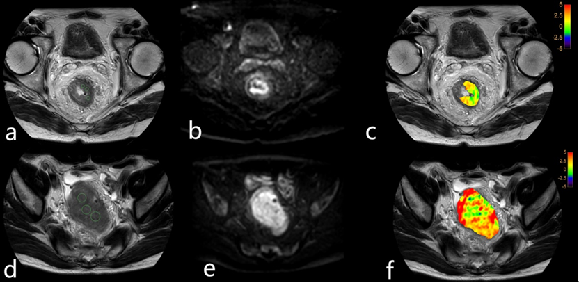
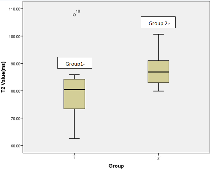
Figure2 APTw values for rectal cancer with (group 1) and without (group 2) lymph node metastasis . A significant difference was observed (p = 0.041).