0373
Noise reduction in diffusion weighted MRI of the pancreas using an L1-regularized iterative SENSE reconstruction1Department of Diagnostic and Interventional Radiology, Technical University of Munich, Munich, Germany, 2Department of Radiology, South Egypt Cancer Institute, Assiut, Egypt, 3Philips Healthcare, Best, Netherlands, 4Philips GmbH, Hamburg, Germany
Synopsis
DWI plays a growing role in the imaging of the pancreas. Current abdominal DWI scans have a large field of view with limited resolution. Approaches for a higher resolution single-shot DW-EPI with SENSE often suffer from g-factor-induced noise band-like artifacts from which the pancreas is significantly affected due to its central location. We use an L1-regularized iterative SENSE reconstruction to obtain high resolution single shot DW-EPI with reduced noise in patients with pancreatic cancer. The employed method reduces the noise band-like artifacts over the pancreas and eliminates the noise bias from the ADC measurements without lengthening scan times.
Purpose:
Diffusion-weighted imaging (DWI) has been playing a growing role in the evaluation of the pancreas1. Several studies proposed a promising role for DWI for lesion detection, characterization and proposed the apparent diffusion coefficient (ADC) as a quantitative biomarker2, 3. For the pancreas, a relatively small organ, abdominal DWI using single-shot echo-planar imaging (EPI) offers limited spatial resolution4. Parallel imaging with sensitivity encoding (SENSE) allows smaller voxel size without increased image distortions. However, SENSE with a high acceleration factor usually suffers from a central noise band-like artifact due to the high geometry factor from the employed RF coils in the abdominal region5. The central location of the pancreas makes it more susceptible to such artifacts. The combination of parallel imaging and compressed sensing (CS), has been successful to reduce the acquisition time while avoiding the increased image artifacts in non-EPI acquisitions6. A wavelet-based denoising filter as in the CS framework has been recently combined with a SENSE-informed iterative reconstruction to reduce noise in single-shot EPI DW images7,8,9. The derived L1-regularized iterative SENSE reconstruction has been effective in reducing the noise artifacts from parallel imaging at high b-value DWI without an increase in scan time. The purpose of the present work was to obtain a high-resolution DWI of the pancreas based on the L1-regularized iterative SENSE reconstruction and assess its performance in comparison to DWI based only on SENSE reconstruction in terms of reducing noise bias in DW images and improving precision in ADC measurements.Methods:
Data were collected from 14 patients with pancreatic adenocarcinoma. Written informed consent was obtained from each patient and the protocol was approved by the institutional ethics committee. The study was performed on clinical 3.0T MR scanner (Philips, Ingenia 3.0T using respiratory-triggered single-shot DWI-EPI scans with the following parameters: TR/TE= 1800/55ms, field-of-view= 420(RL)×350(AP)mm, voxel size= 2.5×2.5×3mm, number of slices=25, EPI factor=29, b-values(averages)= 0(1),50(2),600(7) sec/mm2, Acquisition time = 4:25. The scan was repeated twice with an acceleration factor of R=4 in the A/P axis (regular undersampling in PE direction). One scan was reconstructed online using the standard SENSE reconstruction of the vendor and one scan was reconstructed online using an iterative SENSE reconstruction employing L1 regularization after a wavelet sparsifying transform similar to a CS reconstruction7,8. Retrospective reconstruction of the b600 images with a fewer number of averages (6,5&4) was done using both reconstruction methods. ADC maps were generated using b50 and b600 at the different number of averages for both reconstructions. ROIs were drawn over the pancreatic adenocarcinoma and the normal pancreas by a single abdominal radiologist (4 years’ experience). Mean and standard deviation values were determined over each ROI. Mean ADC measurements and coefficient of variation (CV) of ADC of the tumor and the normal-appearing pancreas were compared for both the SENSE reconstruction and the L1-regularized SENSE reconstruction. A paired t-test was used to compare mean ADC and CVADC between the two methods.Results:
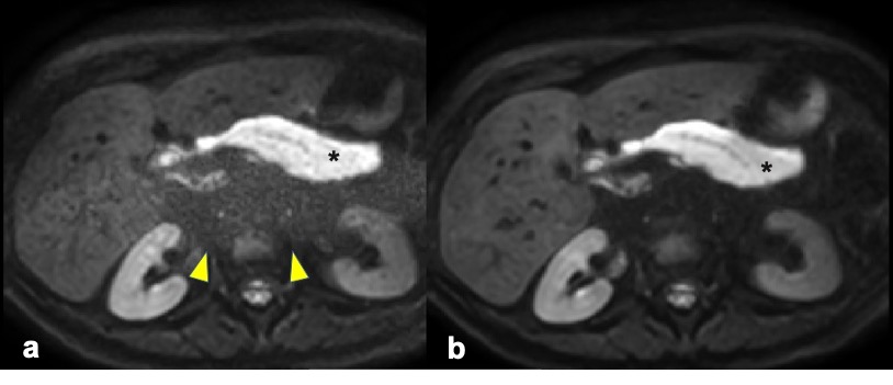
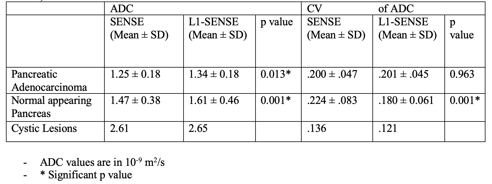
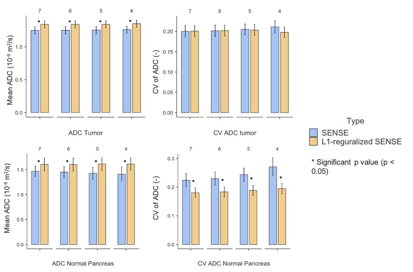
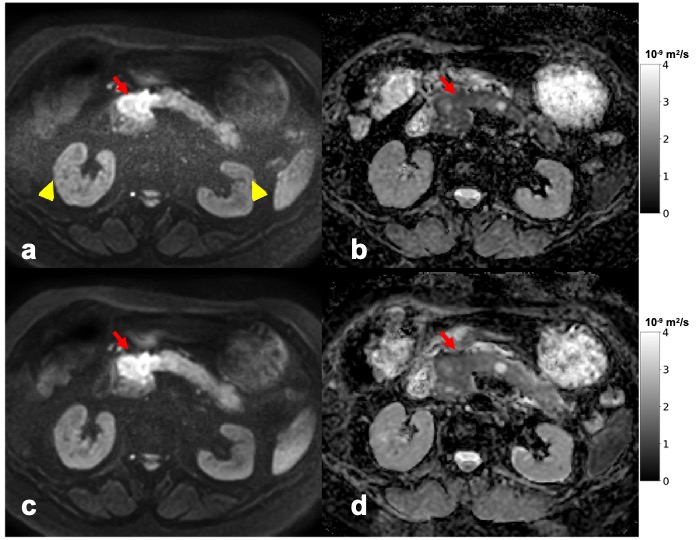
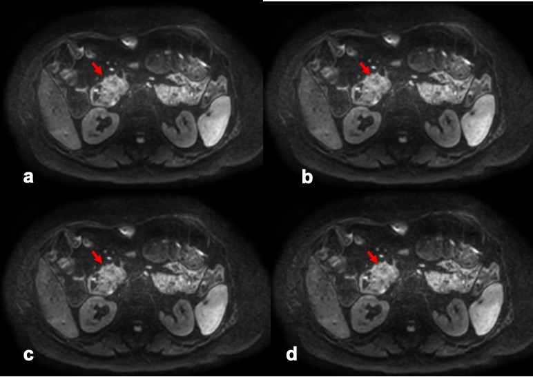
Our study included 14 patients (5 men, 9 women, mean age = 68.9 (range: 56–84). 12 patients had a solid tumor, while in two patients the lesion had an extensive cystic component. Measurements from those patients were listed separately (labeled cystic lesions). Mean ADC values of the tumor and the normal pancreas were significantly higher in L1-regularized SENSE reconstruction compared to SENSE reconstruction at all number of averages (Table1). Figure 1 shows the reduction of the noise band-like artifact in L1-regularized SENSE reconstruction. The mean CV of the ADC of the normal pancreas was significantly lower in L1-regularized SENSE reconstruction compared to SENSE reconstruction at all number of averages. No significant difference in the CV of the ADC within the pancreatic adenocarcinoma was seen (Figure2). Figure 3 shows representative DW images and ADC maps from one patient and Figure 4 shows the DW images quality after reducing the number of averages in L1-regularized SENSE reconstruction.Discussion:
The employed L1-regularized SENSE reconstruction is significantly efficient in getting rid of the noise band-like artifact commonly seen in SENSE at high acceleration factors. The reduction of Rician noise-induced bias can explain the significantly higher ADC measurements of the tumor and normal pancreas. The achieved denoising could also explain the significantly lower CV ADC of the normal pancreas with the L1-regularized SENSE reconstruction compared to the SENSE reconstruction. No difference was observed in the CV ADC of the tumor between the two methods. Such a behavior can be attributed to the heterogeneous nature of the tumor tissues which acts as a confounding factor besides noise in estimating CV based on an ROI and to the higher signal intensity of the tumors in DWI due to diffusion restriction. ADC and CV ADC were stable in the L1-regularized SENSE reconstruction across the different number of reconstructed averages, potentially allowing further scan time reduction.Conclusion:
An L1-regularized iterative SENSE reconstruction effectively reduces central band-like noise and improve the robustness of ADC values of single-shot DW-EPI of the pancreas compared with a standard SENSE reconstruction while using an acceleration factor of 4 with phase encoding in anterior/posterior direction.Acknowledgements
The present work was supported by Philips Healthcare.References
1. Wang, Y., et al., Diffusion-weighted MR Imaging of Solid and Cystic Lesions of the Pancreas. RadioGraphics, 2011. 31(3): p. E47-E64.
2. Hecht, E.M., et al., Can diffusion-weighted imaging serve as a biomarker of fibrosis in pancreatic adenocarcinoma? J Magn Reson Imaging, 2017. 46(2): p. 393-402.
3. Lee, J.H., et al., Usefulness of non-contrast MR imaging in distinguishing pancreatic ductal adenocarcinoma from focal pancreatitis. Clin Imaging, 2019. 55: p. 132-139.
4. Kim, H., et al., Reduced Field-of-View Diffusion-Weighted Magnetic Resonance Imaging of the Pancreas: Comparison with Conventional Single-Shot Echo-Planar Imaging. Korean J Radiol, 2015. 16(6): p. 1216-25.
5. Noel, P., et al., Parallel imaging artifacts in body magnetic resonance imaging. Can Assoc Radiol J, 2009. 60(2): p. 91-8.
6. Feng, L., et al., Compressed sensing for body MRI. J Magn Reson Imaging, 2017. 45(4): p. 966-987.
7. Sprenger P., Morita K., Nakaura T., Yoneyama M., Zhang S., Bode M., Kuhl C.K., Kraemer N.A. 2020, ‘Body Diffusion MR Imaging with Compressed SENSE Based on Single-Shot EPI at 3T and 1.5T: Technical Feasibility and Initial Clinical Experience ’, 28th ISMRM Meeting 2020, 8- 14 August.
8. Yoneyama M., Morita K., Peeters J., Nakaura T., Van Cauteren M. 2019, ‘Noise Reduction in Prostate Single-Shot DW-EPI utilizing Compressed SENSE Framework ’, 27th ISMRM Meeting 2019 Montreal, 11- 16 May.
9. Morita K., Yoneyama M. , Nakaura T., Oda S. , Hatemura M., Yamashita Y. Yamashita Y 2019, ‘Pseudo-3D Diffusion-Weighted Imaging of the Brain using Echo Planar Imaging with Compressed SENSE (EPICS) ’, 27th ISMRM Meeting 2019 Montreal, 11- 16 May.
Figures