0369
Zoomed Diffusion-Weighted Echo-Planar Imaging for the Evaluation of Periampullary Carcinomas1Department of MR Imaging, the First Affiliated Hospital of Zhengzhou University, Zhengzhou, China, 2MR Collaboration, Siemens Healthcare Ltd, Beijing, China
Synopsis
We evaluated the efficacy of a zoomed diffusion-weighted echo-planar imaging (z-EPI) sequence to diagnose periampullary carcinoma compared with a conventional single-shot EPI (c-EPI) sequence. Better delineation of lesion conspicuities and margins were observed, as well as enhanced diagnostic confidence with z-EPI. The diagnostic accuracy increased by combining magnetic resonance cholangiopancreatography (MRCP) and z-EPI together. These findings showed remarkable image quality improvements for periampullary carcinomas using z-EPI. The ability to detect and delineate lesions using z-EPI was superior to that of c-EPI. Diagnostic accuracy was also attained, especially for small lesions.
Abstract
Purpose:Diffusion-weighted imaging (DWI) plays a significant role in diagnosing periampullary carcinomas. DWI images obtained with conventional single-shot echo-planar imaging (EPI) are often degraded by poor spatial resolutions, susceptibility artifacts, and image blurring [1-3]. Therefore, it is often difficult to definitively detect and diagnose these tumors because of their small size. Compared with conventional EPI (c-EPI) DWI, two-dimensional spatially-selective radiofrequency excitation pulses combined with reduced field of view (FOV) (“zoomed”) imaging along the phase-encoding direction could lead to superior image quality with reduced spatial distortions and artifacts, and improved identification of small anatomic structures [4, 5]. Thus, we aimed to evaluate the image quality, and value of a zoomed diffusion-weighted EPI (z-EPI) sequence for periampullary carcinoma detection compared with c-EPI.
Materials and Methods:
Twenty-six patients with periampullary carcinomas confirmed on histopathology were included in this study. All the subjects underwent both c-EPI and z-EPI (b values = 50, 800 sec/mm2). MRCP was also obtained. All the examinations were carried out on a 3T MR system (MAGNETOM Prisma, Siemens Healthcare, Erlangen, Germany). The z-EPI technique was executed with a reduced FOV in the phase-encoding direction by applying two-dimensional spatial-selective RF pulses using an echo-planar transmit trajectory. The acquisition parameters are listed as follows. For c-EPI: TR/TE = 4500 ms/56 ms; FOV=350 × 292 mm2; matrix=158 × 121; slice thickness=5 mm; voxel size reconstructed to 2.2 × 2.2 × 5.0 mm3; parallel acceleration factor=2; b-values=50/800 s/mm2; acquisition time=1 min 52 sec. For z-EPI: TR/TE=2000 ms/61 ms; FOV=230 × 120 mm2; scan matrix=154 × 50; voxel size=1.5 × 1.5 × 5.0 mm3; slice thickness=5 mm; parallel acceleration factor = 2; acquisition time = 1 min 53 sec. Two radiologists reviewed the two image groups for the qualitative analysis independently. Anatomic structural visualizations, artifacts, overall image quality, lesion conspicuities, lesion margins, and diagnostic confidences were evaluated and compared between the two techniques. The image quality of all lesions, as well as small lesions (diameter 20 mm), were compared. DWI signal intensity assessments of the periampullary lesions were observed. Diagnostic accuracy scores were compared between the MRCP and c-EPI DWI combined analysis as well as the MRCP and z-EPI DWI combined analysis. Statistical analyses were performed using the student’s t test and Wilcoxon signed-rank test, and differences were considered significant for P-values 0.05.
Results:
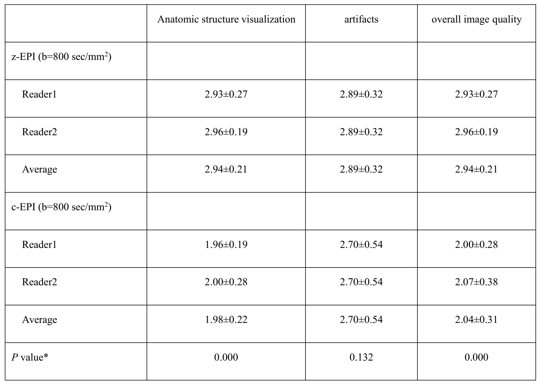
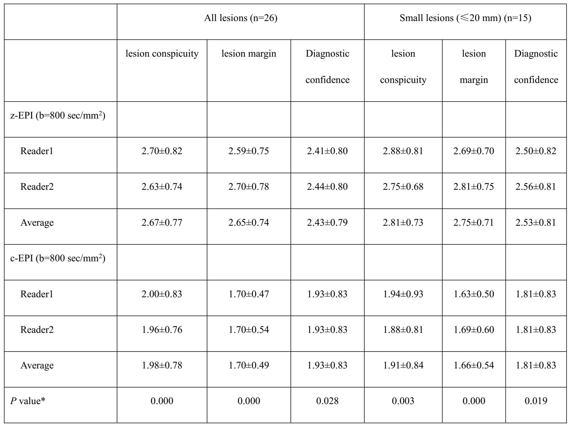
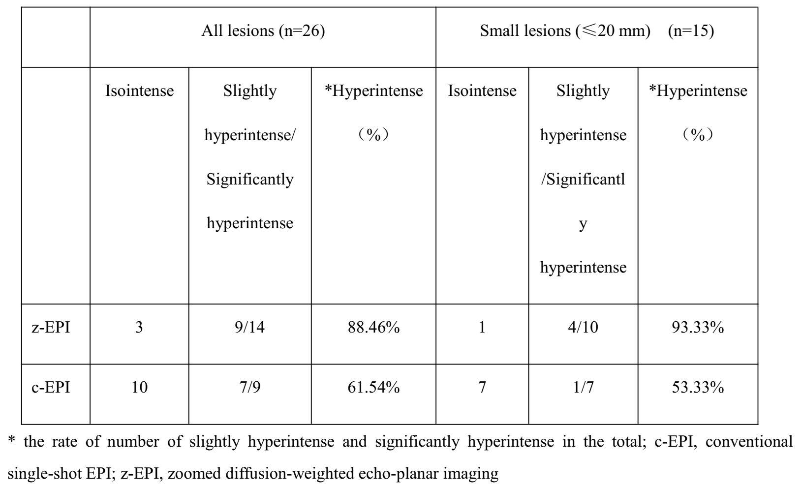
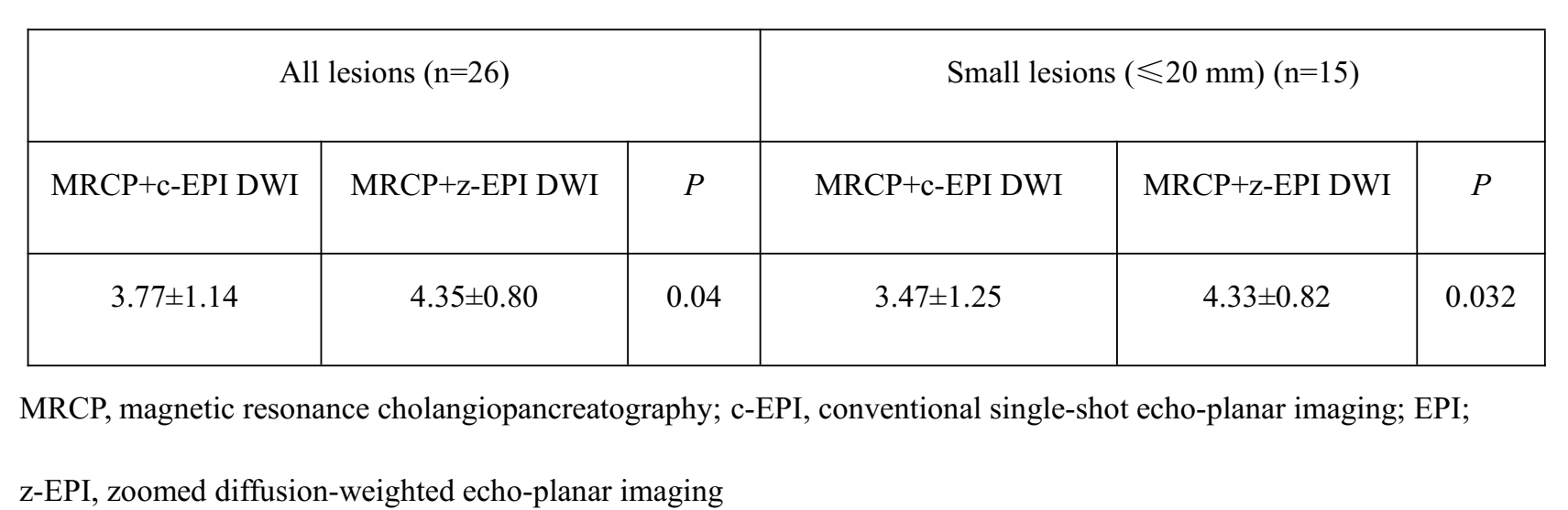
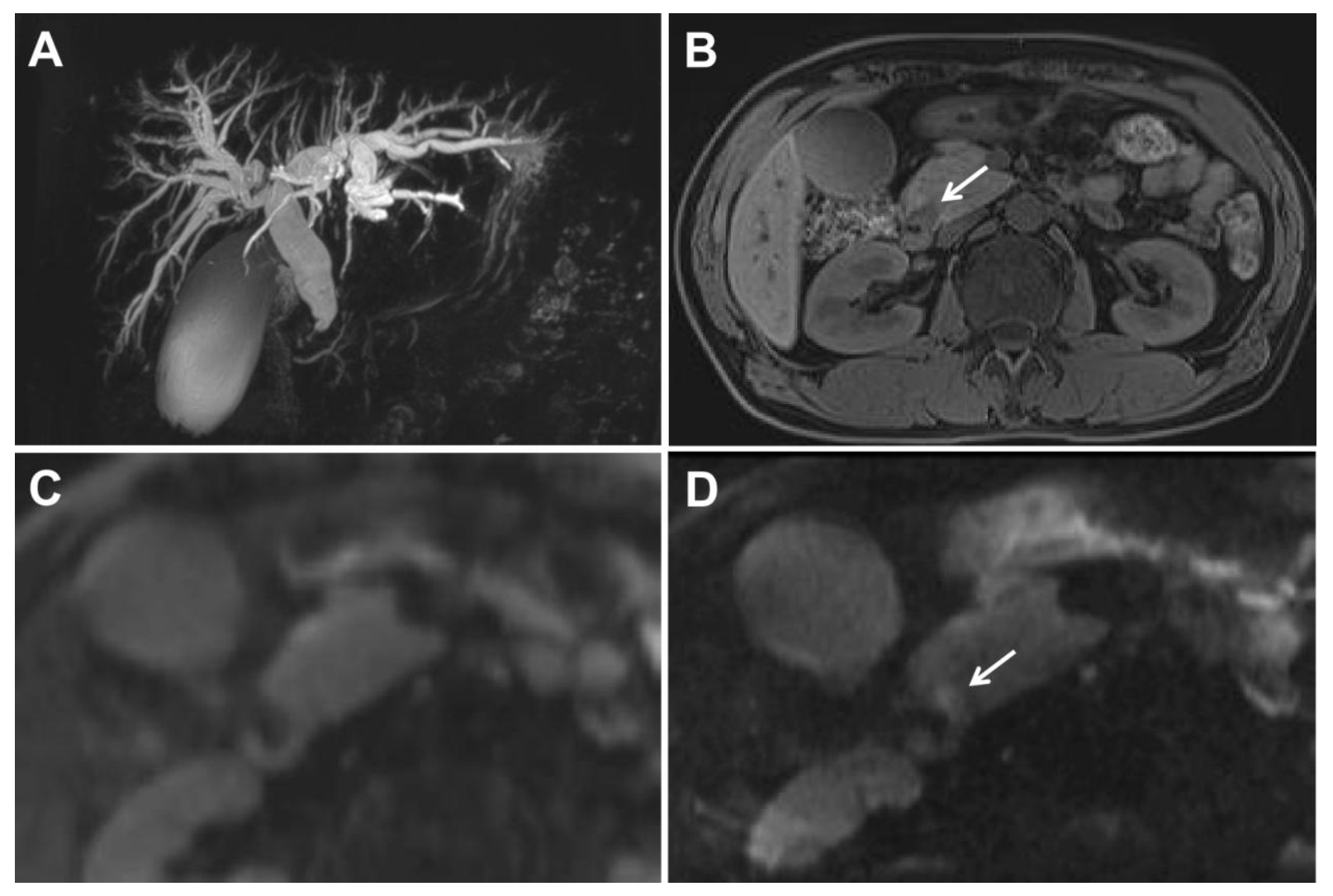
Twenty-six periampullary lesions were detected, and 15 were small lesions. Compared with c-EPI, better delineation of anatomic structural visualizations and overall image qualities were seen in the periampullary regions with z-EPI (all P <0.05). No statistically significant differences were found with artifacts between the two image data sets (P=0.132) (Table 1). Better delineations regarding lesion conspicuities and margins, as well as diagnostic confidences were present with z-EPI compared with c-EPI (all P <0.05) (Table 2). The DWI visual assessments of the periampullary lesions were increased using z-EPI compared with using c-EPI, especially for small lesions with a diameter of 20 mm (Fig. 1) (Table 3). Diagnostic accuracy scores were increased using MRCP/z-EPI DWI combined set compared with using the MRCP/c-EPI DWI combined set (P<0.05) (Table 4).
Discussion:
Abdominal DWI is known to be technically challenging regarding distortion artifacts in the phase-encoding direction. z-EPI has potentially advantages over c-EPI for using two-dimensional spatial-selective radiofrequency pulses to obtain reduced volumes. The results indicated that the periampullary region was better defined with improved anatomic structural visualization, overall image quality, lesion conspicuity, and lesion margin definition with z-EPI than with c-EPI, especially for small lesions. And the diagnostic confidence and accuracy were increased.
Conclusions:
z-EPI led to remarkable image quality improvements and increased lesion visualizations of periampullary carcinoma lesions. z-EPI was also superior to c-EPI in detecting, delineating, and diagnosing lesions, especially small lesions.
Acknowledgements
No acknowledgement found.References
1. Bittencourt LK, Matos C, Coutinho AC Jr. Diffusion-weighted magnetic resonance imaging in the upper abdomen: technical issues and clinical applications. Magn Reson Imaging Clin N Am. 2011;19(1):111-131.
2. Dietrich O, Biffar A, Baur-Melnyk A, Reiser MF. Technical aspects of MR diffusion imaging of the body. Eur J Radiol. 2010;76(3):314-322.
3. Attenberger UI, Morelli J, Budjan J, et al. Fifty Years of Technological Innovation: Potential and Limitations of Current Technologies in Abdominal Magnetic Resonance Imaging and Computed Tomography. Invest Radiol. 2015;50(9):584-593.
4. Riffel P, Michaely HJ, Morelli JN, et al. Zoomed EPI-DWI of the pancreas using two-dimensional spatially-selective radiofrequency excitation pulses. PLoS One. 2014;9(3):e89468.
5. Thierfelder KM, Sommer WH, Dietrich O, et al. Parallel-transmit-accelerated spatially-selective excitation MRI for reduced-FOV diffusion-weighted-imaging of the pancreas. Eur J Radiol. 2014;83(10):1709-1714.
Figures